![]()
![]()
![]()
Use LEFT and RIGHT arrow keys to navigate between flashcards;
Use UP and DOWN arrow keys to flip the card;
H to show hint;
A reads text to speech;
51 Cards in this Set
- Front
- Back
|
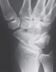
What are the 3 Wrist Views? |
PA: carpalarcuate lines, ulnar variance, radial articular angle Lateral: radiusvolar tilt, scapolunate & capitolunate angle Oblique: radialside carpals & hamate |
|
|
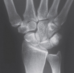
What are the three Hand Views |
PA:phalanges, metacarpals, carpals Lateral:radius, lunate, capitate, 3rdmetacarpal within 10⁰ of each other Oblique:shows hand long bones without superimpositionB |
|
|
Ulnar deviation View |
Shoes scaphpoid and adjacent joint spaces |
|
|
Radial deviation view |
shows ulnar side carpals and intercarpal spaces |
|
|
Capral tunnel view |
Shows arched carpals on palmar |
|
|
Stress views |
Reveal instabilities |
|
|
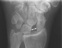
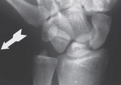
Terry Thomas Sign |
Subluxation of the scaphpoid boe with widening between the scaphoid and lunate |
|
|
What are Gilula arcs |
alignment described on posteroanterior or anteroposterior wrist radiographs and are used to assess normal alignment of the carpus |
|
|
What are the 3 gilula arcs |
First: smooth curve outlining the proximal convexities of the scaphoid lunate and triquetrum Secondarc: tracesthe distal concave surfaces of the same bones Thirdarc: follows the main proximal curvatures of the capitate and hamate |
|
|
What to assess in Oblique wrist view |
Trapezium and articulations
Radial and ulnar styloids |
|
|
Lateral Wrist and Hand assessement |
Long axes of 3rd MC, cap, lun, and radial align within 10 Lunate and radius should be parallel Lunate > 15 toward palm = volar intercalated segmental instability Lunate extended > 10 = Dorsal intercalated segmental instability |
|
|
Normmal Scapholunate angle? |
Average 47 +/- 15
If angle is greater, carpal could be unstable. |
|
|
Normal Capitolunatele angle |
Normally less than 20 Greater than 20 suggests instability |
|
|
Purpose of Tangential inferosuperior view |
View carpal sulcus to identify abnormalities that may be compressing the median nerve or flexor tendons |
|
|
What is advanced imaging used for |
Diagnosing scaphoid fractures |
|
|
Fracture of distal 5th metacarpal |
Boxer's Fracture |
|
|
Common Hand fractures |
Distal Ph - crushing injuries Metacarpal and Phal - Desc by location -Avulsion Fx at tendon/lig attachment Thumb - Freq at base: intra or extra-articular |
|
|
Consquences of Scaphoid fracture |
Blood supply of scaphoid is distal which means that if the Fx is at the middle or proximal part, avascular necrosis may occur |
|
|
What is a colles fx |
extra-articular fx about 1.5 inches proximal to the wrist with dorsal displacement |
|
|
What is a Barton Fx |
Fx dislocation of the distal radius and radiocarpal joint. (pointed tips) |
|
|
What is a Die Punch fracture |
Lunate is driven into the radius causing articular damage at the radiocarpal joint |
|
|
Standard care of a Fx |
Treat it like a Fx if uncertain - Immobilize Radiograph - and if inconclusive - repeat in one week |
|
|
Common Distal Radius Fractures |
Adults - Colles Young Adults - Die Punch Kids - Most common fx - heals well |
|
|
Classifications of Wrist Instability |
–Predynamic:no radiologic abnormalities –Dynamic:seen only on functional radiographs, stress views, or cineradiography –Static:seen on conventional radiographs |
|
|
What is Cineradiography |
a motion study at the wrist and is used to assess dynamic instability Pt moves wrist back and forth between radial and ulnar deviation to reproduce clicking and snappinf |
|
|
What are the 3 types of carpal instabilities |
–Carpalinstability dissociative (CID) –Carpalinstability nondissociative (CIND) –Carpalinstability combined (CIC) |
|
|
What is wrist imaging significant for |
Identify extent of pathology Exclude differential dx Precise dx to direct tx Provide an informed prognosis |
|
|
What can assess the wrist alignment |
Radiographs, Ct, Functional Radiograph |
|
|
What can asses ligamentous lesions |
Arthrography, MRI, MRI -artho, CT -arthro |
|
|
What are the two types of instablity |
Distal radioulnar joint Between Carpal rows/each bones |
|
|
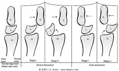
What is a perilunate dislocation |
dislocation of the capitate to the lunate |
|
|
What is the Triangular Fibrocartilage complex? |
Isan important stabilizer of the distal radioulnar joint, and may be torn byitself or in combination with other injuries. |
|
|
How to Dx Carpal tunnel? |
Electromyography and nerve conduction velocity and advanced imaging |
|
|
What is the TFCC |
Major stabilizer of the ulnar carpus and DRUJ Absorbs 20% of the axail load of the wrisst |
|
|
What does the TFCC consist of |
Radiotriquetral lig Articular disk ECU tendon sheath Palmar and radioulnar lig Ulnocarpal lig/meniscus |
|
|
What is Arthritides |
DJD common after age 50 Signs include decreased joint space, sclerosis, and osteophytosis |
|
|
Types of Arthitides |
Heberden's Nodes" Distal Phalanx Bouchards: Proximal phalanx Basal Joint Arthritis at 1st MCP joint RA can be seen at wrist, MCP, and proximal IP Joints |
|
|
Hallmarks for RA |
Uniform joint space narrowing Periarticular rarfaction Articular erosions Synovial cysts Joint deformities (swan and boutonniere) |
|
|
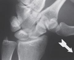
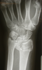
Scapholunate Dislocation - Terry Thomas Sign |
|
|
PA View |
|
|
Oblique View |
|

|
Lateral View |
|
|
Ulnar Deviation |
|
|
Radial Deviation |
|
|
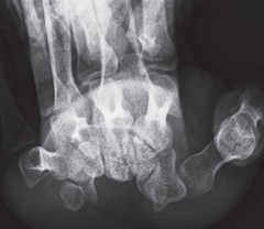
Carpal Tunnel View |
|
|
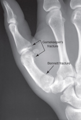
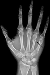
Gamekeepers Bennetts Fracture |
|
|
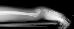
Colles Fracture |
|
|
Die Punch Fracture |
|
|
Boxer's Fracture |
|
|
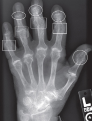
Heberden's Nodes (DIP) Bouchard's Nodes (PIP) |
|
|
Perilunate Dislocation |