![]()
![]()
![]()
Use LEFT and RIGHT arrow keys to navigate between flashcards;
Use UP and DOWN arrow keys to flip the card;
H to show hint;
A reads text to speech;
23 Cards in this Set
- Front
- Back
|
Views of Elbow evaluation |
AP elbow in the anatomical position Lateral with the elbow at 90deg and forearm in neutral Oblique with elbow extended (Pronated forearm - internal oblique // Supinated forearm,: external oblique) |
|
|
What do you see in each elbow projection? |
Distal Humerus, proximal ulna and radius |
|
|
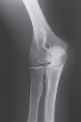
What to Assess in AP Elbow |
ABCS Carrying angle - increase/decrease may be sign of fracture of posttraumatic deformity |
|
|
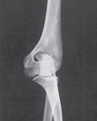
What to Assess in Lateral Elbow |
ABCs Adults-concentric circles Kids normal position of capitulum Fat pads |
|
|
What can you see in Internal and External Oblique Elbow views? |
Internal: Done in pronation allows coronoid process of ulna to be seen External: Done in supination allows radial head to be seen |
|
|
What soft tissue signs can be seen on a radiograph |
Fat Pad Sign: Displaced tissue due to effusion Abnormal supinatior line: blurred supinator muscle in radial head fractures |
|
|
What can the different imaging types show for Elbow Trauma |
Radiograph: Fracture, dislocation, subluxation CT/MRI: Osteochondral fx, complex fx, subtle fx MRI/MRA/US: Soft tissue injuries |
|
|
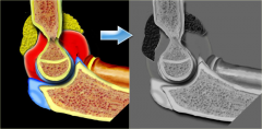
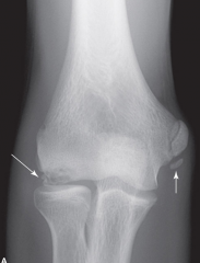
Postive fat pad sign |
Displaced from bony fossa due to joint effusion |
|
|
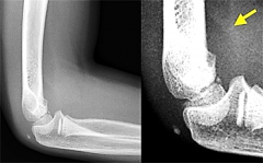
Abnormal supinator line |
Fat plane overlying the muscle will be elevated and widened in all cases of radial head fracture |
|
|
Classifications of elbow fractures? |
Supracondylar (children) Transcondylar Intercondylar (adults) Condylar Articular Epicondylar |
|
|
How are elbow dislocations desccribed? |
By the direction the elbow is displaced relative to the distal humerus Most elbow dislocations involve both the radius and ulna displaced posterior or posterolateral |
|
|
What is epicondylitis |
Overuse characterized by tendinitis folllowed by teninosis |
|
|
Medial vs Lateral Epicondylitis |
ME: Overuse of the wrist flexors LE: Overuse of the Wrist extensors |
|
|
What is Capitellum Osteochondritis Dissecans |
Local joint injury where a cartilage segment and subchondral bone seperate from the articular surface MRI or US to diagnose early stage |
|
|
How is OCD seen in Radiographs |
No Abnormalities in early stages Sclerotic rim Irregular ossification Flattened capitulum Radiolucency due to hyperemia Bony Defect Seperation of osteochondral fragments |
|
|
What is Weissman's Paradox |
Severity of degenerative changes evident on radiographs do not always reflect the severity of clinical symptoms. Excellent f unction may exist with distorted anatomy. |
|
|
Pronated Oblique |
|
|
Supinated Oblique
|
|
|
Hemarthrosis resultsin an upward displacement of the anterior fat pad and a backward displacementthe posterior fat. i |
|
|
Fat pad sail sign suggest occult fracture |
|
|
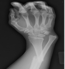
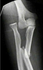
Galeazzi Fx: Fx of radius with dislocation of the distal ulna |
|
|
Monteggia Fx: Fx of the ulna with dislocation of the proximal radius
|
|
|
Capitellum Osteochondritis Dissecans |