![]()
![]()
![]()
Use LEFT and RIGHT arrow keys to navigate between flashcards;
Use UP and DOWN arrow keys to flip the card;
H to show hint;
A reads text to speech;
24 Cards in this Set
- Front
- Back
|
What is the role of bipolar cells and ganglion cells? |
These are the cells which ultimately convert the light ray information into action potentials towards the brain. |
|
|
What kind of action potential response mechanism do the bipolar cells of the visual processing unit use? |
Use graded responses where there is partial activation, as opposed to the all or nothing action potential responses of the rest of the nervous system. |
|
|
Why do bipolar cells in the visual processing unit have 'graded responses'? |
Lack of voltage gated ion channels. |
|
|
What is the difference between a bipolar cell and a ganglion cell? |
Ganglion cells have voltage gated ion channels and thus are the first cells in the visual processing pathway where action potentials can be initiated. |
|
|
In what ways do ganglion cells interaction with bipolar cells and photoreceptor cells? What is the basic biochemical principle behind this? |
1. On pathways & Off pathways 2. Glutamate continually released onto bipolar cells at rest, when ganglion cell becomes hyperpolarised glutamate release decreases. |
|
|
What is the key difference between bipolar cells in the 'on' pathways vs bipolar cells in the 'off' pathway? |
Bipolar cells in the ON pathway spontaneously DEPOLARISE in the absence of light. (deactivated) Bipolar cells in the OFF pathway HYPERPOLARISE in the absence of light. (activated) |
|
|
What is the key difference between glutamate receptors of the 'ON - Pathway' vs glutamate receptors in the 'OFF - Pathway'? |
Glutamate receptors in the ON pathway are INHIBITORY Glutamate receptors in the OFF pathway are EXCITATORY |
|
|
What is the nature of the glutamate receptors in the OFF and ON pathways? |
In the OFF pathway the glutamate receptors are ionotropic and open ion channels. In the ON pathway the glutamate receptors are metabotropic and cause enzymatic breakdown of cGMP via a G protein mechanism. |
|
|
Describe the process which occurs when glutamate is released (absence of light) onto an ON bipolar cell. |
1. Glutamate released binds to metabotropic receptors 2. Metabotropic receptors become activated 3. Through a g protein mechanism the mGluR6 receptor (an Na+ ion channel linked receptor) becomes inhibited 4. Inhibition of passage of Na+ ions into the cell hyperpolarises the bipolar cells. 5. Bipolar cell no longer releases excitatory neurotransmitters onto associated ganglion cells - therefore no action potential. |
|
|
Describe the process which occurs when glutamate is released (absence of light) onto an OFF bipolar cell. |
1. Glutamate binds to ionotropic receptors which become activated 2. Non selective cation channels open allowing ions to rush into the cell causing depolarisation 3. This stimulates the release of excitatory neurotransmitters onto their associated ganglion cell - Simulating AP. |
|
|
What are receptive fields? |
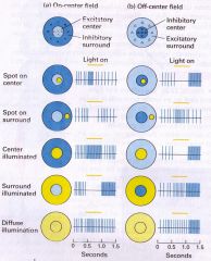
Receptive fields refers to the organisation of OFF and ON bipolar cells within a particular area of the eye. There are 'ON centre' and 'OFF centre' receptive fields. At the centre of each is a collection of ON or OFF bipolar cells respectively and on the surround of the receptive field the opposite. |
|
|
What is the effects of having receptive fields. |
Because there are so many overlapping receptive fields, the presence of receptive fields allows for very high levels of visual acuity. |
|
|
How is lateral inhibition created? |
Created through the presence of horizontal cells and amacrine cells. A ganglion which is very heavily stimulated will be connected to surrounding horizontal and amacrine cells which will inhibit the closely lateral rods to the excited rod. |
|
|
What is the effect of lateral inhibition? |
Lateral inhibition allows mammals to perceive far higher acuity images by narrowing down which rod cells are most affected by light. |
|
|
What do the collective axons of the ganglionic cells form? |
The axons from the output of the retina for the optic nerve - aka the cranial nerve II |
|
|
Where do the optic nerves which leave each eye meet at? |
Each optic nerve leave the eye to a meeting area called the optic chiasm. |
|
|
Through what do optic nerves travel through to reach the visual cortex? |
The optic nerves which go to the opposite side of the brain. |
|
|
How is the information from the central visual field of each eye processed and what does this enable? |
Input from each central field overlaps which is termed an area of binocular vision. The ability to analyse both pieces of information from the central portion of the vision allows for depth perception. |
|
|
What are the synapses of rods and cones called? |
Ribbon synapses |
|
|
How do horizontal cells module responses in photoreceptors? |
Via GABA receptors - GABA is released by the horizontal cell to hyperpolarise the target cell (hyperpolarisation = inhibition = lateral inhibition by horizontal cell) The GABA receptors are ionotropic and cause influx of Cl- ions. |
|
|
What connects rods to ganglion cells? |
AII and A17 amacrine cells |
|
|
What levels of visual processing are there? Give examples of types of visual information processed at each level. |
1. Low level - orientation, colour etc 2. Intermediate level - contour integration, surface depth, shape discrimination 3. High level processing - object identification & higher integration |
|
|
What is the difference between the magnocellular neurons and parvocellular neurons? What are the pathways which lead to each? |
Magnocellular -> visual detection. They operate at a very high speed at the expense of visual acuity define 'where' an object is. - Magnocellular channel Parvocellular -> visual acuity & colour. Slower but define 'what' an object is. - Parvocellular channel |
|
|
What is the fovea dominated by? What is the eye periphery dominated by? |
1. Cone cells 2. Rod cells |