![]()
![]()
![]()
Use LEFT and RIGHT arrow keys to navigate between flashcards;
Use UP and DOWN arrow keys to flip the card;
H to show hint;
A reads text to speech;
45 Cards in this Set
- Front
- Back
|
Describe the following parts of a virus particle: Capsid Envelope Glycoproteins |
Capsid- proteins surrounding a virus Envelope- cellular in origin Glycoproteins, located on the envelope, interacts with the host tissue |
|
|
Describe the Direct fluorescent antibody test- procedure and what does a (+) test look like? |
1. the tissue sample is frozen 2. the cryostat is used to section the tissue slices 3. The sections are put on a slide with a known antibody for the suspected virus 4. If a (+) test results, you will have a antigen-antibody complex made and the complex will fluoresce |
|
|
What is immunohistochemistry? How is it done and how is it different than the Direct fluorescent antibody test? |
A test very similar to direct fluorescent antibody test but the sample is formalin fixed and paraffin embedded. The process is the same as direct DFA but the result is a slide that is better in morphology but the process takes longer |
|
|
Describe Virus Isolation in how it is done and what a (+) test would look like If you are isolating Lentivirus, how does the results look different? |
1. an infectious sample has to be collected, ideally early in the infection 2. the sample is homogenized 3. The fluid with the virus is isolated and inoculated with a known antibody 4. the sample is examined daily looking for cytopathic effect, a (+) test If you are looking for lentivirus, look for giant cells rather than cytopathic effect |
|
|
You can also do virus isolation in ________________ to isolate the virus in question. |
Viable eggs |
|
|
If you are doing an ELISA test, how would you do it? How would you know the test was (+)? |
1. You place your unknown sample in a plate well that is coated with antibody. 2. If you have a (+) test, an antibody-antigen complex would have formed in the plate well 3. You add a chromagen to the well and look for a color change, which would signify a (+) test |
|
|
Define: chromagen |
Colorless marker that, when it undergoes an enzymatic reaction, it changes color |
|
|
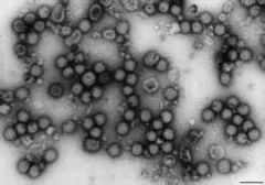
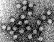
What is a Negative Staining EM? |
A test done on a completely unknown virus. This is done to identify the family that the virus belongs to in order to get an idea of what you're dealing with. Once you identify the family, you can follow up with another test to get a more specific (+) test. |
|
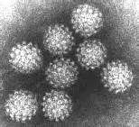
Name this family of virus |
Togaviridae |
|
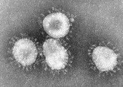
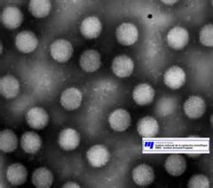
Name this family of virus |
Coronaviridae |
|
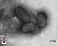
Name this Family of virus |
Poxviridae |
|
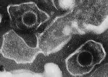
Name this family of virus |
Herpesviridae |
|
Name this family of virus |
Papoviridae |
|
Name this family of virus |
Adenoviridae |
|
Name this family of virus |
Parvoviridae |
|
|
What are the tree steps of PCR? |
1. Denaturing 2. Annealing 3. Extension (duplicate these three steps to get many cycles of PCR) |
|
|
What is the difference between PCR and Realtime PCR |
There is a a probe in Real-time PCR that will show you that the cycle is working by fluorescing |
|
|
Define Hemagglutination Name two examples of viruses that are capable of doing this |
A virus has the capability to make a carpet of RBCs Canine Parvovirus, Influenza |
|
|
Describe the Hemagglutination test. |
1. You plate the unknown sample with a known antibody 2. If there is antigen present in your unknown, you get a carpet of cells, which is a (+) test (A negative test looks like just a red dot in the middle of the well) *you can also do this in serial dilution to determine the concentration of virus in an unknown |
|
|
Describe the Restriction Endonuclease (RE) analysis |
1. put an enzyme in with an unknown sample that breaks up the DNA into pre-determined sections 2. Run the unknown with a known sample through electrophoresis 3. Compare the known with the unknown. If your known is a vaccine and the vaccine matches the unknown, the vaccine is causing the disease. If it is different, it is a different strand causing disease. |
|
|
Describe the Indirect fluorescent antibody test |
Similar test to the direct antibody test but you put in an unknown looking for antibody. So, the well is coated with antigen. You want to only put enough virus in to get a PARTIAL infection, to test the specificity A positive test is still documented by fluorescence |
|
|
How do you make an antibody prep for an indirect fluorescence test? |
You inoculate susceptible cells with a virus so that all of the cells make antibody and get infected with the virus. This is your antibody prep that can be stored until it is needed to test an unknown sample. |
|
|
Describe the Antibody detection ELISA test What can it be used to test? What doesn't it work for? |
Same as the ELISA test already described but the antigen is in the wells, not the antibody. It is not good for distinguishing between vaccinated animals and not vaccinated animals (unless you test for a section that is only found in the field virus) |
|
|
how do you perform a Hemagglutination inhibition test? What does a (+) test look like? |
1. Combine an unknown serum with a hemagglutination virus. If the antibody for that virus is present, the virus will be coated, if the antibody is not present, the virus will remain intact. 2. Then you put RBCs in the complex. If the virus is coated, you get a red dot, this is a (+) test. If the virus is still intact, you get hemagglutination and a carpet of RBCs will happen. |
|
|
Describe the Agar Gel Immunodiffusion Test (aka Coggins test) |
1. Antigen is placed in the center well 2. 3 unknowns and 3 knowns are placed in the well surrounding the antigen, in an alternating pattern 3. When allowed to diffuse out and the antigen and antibody complex, there will be a line that forms (+ test). The location of this line is the ideal concentration between antigen and antibody (ie. tells you an idea of the [ab] in the unknown). |
|
|
If you have a faint positive on a AGID test, how do the lines on either side of the faint positive look? |
The lines on either side of the faintly positive unknown will be curved. If it is a true negative, the lines on either side of the unknown will be straight and run right into the unknown rather than being curved and then stopping. |
|
|
If you were performing a virus neutralization test, what would you be testing for? How is the test performed? |
You are testing for vaccine titers 1. You make a two-fold dilation of the unknown and place those on plates. 2. Place equal amounts of virus on each of the plates 3. The titer is the last dilution that results in NO cytopathic effect (meaning that the virus is neutralized). Once a cytopathic effect is seen, there is not enough antibody in the serum to neutralize the virus. |
|
|
What is the name of the confirmatory test that we talked about in class? Used to confirm any of the other tests? |
Western/Immunoblotting test |
|
|
Feline herpesvirus: Is it enveloped or not? What does this mean for disinfection? Is it an RNA or a DNA virus? What is its host range? Is it a large or small player in respiratory disease in its host? |
Enveloped- disinfection is easy with detergents DNA virus Host is cats only Plays a large role, 45% of cases of respiratory disease in cats have this disease as one of its players. |
|
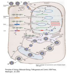
How does a virus get into a host and replicate? Know this diagram |
1. the binding of one glycoprotein stimulates the binding of a second glycoprotein, resulting in a more permanent binding. 2. The membranes fuse and the virus gets into the nucleus 3. The glycoprotein parts (gamma proteins) are made last with the parts that regulate the glycoproteins made first (alpha and beta) |
|
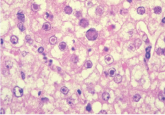
What are these in this picture? What is this a sign of? |
Inclusion bodies Sign of nuclear replication |
|
|
What are the ways that feline herpesvirus spreads? What are the more popular ways? |
1. Aerosol* important- can spread 7-21 days after being infected 2. Fomites 3. Latent Asymptomatic carriers (once infected, all become latent carriers, and will carry it forever) |
|
|
What are some of the ways that reactivation of a latent infection can occur? |
Moving, use of corticosteroids, nursing (4-6 weeks after parturition), boarding, parturition |
|
|
Where does herpesvirus sit and replicate in the host? What does it do to the cat systemically? |
Replicates in the upper airway. It normally cannot get into the body to cause infection because of the temperature. The herpesviruses prefer cooler temperatures to replicate. Systemically, it can cause disease in kittens that become hypothermic. |
|
|
What are the 4 very prominant (+++) symptoms seen with Feline Herpesvirus? |
Lethargy Sneezing Ocular Discharge Nasal Discharge |
|
|
What are the 2 somewhat prominent (but still important) symptoms (++) for feline herpesvirus? |
Conjunctivitis (especially seen with recurrent infection) Keratitis |
|
|
What are the 5 rarer symptoms (+) for feline herpesvirus? |
Hypersalivation Oral ulceration Coughing Dyspnea Pneumonia |
|
|
If you had an Atypical presentation of feline herpesvirus, what 3 signs could you see? |
Skin lesions Viraemia (seen in neonates) Pneumonia |
|
|
If feline herpesvirus was chronic in an animal, what symptoms would you see? |
Stomal keratitis Chronic rhinosinusitis |
|
|
Regarding pregnant queens and young kittens, what symptoms can herpesvirus cause? |
Abortions in queens Birth defects/encephalitis in neonates Death of kittens Lung lesions |
|
|
Is lameness associated with feline herpesvirus? |
Nope |
|
|
In a latent infection, where would you find feline herpesvirus? |
In the nerve endings of the trigeminal nerve There is a small portion that continues to replicate but major re-infection does not occur until stress allows the virus to get out. |
|
|
How would you diagnose feline herpesvirus? what is the test of choice to detect this virus? |
Clinical Signs Virus Isolation Real time PCR- test option of choice |
|
|
What are the treatments of choice for feline herpesvirus? What is one drug that is commonly used but has been shown to not be very effective in treating this disease? |
Acyclovir and Famciclovir L-Lysine |
|
|
How do you prevent the infection of feline herpesvirus? |
MLV administered subcutaneously (will then be rendered avirulent from the body temperature of the animal) There is also a MLV given intranasally but it is not as commonly used |