Reading...
![]()
Play button
![]()
Play button
![]()
Use LEFT and RIGHT arrow keys to navigate between flashcards;
Use UP and DOWN arrow keys to flip the card;
H to show hint;
A reads text to speech;
32 Cards in this Set
- Front
- Back
|
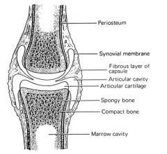
Synovial joint basic structure
|
Synovial membrane
Synovial fluid Articular cartilage Articular space |
|
|
Types of joints (3)
|
Synovial
Fibrous Cartilagenous |
|
|
2 features of synovial membranes that contribute to function and pathology
|
well vascularised
no basement membrane |
|
|
synoviocentesis
|
collection of synovial fluid from synovial joint
|
|
|
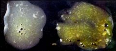
Synovial fluid
- normal appearance |
Clear fluid
Low cell count |
|
|
Articular cartilage
- normal appearance |
White-bluish
Glistening (spots) |
|
|
OCD
|
Osteochondritis dissecans
|
|
|
OCD
- basic description |
Split in epiphyseal growth cartilage (under the articular cartilage)
A form of osteochondrosis |
|
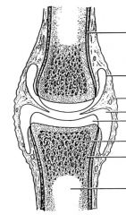
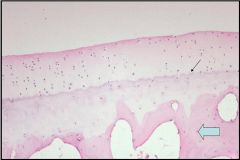
Name the different parts of a synovial joint
|
|
|
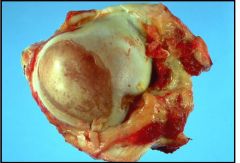
Name this condition. What is the pathogenesis
|

|
|
What is this condition & likely predisposing conditions and aetiology?
|

Osteochondritis dissecans
Rapid growth, large breed dogs, conformation, increased physical activity, gender (males) Vigorous exercise -> trauma -> fissue in epiphyseal growth cartilage -> flap -> detach -> joint mice -> DJD |
|
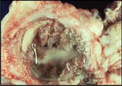
Name the different structures in this synovial joint
|
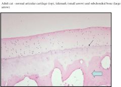
|
|
What is this material likely to be?
What is it's source and why is it significant? |

Pannus = fibrovascular granulation tissue
Derived form marrow spaces of subchondral bone when damage penetrates the tide mark -> fills overlying cartilage. |
|
|
What are potential consequences of pannus?
|
Fibrocartilaginous scar -> interferes with diffusion of nutrients into articular cartilage
-> erosion dt collenagse activity -> DJD if joint is injured or inflammed (dt mediators) |
|
|
What is the common sequence of degeneration in injured synovial joints?
|
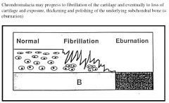
|
|

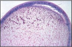
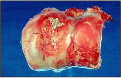
What processes are seen in this ox femoral condyle?
|
Erosion and fibrillation
|
|

What process is most event in this hose humeral head?
|
Eburnation of subchondral bone
|
|
What process is evident in his opened joint under water?
What is the pathogenesis? |

Synovial villous hyperplasia
Hypertrophy of synoviocytes commonly occurs in diseased joints |
|
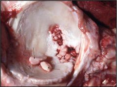
What are the arrows indicating?
How can this arise? |

Joint mice
Detached articular flaps in OCD Detached osteophytes Detach chrondromas /osteochrondromas Bone fragments after fracture? |
|
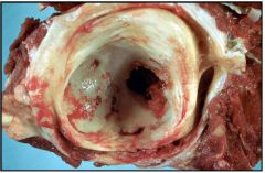
What is the likely aetiology for this acetabulum from an ox?
In other words, what are the key abnormalities? |
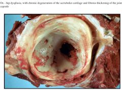
Hip dysplasia related to
- small pelvic mm mass relative to pelvis size - subluxation of the femoral head - chronic degeneration of cartilage - remodelling of the femoral neck - flattening of the acetabulum (shown here) - fibrous thickening of the joint capsule |
|
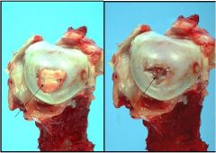
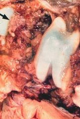
What is illustrated for this canine humeral head?
What are the likely predisposing factors? |

Osteochondritis dissecans with the articular flap intact and removed.
Predisposing: large breed dogs undergoing rapid growth and much physical exercise or trauma |
|

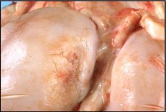
These pigs have inflamed joints.
What's the condition and likely cause? Give 2 possible sequelae |
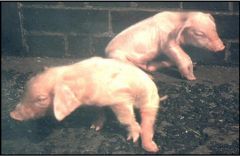
Polyarthritis
Erysipelothrix rhusiopathiae Meningitis, valvular endocarditis |
|
What's the likely diagnoses of these exudates from infected synovial joints?
|

Fibrinous arthritis - cloudy & turbid dt fibrin formation
Suppurative arthritis - cloudy and yellow dt infection with pyogenic bacteria. Note: both are infectious arthriditis but suppurative is dt pyogenic bacteria. |
|

Give a morphological dx for this pig joint
|
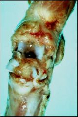
Acute severe fibrinous septic arthritis
- see excess synovial fluid and marked synovial oedema and hyperaemia |
|

Give three ddx
|
Severe septic arthritis
Synovial sarcoma Osteosarcoma |
|

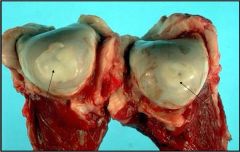
What's this?
Why does it occur? What other species can be affected? |
Articular goat
Inflammatory reaction dt deposited urate crystals (tophi) Humans and reptiles also lack the uricase enzyme |
|
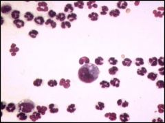
Synovial fluid cytology
What's the likey cause and why? |

Immune-mediated arthritis
Predominance of neutrophils Note: expect to see predominance of neutrophils in suppurative arthritis but would also expect to see bacteria? |
|

What's this?
What are some likely consequences? |
Transverse section through an IV disc in which the nucleus pulposus has degenerated and prolapsed dorsally (hansen's type I)
Consequences depend on site in spinal cord but often localised affect of 2-4 spinal segments leading to direct cord compression causing vascular injury -> focal myelomalacia, diffuse demyelination -> pain, paresis or paralysis |
|
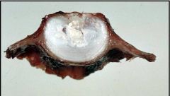
What's happening to this dog vertebral column and spinal cord?
|

Hansen's type I (degen of nucl pulposis causing degen of annual fibrosis) intervertebral disc protrusion
|
|
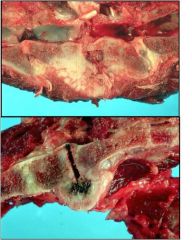
What's the difference between these two images?
|

Top: chronic discospondylitis (inflammation of the intervertebral disc with osteomyelitis of contiguous discs) - often dt bacterial localisation
Below: ankylosing spondylosis (spondylosis deformans) - formation of osteophytes -> ventrolateral spurs -> bridge and restrict movement. Often seen in AI stud bulls. |
|

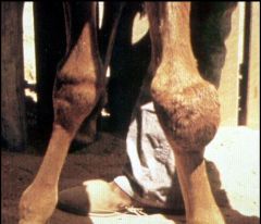
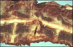
Dog carpus - ddx?
|
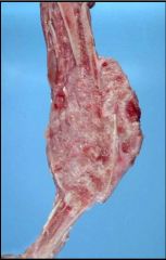
Synovial sarcoma
Prob any other invasive sarcoma originating from the joint or subchondral bone - osteosarcoma - chondrosarcoma? |
|

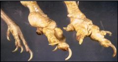
Stifle joint from a severely lame 3 week old pig
What two features are apparent? Give a morphological diagnosis? Likely causative agent? |
Severe synovial villous hyperplasia
Synovial hyperaemia Moderately severe, chronic active, diffuse, fibrinous (or fibrinosuppurative) arthritis of the stifle joint Erysipelothrix rhusiopathiae |