![]()
![]()
![]()
Use LEFT and RIGHT arrow keys to navigate between flashcards;
Use UP and DOWN arrow keys to flip the card;
H to show hint;
A reads text to speech;
33 Cards in this Set
- Front
- Back
|
where is the thorax located |
between the neck and the diaphragm |
|
|
what are the two components of the thorax |
thoracic wall and thoracic cavity |
|
|
what are the boundaries on the thoracic wall |
posterior: vertebrae and intervertebral discs from T1-T12 anterior: sternum lateral: ribs, costal cartiledge superior: thoracic inlet (opening bounded by vertebrae, manubrium of sternum and ribs), plane at an angle, apices of lungs project into neck inferior: thoracic outlet, demarcated by diaphragm. separates from abdominal cavity, lower posteriorly |
|
|
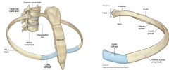
describe the 3 parts of the sternum |
![superior: manubrium (jugular notch, clavicular notch, 1st sternocostal joint [no movement with cartiledge to fix with rib cage])
Body, meets manumbrium at manubriosternal joint (sternal angle), some movement, 2nd rib at sternal angle. Ribs 3-7 art...](https://images.cram.com/images/upload-flashcards/80/86/11/11808611_m.png)
superior: manubrium (jugular notch, clavicular notch, 1st sternocostal joint [no movement with cartiledge to fix with rib cage]) Body, meets manumbrium at manubriosternal joint (sternal angle), some movement, 2nd rib at sternal angle. Ribs 3-7 articulate body, synovial (movable joints) inferior: xiphoid process, articulates with body at xiphisternal joint |
|
|
which rib do you start palpating and why |
start with 2nd rib (1st rib is too deep to clavical), use the sternal angle to find it |
|
|
describe the intercostal spaces
|
spaces between ribs, there are 11, named ICS whatever rib number they are below |
|
|
what is the transverse thoracic plane |
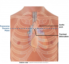
transverse plane from the sternal angle back to the intervertebral disk between T4/T5 marks location of bifurcation of trachea into bronchi, the aortic arch (beginning and end) |
|
|
what are the two ways of classifying ribs and how is each rib classified |
1. how ribs attach to sternum true ribs-- attach directly to sternum with costal cartilage (1-7) false ribs-- indirectly attach to sternum, cartilage attaches to cartilage above it (8-10) floating ribs-- have no connection to sternum 2. features typical ribs (3-9) 6 rules for typical ribs: 1. head of rib articulates with vertebral body and intervening intervertebral disk (costovertebral joint, synovial) 2. neck is between head and tubercle 3. articular tubercle articulates with transverse process of vertebra 4. angle (region of maximum curvature) 5. costal groove for VAN 6. shaft ends in cup shaped depression for costal cartilage atypical ribs (1,2, 10-12) lack one or more of defining characteristics from above |
|
|
define the costal margin |
the line created by the costal cartilage of ribs 7-10 |
|
|
what are the 2 types of thoracic wall movements? |
inspiratory: increase thoracic cavity size, decrease pressure, air in expiratory: decrease size, increase pressure, air out |
|
|
describe the process of diaphragm contraction |
diaphragm contracts, moves down, increases vertical dimension of thoracic cavity |
|
|
describe the rib elevation in inspiratory movements |
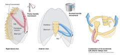
1. pump-handle: sternum out, rib up (upper ribs) 2. bucket handle: lateral ribs out and up (lower ribs) increase antero-posterior dimension of thoracic cavity |
|
|
describe the intercostal muscles location, function and arrangment
|
located between ribs
function to keep taught intercostal space (no bulging when breathing) during relaxed breathing 3 layers: external, internal and innermost |
|
|
describe the external intercostal muscles |
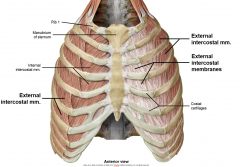
from above rib to below, hand in pocket orientation (lateral to medial down) don't extend to sternum, replaced by thin external intercostal membrane raise ribs in forced inspiration |
|
|
describe internal intercostal muscles |
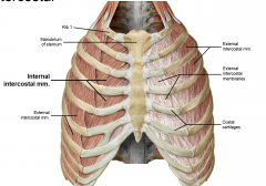
deep to external, opposite direction of fibers forced experiation wrap all the way around from sternum to spine |
|
|
describe the innermost intercostal muscles |
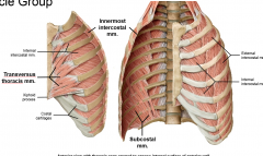
3 muscle groups connected by membrane anterior group: transversus thoracis laterally: innermost intercostal muscles posterior group: subcostal muscles (forced expiration) |
|
|
describe the location and contents of the neurovascular plane |
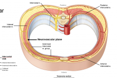
located between internal and innermost intercostal muscles contains intercostal nerve and vessels (VAN in order) collateral branches in upper border of rib below posterior and anterior intercostal arteries and veins sheltered by costal groove of rib |
|
|
describe the intercostal nerves |
ventral rami of T1-T11 muscular branches throughout at midaxillary line, lateral cutaneous branch just lateral to sternum, anterior cutaneous branch motor fibers for intercostal muscles, sensory fibers for specific dermatome, sympathetic fibers of blood vessels and sweat glands no parasympathetic supply to body wall travel with branches and tributaries of arteries and veins |
|
|
describe the thoracoabdominal nerves |
intercostal nerves 7-11
travel normally with thoracic neurovascular plane but then exit and continue into abdominal neurovascular plane (between transversus abdominal and internal oblique muscles) transmit motor, sensory and sympathetic fibers to thoracic and abdominal walls |
|
|
describe the intercostal arteries |
posterior intercostal artery (most of supply, right off aorta) anterior intercostal arteries (off internal thoracic artery, not vital) anastomosis between |
|
|
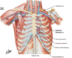
describe the internal thoracic artery |
branch of subclavean artery, parallels sternum in descent around 6th intercostal space, divides into terminal branches |
|
|
describe the musculophrenic artery |
one of the terminal branches of internal thoracic artery hugs intercostal margin, lowest branch |
|
|
describe the anterior intercostal veins |
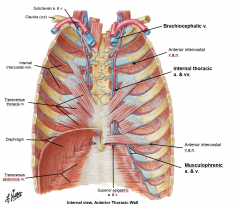
drain oxygen poor blood away from thoracic wall mirrors artery, same on both sides musculophrenic vein, internal thoracic veins, ultimately to brachiocephalic vein |
|
|
describe the posterior intercostal veins |
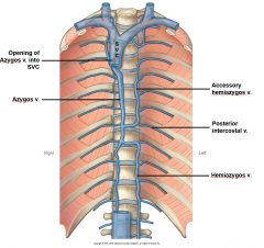
different on left/right side, drain most of thoracic wall Left: to accessory hemiazygos vein or hemiazygos vein, cross midline on vertebra Right: azygos vein, drains into superior vena cava |
|
|
describe the thoracic cavity organization |
3 compartments: 2 lateral (contain lungs and pleural sacs, majority of space) and mediastinum |
|
|
what are the subdivisions of the mediastinum |
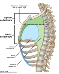
superior: thoracic inlet to transverse thoracic plane (sternal angle to intervertebral disk of T4/T5) inferior: transverse thoracic plane to the diaphragm |
|
|
what are the components of the inferior mediastinum |
anterior (to heart) middle (contains heart) posterior (to heart) |
|
|
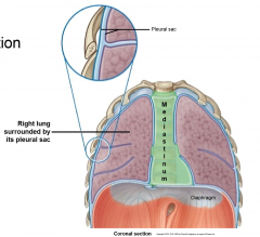
describe the layers of the pleural sac |
visceral pleura: innermost layer, intimately attached to surface of lung tissue, follows fissures parietal pleura: outer layer continuous together at point called hilum (doorway into lung) space between layers is pleural cavity (pleural film for lubrication and surface tension) |
|
|
describe the regions of the parietal pleura |
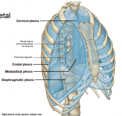
cervical-- arches over apecis of lungs costal-- lines inner wall of thorax mediastinal-- lateral wall of mediastinum (form pleural sleeve for root of lung, pulmonary ligament) diaphragmatic-- superior surface of diaphragm |
|
|
what are the lines of pleural reflection |
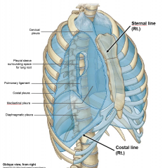
where the parietal pleura bends/changes (4 important ones) R/L sternal line (where mediastinal becomes costal) R/L costal line (diaphragmatic becomes costal) right sternal line down to 6th costal cartilage left has lateral excursion (exposes bare area of pericardium) then down to 6th costal lines begin at 6th |
|
|
define pleural recesses |
when lungs are exhaled, space between the visceral and parietal pleura creates recesses R/L costomediastinal recesses R/L costodiaphragmatic recesses (lowest points of thoracic cavity (blood or pus settles here) |
|
|
describe the extent of the pleural cavities with reference to lines |
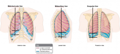
midclavicular line (visceral to 6, parietal to 8) midaxillary line (visceral to 8, parietal to 10) scapular line (visceral to 10, parietal to 12) 2 rib difference at inspiration, diaphragm moves down, lungs increase, apparent difference decreases |
|
|
define pneumothorax |
air in the pleural cavity most common cause: bullet to chest result is lung collapse tube thoracotomy (chest tube) |