![]()
![]()
![]()
Use LEFT and RIGHT arrow keys to navigate between flashcards;
Use UP and DOWN arrow keys to flip the card;
H to show hint;
A reads text to speech;
134 Cards in this Set
- Front
- Back
- 3rd side (hint)
|
Which skeleton includes the skull? |
Axial Skeleton |
|
|
|
How many bones are found in the skull? How many make up the braincase? How many make up the face? |
22 Bones in the Skull; 8 in the Braincase; 14 in the Face |
|
|
Name the 8 bones of the cranium (a.k.a. braincase). Remember two bones are paired. |
(1) Occipital Bone (2) Parietal Bones X2 (3) Frontal Bone (4) Temporal Bone X2 (5) Sphenoid Bone (6) Ethmoid Bone |
F PESTO |
|
Name the 14 bones of the face. Remember, all but two are paired. |
(1)Maxillae (2) Palatine Bones (3) Nasal Bones (4) Inferior Nasal Conchae (5) Zygomatic Bones (6) Lacrimal Bones (7) Vomer (Unpaired) (8) Mandible (Unpaired) |
Nasty Vicious Zombies LIMMP |
|
|
Name the 4 bones of the skull cap (a.k.a. the claveria). |
(1) Frontal Bone (2) Parietal Bones X2 (3) Occipital Bone |
|
|
|
The bones of the skull are held together by this type of joint. It is a kind of fibrous joint that is synarthrotic. |
Sutures |
|
|
|
True or false. The skull joints allow for a decent amount of movement so that the brain can expand and contract during times of injury. |
False. The Suture joints of the skull are synarthrotic and as such only allow for a tiny amount of movement. For our purposes they are considered immovable. |
|
|

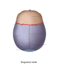
Name the suture. |
Coronal Suture occurs between the Frontal Bone and the Parietal Bones. |

Superior View |
|
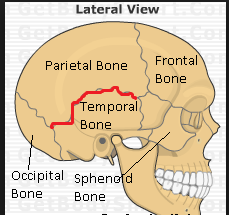
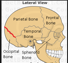
Name the suture. |
Squamosal Suture occurs between a Parietal Bone and a Temporal Bone. |
|
|

Name the suture. |
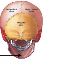
Lambdoidal Suture occurs between the Parietal Bones and the Occipital Bone. |

Superior View |
|

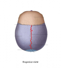
Name the suture. |
Sagittal Suture connects Right and Left Parietal Bones. |
|
|
|
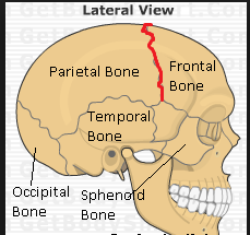
Between which three bones does the Coronal Suture occur? |
Coronal Suture occurs between the Frontal Bone and the Parietal Bones. |

|
|
|
Between which two bones does the Squamosal Suture occur? |
Squamosal Suture occurs between a Parietal Bone and a Temporal Bone. |

|
|
|
Between which three bones does the Lambdoidal Suture occur? |
Lambdoidal Suture occurs between the Parietal Bones and the Occipital Bone. |

|
|
|
Between which two bones does the Sagittal Suture occur? |
Sagittal Suture occurs between the Right and Left Parietal Bones. |
|
|
Name the suture. |
Coronal Suture (Superior View) |
|
|

Name the Suture. |
Lambdoidal Suture (Superior View) |
|
|
|
What is the term for where the Sagittal Suture meets the Coronal Suture? |

Bregma |
|
|
|
What is the term for where the Sagittal Suture meets the Lambdoidal Suture? |
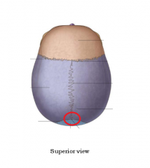
Lambda |
|
|
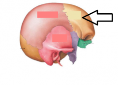
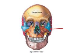
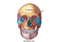
Name this bone. |
Frontal Bone |
|
|
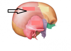
Name this bone. |
Parietal Bone |
Lateral Exterior View |
|
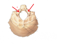
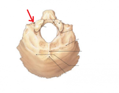
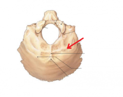
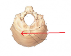
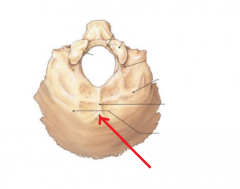
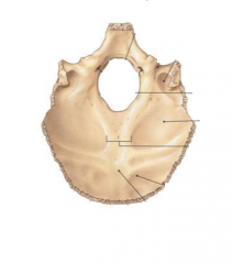
Name the bone. |

Occipital Bone |

Interior Superior View |
|
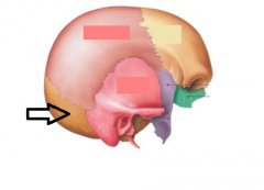
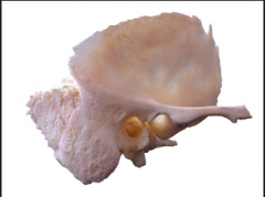
Name this bone. |
Temporal Bone |
|
|
Name this bone. |
Sphenoid Bone |
|
|
Name this bone. |
Ethmoid Bone |
|
|
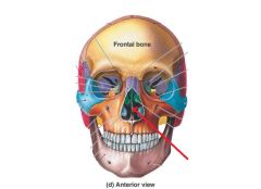
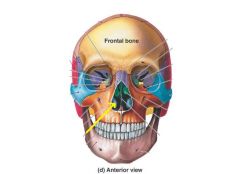
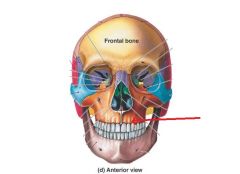
Name this bony marking. It falls just below the eyebrows and helps to form the orbit. |
Superciliary Arch of the Frontal Bone |
|
|
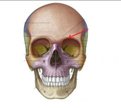
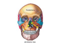
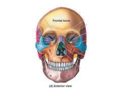
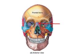
Name this bony marking. Along with tissue, this creates the foramen through which the sensory branch of the ophthalamic n. (CN V1) passes. |
Supra-Orbital Notch (Foramen) of the Frontal Bone |
|
|
|
These are spaces within bones. They help to keep the skull lighter, help to resonate the voice, and are lined with mucous membranes that produce mucous when you're sick. There are four of these in the skull, each named after the bone in which they're found. |
Paranasal Sinuses |
|
|
|
Name the four types of paranasal sinuses. |
(1) Frontal Sinus (2) Sphenoid Sinus (4) Maxillary Sinus |
|
|
Name the bony marking. |
Frontal Sinus (in Frontal Bone) |
|
|

Which side of which bone are you looking at? |
Exterior Lateral View of Parietal Bone |
|
|
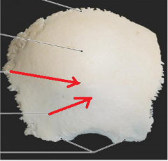
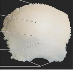
Name the bony markings. These are the origin of the temporalis m. |
Superior and Inferior Temporal Lines of the Parietal Bone |
|
|
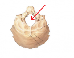
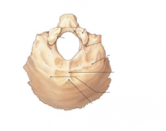
Name the bony marking. The spinal cord and vertebral aa. enter the skull here. |
Foramen Magnum |
|
|
Name the bony markings. The skull articulates with C1 here, allowing us to make the "yes" motion. |
Occipital Condyles (A good indicator that you're looking at the exterior Occipital Bone as they don't exist on the interior) |
|
|
Name the bony marking. CN XII runs through here. |
Hypoglossal Canal |
|
|
Name the bony marking. This is one of two points of origination for the ligamentum nuchae which is where the trapezius m. originates. |
Inferior Nuchal Line |
Inferior Nuchal Line is closer to foramen magnum. |
|
Name the bony marking. This is one of two points of origination for the ligamentum nuchae which is where the trapezius m. originates. |
Superior Nuchal Line |
Superior Nuchal Line is the one farther away from the foramen magnum. |
|
Name the bony marking. This is the bump you can feel on the back of your head. |
External Occipital Protuberance |
|
|
Name the bony marking. The Internal Jugular v., CN IX, X, & XI pass through here. |
Jugular Notch |
|
|
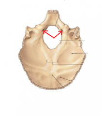
Name the bony marking. CN XII passes through the occipital condyle through this foramen to enter the skull. |
Entrance to Hypoglossal Canal |
|
|
Which side of which bone are you looking at? |
Exterior Inferior View of Occipital Bone |
|
|
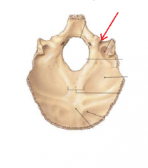
Which side of which bone are you looking at? |
Interior Superior View of Occipital Bone |
|
|
Which side of which bone are you looking at? |
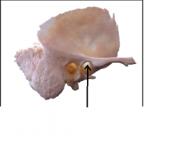
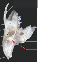
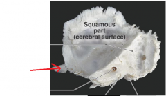
Lateral View of Exterior Temporal Bone |
|
|

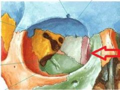
To what part of the temporal bone does the arrow point? What are the characteristics of this area? |
The Squamous Area of the Temporal Bone is a Thin, Flat Area of Bone |
Squamous means thin/flat |
|

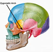
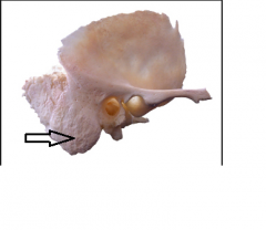
Name the bony marking. It joins with the temporal process of the zygomatic bone to form the zygomatic arch. |
Zygomatic Process of the Temporal Bone (Can also be seen on interior view) |

Interior View |
|

Name the bony marking. It forms the temporalmandibular joint with the head of the mandible. |
Mandibular Fossa of the Temporal Bone (Can also be seen on inferior view) |

Inferior View |
|

Name the bony marking. This is the site of attachment for the "swallowing" muscles. |
Styloid Process of the Temporal Bone (Can also be seen on interior and inferior views) |

Inferior View |
|

Name the bony marking. This is a site of muscle attachment. |
Mastoid Process of the Temporal Bone (Can also be seen on interior and inferior views) |

Inferior View |
|
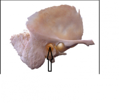
Name the bony marking. It carries sound waves to the hearing organs and makes up the most outer bony portion of the ear. |
External Acoustic Meatus of the Temporal Bone (Can also be seen on inferior view) |

Inferior View |
|
|
What does meatus mean? |
Large Foramen |
|
|
|
Name the two components of the zygomatic arch. |
(1) Zygomatic Process of the Temporal Bone (2) Temporal Process of the Zygomatic Bone |
|
|
|
This is the area superior and deep to the zygomatic arch. Many nerves and arteries are found here. |
Temporal Fossa |
|
|
|
This is the area inferior and deep to the zygomatic arch. Many nerves and arteries are found here. |
Infratemporal Fossa |
|
|
Which view of which bone are you looking at? |
Inferior View of the Temporal Bone |
|
|

Name the bony marking. |
Styloid Process of the Temporal Bone (Inferior View) |
Lateral Exterior View |
|
Name the bony marking. |
Mandibular Fossa of the Temporal Bone (Inferior View) |
Lateral Exterior View |
|
Name the bony marking. |
External Acoustic Meatus of the Temporal Bone (Inferior View) |

Lateral Exterior View |
|

Name the bony marking. |
Mastoid Process of the Temporal Bone (Inferior View) |
Lateral Exterior View |
|
Name the bony marking. The cartoid a. passes through here to supply anterior brain circulation. |
Carotid Canal of the Temporal Bone |
|
|
Name the bony marking. The motor branch of the Facial n. (CN VII) (responsible for facial expression) exits the skull here. |
Stylomastoid Foramen of the Temporal Bone |
|
|
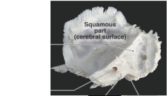
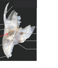
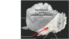
Which side of which bone are you looking at? |
Lateral View of Interior (Cerebral) Side of Temporal Bone
|
|
|
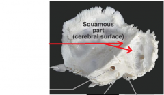
Name the bony marking. The auditory ossicles are found just deep to this. |
Petrous Part of the Temporal Bone |
Petrous means rocky |
|
Name the bony marking. |
Mastoid Process of the Temporal Bone (Interior View) |

Inferior View |
|
Name the bony marking. |
Zygomatic Process of the Temporal Bone (Interior View) |

Exterior Lateral View |
|
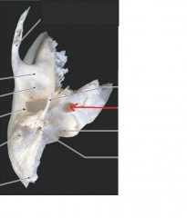
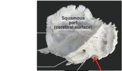
Name the bony marking. The branches of CN VII & VIII pass through here. There is no clear pass from the exterior portion of this to this. |
Internal Acoustic Meatus of the Temporal Bone |
|
|
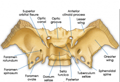
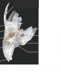
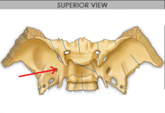
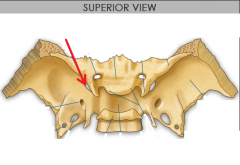
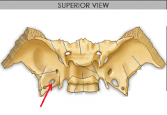
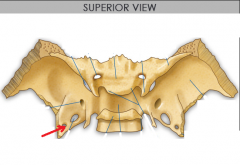
Which view of which bone are you looking at? |
Superior View of the Sphenoid Bone |
|
|
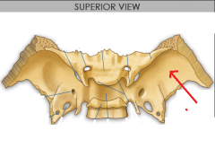
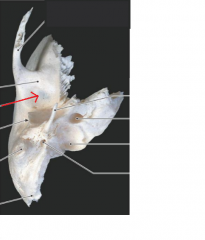
Name the bony marking. |
Greater Wing of the Sphenoid Bone |
|
|
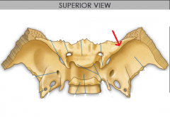
Name the bony marking. |
Lesser Wing of the Sphenoid Bone |
|
|
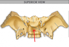
Name the bony marking. The Pituitary Gland sits here. |
Sella Turcica of the Sphenoid Bone |
Means "Turkish Saddle" |
|
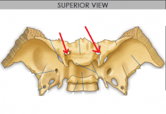
Name the foramen. CN II and the ophthalamic a. pass through here. |
Optic Canal of the Sphenoid Bone (Paired) |
CN II = Optic n. |
|
Name the foramen. CN V2 passes through here. |
Foramen Rotundum of the Sphenoid Bone (Paired) |
|
|
Name the foramen. CN III, IV, V 1, & VI enter the skull here. |
Superior Orbital Fissure of the Sphenoid Bone (Paired) |
|
|
Name the foramen. CN V3 passes through here. |
Foramen Ovale of the Sphenoid Bone (Paired) |
|
|
Name the foramen. The Middle Meningeal a. passes through here. |
Foramen Spinosum of the Sphenoid Bone (Paired) |
|
|
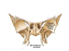
Which side of which bone are you looking at? |
Anterior View of the Sphenoid Bone |
|
|
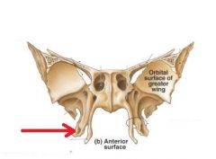
Name the bony marking. |
Lateral Plate of the Sphenoid Bone |
|
|
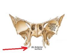
Name the bony marking. |
Medial Plate of the Sphenoid Bone |
|
|
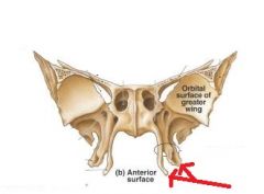
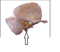
Name the bony marking. The space between the two plates is an important region for many ganglia and nerves. |
Pterygoid Process of the Sphenoid Bone |
|
|
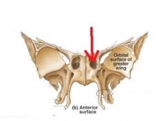
Name the bony marking. |
Sphenoid Sinus |
|
|
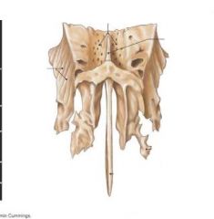
Which view of which bone are you looking at? |
Superior View of the Ethmoid Bone |
|
|
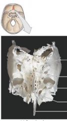
Which view of which bone are you looking at? |
Anterior View of the Ethmoid Bone |
|
|
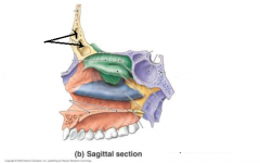
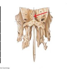
Name the bony marking. CN I passes through here to reach the nasal cavity. Contains many small foramina named after this region. |
Cribriform Plate of the Ethmoid Bone |
|
|
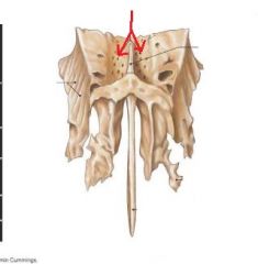
Name the bony marking. The falx cerebri (meningeal membrane that separates the R & L side of the brain) inserts here. |
Crista Galli of the Ethmoid Bone |
|
|
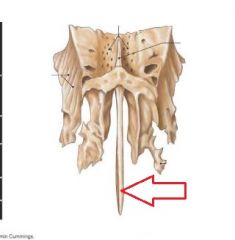
Name the bony marking. This forms the superior aspect of the nasal septum. |
Perpendicular Plate of the Ethmoid Bone |
|
|
|
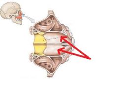
These two components of the ethmoid bone along with another separate bone (most superior component cannot be seen) help us to move air around internally before it enters the lungs. This helps to warm cold air, moistens dry air, and removes large particles from the air (gets caught in mucous). |
Superior Nasal Concha, Middle Nasal Concha, & Inferior Nasal Concha |
|
|
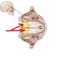
Name the bony marking. Is this its own bone or part of a bone? |
Middle Nasal Concha of the Ethmoid Bone |
|
|
Name the bony marking. Is this its own bone or part of a bone? |
Inferior Nasal Concha is its OWN Bone |
|
|

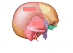
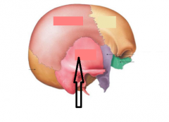
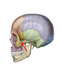
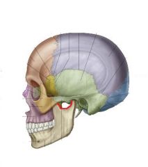
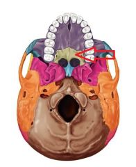
Name the region of the skull highlighted in pink. What parts of the brain can be found here? |
Anterior Cranial Fossa houses the Frontal Lobes |
|
|
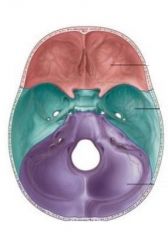
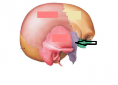
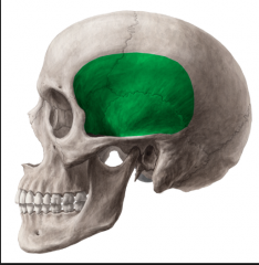
Name the region of the skull highlighted in green. What parts of the brain can be found here? |
Middle Cranial Fossa houses the Anterior Portion of the Temporal Lobes |
|
|

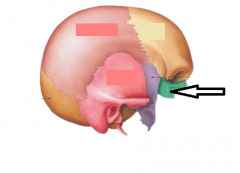
Name the region of the skull highlighted in purple. What parts of the brain can be found here? |
Posterior Cranial Fossa houses the Brain Stem and Cerebellum |
|
|
|
What is the function of the cranial fossae? (Anterior, Middle, & Posterior) |
They provide support and protection to certain brain areas. |
|
|
Name the only two Facial Bones that are unpaired. |
(1) Vomer (2) Mandible |
|
|
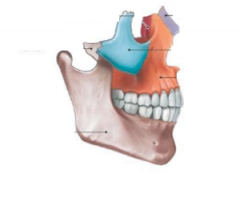
Name the Facial Bone. |
Maxillary Bone (Paired) |
|
|
Name the bony marking. This is where the upper teeth attach to the skull. |
Aveolar Process of the Maxillary Bone |
|
|

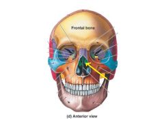
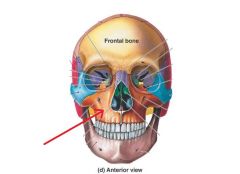
Name the bony marking. The sesnory branch of the Maxillary n. (CN V2) exits the skull here to provide sensation to the cheek. |
Infra-Orbital Foramen of the Maxillary Bone |
|
|
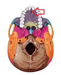
Which bones compose the anterior 2/3rds of the hard palate? |
Palatal Process of Right and Left Maxilla |
|
|
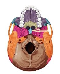
Which bones compose the posterior 1/3rd of the hard palate? |
Horizontal Plates of Right and Left Palantine Bones |
|
|
|
Which paranasal sinus is most superior? |
Frontal Sinus |
|
|
|
Which paranasal sinus can be found just inferior to the Frontal Sinus? This sinus and one other are in the best positions to drain. |
Ethmoid Sinus |
|
|
|
Which sinus is the most inferior and is situated such that the it must fill entirely in order to drain? |
Maxillary Sinus |
|
|
|
Which sinus is found just superior to the Maxillary Sinus? This and one other sinus are in the best positions to drain. |
Sphenoid Sinus |
|
|
Name the Facial Bone. It forms the bony portion of the top of the nose and the cartilage of the nose attaches here. |
Nasal Bone (Paired) |
|
|
Name the Facial Bone. |
Zygomatic Bone (Paired) |
|
|
Name the bony marking (not the bone). This joins to the zygomatic process of the temporal bone to form the zygomatic arch. |
Temporal Process of the Zygomatic Bone |
|
|

This facial bone forms the medial most aspect of the orbit. |
Lacrimal Bone (Paired) |
|
|
|
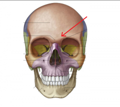
Name the seven bones that make up the orbit. |
(1) Maxillary Bone (2) Frontal Bone (3) Zygomatic Bone (4) Ethmoid Bone (5) Lacrimal Bone (6) Sphenoid Bone (7) Palatine Bone |
Many Friendly Zebras Enjoy Lazy Summer Picnics |
|

Name the Facial Bone. It forms the inferior posterior portion of the nasal septum. |
Vomer Bone (Unpaired) |
|
|
|
Name the portion of bone that makes up the superior posterior portion of the nasal septum. |
Perpendicular Plate of the Ethmoid Bone |
|
|
|
What makes up the anterior portion of the nasal septum? This is made of hyaline cartilage. |
Septal Cartilage |
|
|
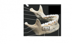
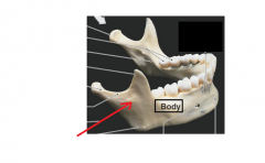
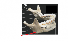
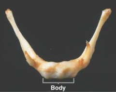
Name the three main pieces of the mandible. |
(1) Body (2) Angle (3) Ramus |
|
|
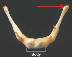
Name the part of the mandible. It is the most superior and posterior portion of the mandible. It is verticle and contains the projection which articulates with the mandibular fossa of the temporal bone. |
Ramus of Mandible |
|
|
Name the part of the mandible. It connects the ramus to the body. |
Angle of the Mandible |
|
|

Name the bony marking. This is the anterior portion of the ramus. The temporalis m. inserts here. |
Coronoid Process of the Mandible |
|
|

Name the bony marking. This is the posterior portion of the ramus & the site of articulation with mandibular fossa of the temporal bone (forms temperomandibular joint). |
Condylar Process of the Mandible |
|
|
Name the bony marking. It is a paired foramen through which the sensory branch of CN V3 exits the skull. |
Mental Foramen of Mandible |
Sensory Branch of CN V3 is called the Mental n. |
|

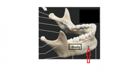
Name the bony marking. This is what one might commonly refer to as their chin. |
Mental Protuberance of the Mandible |
In Anatomy, Mental means Chin |
|
|
What is the name for the joint between the condylar process of the mandible and the mandibular fossa of the temporal bone? |
Temperomandibular Joint |
|
|
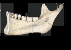
Which side of which bone are you looking at? |
Interior View of 1/2 of a Mandible |
Mandibular Foramen indicates that you are looking at the inside and not the outside |
|
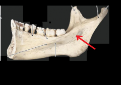
Name the bony marking. The inferior aveolar n. passes through here to give sensation to the lower teeth (dentist numbs this). |
Mandibular Foramen of the Mandible (good way to tell you're looking at the interior side of the Mandible) |
|
|
Name the bony marking. Many tongue-associated muscles insert here. |
Genoid Tubercle of the Mandible |
Genu means bend |
|
|
This is the term for the soft fibrous areas in a <4 y.o. child's skull where several sutures unite. They allow for molding and remodeling of the skull during development. There are 6 total in a normal child's skull. |
Fontanelles (a.k.a. Soft Spots) |
|
|
|
During birth, an infant's head must squeeze through the birth canal. In order to do this, it must change shape. Fontanelles allow the skull to do this. What is this process called? |
Molding |
|
|
|
As a child grows and develops, so does their brain and skull. Fontanelles allow the skull to do this. What is this process called? |
Remodeling |
|
|
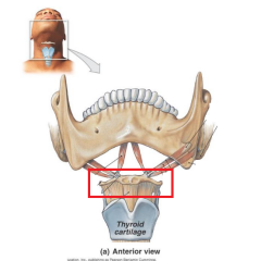
Name the bone. It does not actually articulate with any of the bones of the skull and is only attached by muscles from the mandible to it and from the Adam's Apple to it. This is commonly damaged when someone is strangled. |
Hyoid Bone (Not a bone of the Skull!!) |
|
|
Name the bony marking. |
Greater Horn of the Hyoid Bone |
|
|

Name the bony marking. |
Lesser Horn of the Hyoid Bone |
|
|
Name the bony marking. |
Mandibular Notch of the Mandible |
|
|

Name the bony marking. |
Superior Orbital Fissure of the Sphenoid Bone |
|
|

Name the bony marking. |
Inferior Orbital Fissure (between the Sphenoid Bone & Maxilla) |
|
|
Name the bony marking. |
Incisive Foramen of the Maxillae |
|
|
Name the bony marking (not the bone). |
Lacrimal Groove |
|
|
Name the bone. |
Vomer |
|
|
Name the bone. |
Palatine Bone (Here we specifically see the horizontal process). |
|
|
Name the bony marking. |
Optic Canal of the Sphenoid Bone |
|