![]()
![]()
![]()
Use LEFT and RIGHT arrow keys to navigate between flashcards;
Use UP and DOWN arrow keys to flip the card;
H to show hint;
A reads text to speech;
61 Cards in this Set
- Front
- Back
|
the integumentary system |
the largest system in your body 17-20 sq. feet if a person was skinned makes up about 16%of your body weight has the most diseases associated with this system vs. any other system in the body about 5% of your blood is ass. with this system |
|
|
dermatology |
the study of skin |
|
|
functions of the skin |
protection water barrier: prevents desiccation and provides waterproofing protects from UV light protects you from microbes, environmental things, and poisons first line of defense against disease regulates body heat tactile receptors separates you from the outside world allows for movement important in electrolyte balance helps make vitamin D through vitamin D synthesis can be important in absorption such as trans-dermal patches
|
|
|
epidermis |
the outside of the skin |
|
|
keratinocyte |
most numerous cells of the skin. hardened by keratin. important in waterproofing and immunity
|
|
|
melanocytes |
very distinct cells located at the base of the epidermis. associated with the brown pigment melanin. melanin is responsible for protection against UV light. |
|
|
melanin |
responsible for the color of skin. in Caucasians: melanin would be located in stratum basale in a lower place in brown skin; melanin would be located higher up in the stratum basale |
|
|
albinism |
have the same number of melanocytes but they are not working properly; such as problems in tyrosine. |
|
|
vitiligo |
have patchy white or colorless spots on the skin. results of melanin mal-metabolic functions |
|
|
tyrosine |
is important in the formation of melanin |
|
|
langerhans cells |
rise from the bone marrow and migrate to the epidermis. these cells are important in immunity. work in conjunction with a type of lymphocyte called a killer T. |
|
|
grinstein cells |
these cells help in protection of UV light. They help with suppressor cells made up of stratified squamous tissue. |
|
|
Thick skin |
type of skin that has 5 layers ex: soles of the feet,palms of your hands more involvement of the dermis |
|
|
thin skin |
type of skin made up of 4 layers
|
|
|
stratum corneum |
outermost layer of skin located in thick and thin skin flat, dead, filled with keratin good for waterproofing you shed this layer of the skin constantly associated with perspiration |
|
|
insensible perspiration |
... |
|
|
sensible perspiration |
... |
|
|
stratum lucidum |
found in thick skin only ex: the palms of your hand and soles of your feet contains droplets of eliden, which is important informing keratin |
|
|
stratum granulosum |
flat in cells about 5 layers thick contains the chemical keratohylin the nucleus is in the process of dying keratinzation is still occurring tonodilaments: help in slowing down water loss |
|
|
stratum spinosum |
contains spine-like stuctures called prickle cells langerhans cells demosomes shape varies these cells are alive |
|
|
stratum basale or germinativum |
the innermost layer of the skin cubodial or columnar shaped undergo cell division 25%of the cells are melanocytes associated with epidermal ridges mekel cells: sensitive to touch |
|
|
pallor |
a type of paleness of the skin that may be due to several things
|
|
|
bronze |
a bronzing of the skin, almost metallic; due to addison's disease |
|
|
jaundice |
a yellowing of the skin or sometimes the whites of the eyes. can be cause by alcoholism or a bacterial infection such as herpes. |
|
|
hematoma |
bruising of the skin |
|
|
erythema |
a redness of the skin such as being due to embarrassment, fever, hypertension |
|
|
dermis |
very thick in some areas such as the feet and the palms of the hands contains numerous blood vessels associated with hair follicles have a lot of nerve cells |
|
|
papillary area |
the outer region of the dermis, such as the fingerprints. you see numerous ridges called dermal papillae |
|
|
reticular area |
under the papillary area the innermost portion of the dermis it's tougher, more dense, and has a lot of collagen a lot of fat and blood vessels associated with this area contains hair follicles and macrophages associated with wrinkles allows for swelling |
|
|
striae |
stretch marks |
|
|
hypo-dermis |
attached to the dermis on its upper end and a membrane on its lower en made up of connective tissue and fat |
|
|
hair |
the most obvious accesory organ in mammals found all over the body it's ornamental it can be used for heat regulation can be used for touch provides protection reduces friction |
|
|
lanugo hair |
fine hair that we usually loose after birth. hormones contribute to the loss of this hair |
|
|
vellus hair |
fine hair you may see this on the legs and arms of little kids |
|
|
terminal hair |
the hair on you hair or eyebrows |
|
|
hypertichosis |
wolf man; excessive hair |
|
|
atrichosis |
lack of hair |
|
|
hirsutism |
excessive hair on females such as the bearded lady, cause by a tumor on the adrenal gland |
|
|
3 distinct regions of hair |
1. cuticle: outermost region of the hair. highly keratinized. 2. cortex: the middle part of the hair. containd the pigments of hair. 3. Medulla: the innermost region of the hair. has a lot of air spaces. |
|
|
sebum |
produced by sebaceous glands; keep hair healthy and shiny |
|
|
alopecia |
balding (more common in males) |
|
|
nails |
highly keratinized regions in the terminal portions of toes and hands help to protect the tops of our hands |
|
|
3 parts of the nail |
1. nail body: most of your nail 2. the free edge: the end of your nail 3. lunular: the cuticle, the outermost region of the nail |
|
|
sudoriferous glands |
sweat glands important in thermo regulation |
|
|
cystic fibrosis |
sweat excessively |
|
|
mammary glands |
a type of sweat gland that is designed to produce milk this gland destroys itself in the act of making milk the number of these glands is set genetically (ex:humans have 2) |
|
|
ceruminous glands |
wax glands of the ear trap dirt and other foreign objects |
|
|
lacrimal glands |
tear glands |
|
|
meiobian glands |
modified sebaceous glands; they secrete oil onto the eyeball |
|
|
epidermal healing |
what happens when your cat nicks you cell division increases and heals in a couple of days |
|
|
dermal healing |
takes longer to heal causes scarring |
|
|
sepsis |
A life-threatening complication of an infection. |
|
|
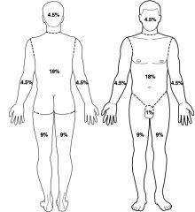
the rule of nine |
|
|
|
1st degree burns |
superficial burn just makes your skin red and swelling is painful usually heals in 2-3 days without special attention may peel |
|
|
2nd degree burns |
partial thickness burn blistered pink region around the burn red color and swelling effects the epidermis and the upper dermis painful heals well in a couple of weeks usually hair and glands with repair themselves |
|
|
3rd degree burn |
full thickness tissue damage is extensive Involves massive necrosis Can vary in color (white, black red) You can smell the tissues burning They can be dried out or they can be wetEschar: the crusty appearance around 3rd degreeburns Involve intensive medical care. Make require skin grafts |
|
|
neoplasm |
an abnormal growth of cells; mostly benign and not cancerous |
|
|
basal cell carcinoma |
the most common type of skin cancer due to UV exposure Have lesions in the skin exposed portion of the body such asthe face Startsoff shiny then it starts becoming dome shaped and in the middle of the domethere will be an ulcer. Thelayer of the skin effected the most with this type of skin cancer is the Basalelayer. Can be cut out Itwon’t spread (metastasis) for the most part. |
|
|
squamous cell carcinoma |
Develops in the stratum spinosum and the keratinocytes
Scaly and red lesions Appear on the scalp, back of hand, or ear.Requires a lot of incisions or chemotherapy for the most part recovery is okay. |
|
|
ABCDE rule of skin moles |
A: asymmetry- A benign mole has smooth, even borders, unlike melanomas. B: border- A benign mole has smooth, even borders, unlike melanomas. C: color-Most benign moles are all one color D: diameter-Benign moles usually have a smaller diameter than malignant ones. E: evolving-Common, benign moles look the same over time. |
|
|
aging and integument |
The skin thinsBruising will increase
The immune response to the skin decreases Melanocyte production fails The hair will lose pigment or fall out The skin will take longer to heal The dermis will thin andlose its elastic ability The circulation of blooddecreases within the skin Thermos regulatory problems. |