![]()
![]()
![]()
Use LEFT and RIGHT arrow keys to navigate between flashcards;
Use UP and DOWN arrow keys to flip the card;
H to show hint;
A reads text to speech;
33 Cards in this Set
- Front
- Back
- 3rd side (hint)
|
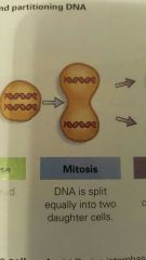
Cell Division |
The distribution of identical genetic material (DNA) to two clones... daughter cells. Purpose of CELL DIVISION is to grow and repair... divide. |
|
|
|
Cellular Organization of Genetic Material |
DNA molecules are packaged into CHROMOSOMES... EUKARYOTIC CHROMOSOMES are made of CHROMATIN (DNA plus PROTEIN). GENES are located, found on CHROMOSOMES. Sequences of DNA are found on CHROMOSOMES that code for PROTEIN. |
|
|
|
Cancer |
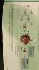
When cell process goes uncontrolled, resulting in a mass or tumor. Uncontrolled and exponential cell growth and division. Cancers do not respond to normal signals of the cell cycle, like Density-Dependent Inhibition or continued growth even in the absence of growth factors. |
|
|
|
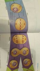
Distribution of Chromosomes during Cell Division |
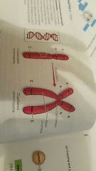
After DNA duplication, CHROMOSOMES condense... densley coiled and folded. Each duplicate CHROMOSOME has two sister CHROMATIDS containing an identical DNA molecule. Narrow waist region is called a CENTROMERE where the two CHROMATIDS are most closely bound. |
|
|
|
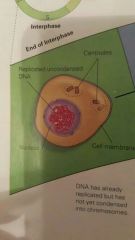
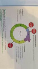
1.) Interphase |
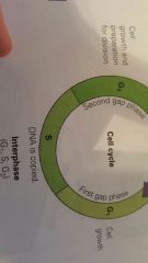
Interphase: 1.) G1 Phase (first gap): Cell growth and production of cell oganelles... roughly doubles. 2.) S Phase (synthesis): Duplication of Chromosomes and continued growth. 3.) G2 Phase (second gap): Continued growth. |
|
|
|
2. Mitotic Phase |
1.) Prophase. 2.) Prometaphase. 3.) Metaphase. 4.) Anaphase. 5.) Telophase. |
|
|
|
Phases of the Cell Cycle |
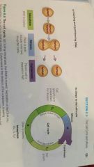
1.) Interphase (G1. S. G2.). 2.) Mitotic Phase (Prophase, Prometaphase, Metaphase, Anaphase, and Telophase). 3.) Cytokinesis. |
|
|
|
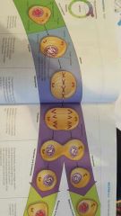
Prophase |
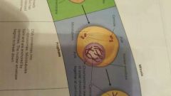
Appearance of duplicated CHROMOSOMES. Formation of a MITOTIC SPINDLE. CENTROSOMES/CENTRIOLES spread apart. |
|
|
|
Prometaphase |
CHROMOSOMES link up in center of cell. MICROTUBULES extend from each CENTROSOME toward the middle of the cell. MICROTUBULES attach to KINETICORE (CENTROMERE) of each CHROMOSOME. |
|
|
|
Metaphase |
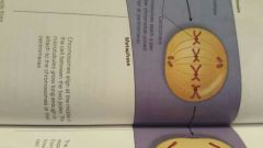
CHROMOSOMES align along METAPHASE PLATE. MITOTIC SPINDLE is now fully formed. |
|
|
|
Anaphase |
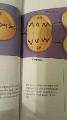
Seperation of sister CHROMATIDS forming CHROMOSOMES. CHROMOSOMES begin moving toward opposite ends of the cell. Each end of cell has identical collection of CHROMOSOMES. |
|
|
|
Telophase |
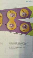
First appearance of CLEAVAGE FURROW. NUCLEAR ENVELOPE begins to form. CHROMOSOMES are less condensed (begin to unwind). NUCLEUS appearance. |
|
|
|
3.) Cytokinesis |
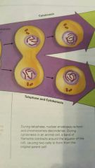
CLEAVAGE FURROW appears in animal cells. Plants, no CLEAVAGE FURROW.
Note... CELL PLATE formed during TELOPHASE. |
|
|
|
Checkpoints in the Cell Cycle |
Critical points that stop or allow the cell cycle continuance. Checkpoints controlled by signals. W/ cancer, they no longer function or are overroad.
1.) G1 CHECKPOINT (restriction point). 2.) G2 CHECKPOINT. 3.) METAPHASE CHECKPOINT. |
|
|
|
G1 Checkpoint (restriction point) |
If allows cycle to continue, the cycle usually completes the other phases and divides. If prevents cycle from continuing, the cell enters the GO PHASE (anon-dividing state). Checks to see if cell is getting bigger and enough nutrients are available for division. |
|
|
|
G2 Checkpoint |
Checks to see if DNA replicated correctly. Checks to see if cell is large enough to divide. Checks for mutations. |
|
|
|
Metaphase Checkpoint |
Are all the CHROMOSOMES attached to MICROTUBULES. |
|
|
|
Density-Dependent (CONTACT) Inhibition |
Likely due to nutrient availability. Think gash on hand... when sides of gash heal towards eachother and finally meet, they touch and CONTACT stops cell division due to this INHIBITOR. |
|
|
|
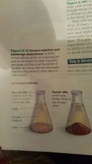
Anchorage Dependence |
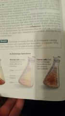
Cells must be attached to something (SUBSTRATE) B-4 they can divide. Seems to involve plasma membrane proteins and their affect on signaling the cell cycle control system. |
|
|
|
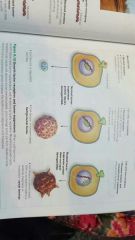
Metastatic Cancers |
Individual cancer cells break away and start a new tumor elsewhere (cancerous). |
|
|
|
Malignant Tumors |
If TUMOR invades surrounding tissue (cancerous). |
|
|
|
Benign Tumors |
If TUMOR has no effect on surrounding tissue (non-cancerous). |
|
|
|
Helacells |
Henrietta Lax story. 'Immortal' cells... grow/divide forever. |
|
|
|
Proto-Oncogenes |
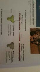
Genes thst code for proteins (growth factors) that regulate the cell cycle. 'Prior to' 'cancer'. |
|
|
|
Oncogenes |
When PROTO-ONCOGENES become mutated, they produce excessive amounts of growth factors. |
|
|
|
HER2 |
Carries instructions for building a receptor protein. If shape of receptor cell on surface is mutated, then the receptor protein could function as if many growth factors are present, even when there arn't many, thus causing cancer growth. If was normal, it would just signal inside of cell to allow normal division to occur. |
|
|
|
Run Down in Size |
CHROMOSOME - GENE - DNA - PROTEIN EX) Suppressor Genes made of protein. |
|
|
|
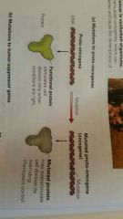
Tumor Suppressor Genes |
Genes may also encode for proteins that prevent or suppress cell division.. called TUMOR SUPPRESSOR GENES... which repair DNA. If these are damaged, then you are ******. If DNA REPAIRER IS BROKEN, OR MUTATED, THEN YOU CANT REPAIR ANY OF OTHER DNA!!! Tumor Suppressor Genes not only stop cell division, but they may also act to detect and repair damage to DNA that may have led to the mutation. |
|
|
|
P53 and BRCA2 |
BRCA2... a tumor suppressor gene... if this protein is damaged, you cant fix damaged DNA in cancerous cells. P53... a tumor suppressor gene... determines if cell will repair damaged DNA or commit cellular suicide if the damage is too severe. Of damaged, damaged DNA can proceed through MITOSIS, thereby passing on more mutations! |
|
|
|
Angiogenesis |
'Making of' 'Blood Vessels'. Cancer brings in blood that carries nutrients, so growth factors become present and tumor will begin to grow fast because of so much blood/nutrients flowing to cancerous area. Sign of cancerous area... bleeds a lot... lots of blood flowing to spot due to ANGIOGENESIS. |
|
|
|
Risk Factors for Cancer |
1.) Tobacco Use... carcinogens contain cancer causing compounds. These compounds inhibit the ability of the cell to repair damaged DNA leading to mutations. Produce FREE RADICALS that damage DNA. 2.) High-Fat, Low-Fiber Diet... eat antioxidants to avoid this. 3.) Lack of Exercise... exercise boosts immune system. 4.) Obesity... abundance of fatty tissue produces hormones that may stimulate hormone-sensitive cancers such as breast, uterine, ovarian, and prostate cancer. 5.) Excess Alcohol Consumption... alcohol usage combined with smoking multiplies the risk of certain cancers. 6.) Increasing Age... with increasing age, the immune system weakens due to accumulation of life time of damages. PREVENT WITH CONSUMPTION OF ANTIOXIDANTS LIKE FRUITS AND VEGGIES... ESPECIALLY BERRIES!!! |
|
|
|
CAUTION |
C – Change in bowel or bladder habits. This is a common sign of colorectal cancer.A – A sore that does not heal in a normal amount of time. If located on the skin or in the mouth, skin cancer or oral cancer could be the cause.U – Unusual bleeding or discharge. Any bleeding from the bladder, vagina, or rectum could mean prostate, cervical, or colorectal cancer.T – Thickening of breast tissue or a lump. Any thickening of tissue or a lump on the breast can be a sign of cancer. A lump on a testicle can mean testicular cancer.I – Indigestion. Indigestion and/or difficulty swallowing can be a symptom of stomach, throat, esophagus, or mouth cancer.O – Obvious changes to moles or warts. This is the most common sign of skin cancer.N – Nagging cough. A cough that lasts for four weeks or longer can be a symptom of lung and/or throat cancer. |
|
|
|
Fetal Bourne Syndrome |
Cells encourage growth in fetus more so than adult cells because they arnt slightly differentiated yet. Unlike adult cells, which maintain stasis, fetal bournw syndrome infers that fetal cells encouraged growth... not 'locked-in' yet. |
|