Reading...
![]()
Play button
![]()
Play button
![]()
Use LEFT and RIGHT arrow keys to navigate between flashcards;
Use UP and DOWN arrow keys to flip the card;
H to show hint;
A reads text to speech;
78 Cards in this Set
- Front
- Back
|
What are the 4 types of tissues
|
1. Epithelial-makes up the layers
2. Nervous- innervate 3. Connective- holds in place 4. Muscle-helps movement of organ |
|
|
3 main types of specialized junctions
|
1. Tight Junctions- Permiability
2. Desmosomes- Secure position 3. Gap junctions- pore like structures that allows H20 and solution molecules to equlibrate. |
|
|
Define: Endo-
|
Endo means directly released into blood
|
|
|
Define: Exo-
|
Exo means released via a duct
|
|
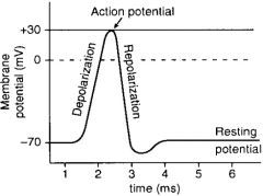
5 main steps of conducting a nerve impulse
|
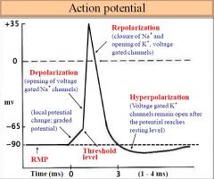
1. Stimulus: Resting nerve is -80m. There is a THRESHOLD that must be reached as an ALL OR NOTHING signal
2. Depolarization: Na+ flows in 3. Repolarization: K flows out. Nerve is -60 4. Hyperpolarization: K flows in 5. Refractory: Na+ chaneels close- no other AP |
|
|
Name to 5 actions that take place at the POST synaptic cleft to propagate an action potential
|
1. The action potential opens the Ca2+ inonophore
2. Ach binds post synaptic cleft and released into the cleft 3. Ach goes to membrane and binds Pre synaptic cleft 4. New action potential is propagated 5. Ach-esterase breakes Ach back to Acetyl- Co |
|
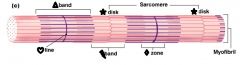
What are the following:
1. Triangle 2. Star 3. Heart 4. Lightening Bolt 5. Diamond |
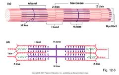
1. Triangle: A band- Myosin (thick)
2. Star: Z disc 3. Heart: M line 4. Lightening Bolt: I band- Actin (thin) 5. Diamond: H zone |
|
|
Describe how muscle contractions work
|
1. Relaxed- Myosin is 45degrees. Troponin hiding the myosin heads.
2. ATP binds and myosin releases the Actin 3. ATP is hydrolyzed to ADP and Pi 4. Ca2+ floods our from the Sarcoplasmic reticulum and shifts the tropomyosin into place 5. Myosin and actin bind 6. Inorganic phosphate leaves and ADP leaves= POWERSTROKE and moves it forward 7. ATP binds and causes myosin to release the actin again |
|
|
Describe the following part of the brain:
1. Paretial 2. Temporal 3. Frontal 4. Occipital |
1. Paretial- touch
2. Temporal- Hearing 3. Frontal- Personality 4. Occipital- Vision |
|
|
Components of Forebrain
|
1. Cerebrum- Cerebral cortex
2. Thalamus- Visual and auditory 3. Hypothalamus- Visceral 4. Pituitary- Master Endocrine |
|
|
Components of Brainstem
|
Midbrain: Movement and pleasure
Cerebellum: Coordination Pons/Medula: Visceral Reticular Formation: alerts and shut off to sleep |
|
|
Example of a Monosynaptic Pathway
|
Extensor Muscles
|
|
|
Example of Polysnaptic Pathway
|
Flexor
|
|
|
starch
|
glucose polymer w/alpha linkages, found in plants
|
|
|
glycogen
|
branched polymer of glucose with alpha linkages
|
|
|
cellulose
|
glucose polymer w/beta linkages, found in plants. not digestable by animals.
|
|
|
competitive inhibitors
|
![take up active site [Efree] but not permenantly. can be overcome by high conc of substrate
Vmax same
Km looks to increase](https://images.cram.com/images/upload-flashcards/939318/683359_m.gif)
take up active site [Efree] but not permenantly. can be overcome by high conc of substrate
Vmax same Km looks to increase |
|
|
Noncompetitive inhibitors
|
![bond to enzyme at nonactive site [Efree] and [ES], change its shape to make it less effective.
Vmax decreases
Km stays the same](https://images.cram.com/images/upload-flashcards/939318/683360_m.gif)
bond to enzyme at nonactive site [Efree] and [ES], change its shape to make it less effective.
Vmax decreases Km stays the same |
|
|
glycolysis
|
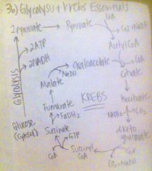
breaks 6-carbon glucose into 2 3-carbon pyruvates, 2 molecules ATP and 2 NADH. occus in cytosol.
|
|
|
aerobic respiration
|
produces 36 ATP incl glycolysis. 1 NADH = 3ATP, 1FADH2 = 2ATP.
|
|
|
krebs cycle
|
pyruvate->acetyl CoA turns cycle 2x. Cycle produces 1ATP, 3NADH, 1FADH2.
|
|
|
Enzymes exhibit saturation kinetics.. =
|
as the conc of substrate increases, the rate of the rxn also increases until Vmax (max rate). Vmax is proportional to enzyme concentration.
|
|
|
Protein Structure:
Primary Secondary Tertiary Quaternary |
Primary: sequence of amino acids that determines basic secondary structure of protein (B mercampenol experiment)
Secondary: α helix, β sheet Tertiary: Twisted polypeptide, interactions within itself (Zinc Finger and Leucine Zippers) Quaternary: multiple polypeptide, interactions between subunits |
|
|
Factors that effect enzymes
|
Temperature
PH |
|
|
Where does most glycolysis occur in humans?
|
Cytosol of Liver Cell
|
|
|
Aerobic Respiration occurs where?
|
In the mitochondria
|
|
|
Which are the Purines
Which are the Pyrimidines |
Adenine, Guanine
Cytosine, Thymine, Uracil CUT the Py |
|
|
Pathway of Eye
|
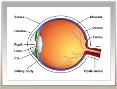
Cornea
Anterior Cavity Pupil Iris Lens (connected to ciliary muscle) Retina (rods and cones) |
|
|
Pathway of Ear
|
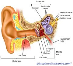
Tympanic Membrane
Malleus, Incus, Stapes Oval Window Cochlea Hair cells |
|
|
Stomach (4 major cell types)
|
Mucous Cells- makes mucous
Chief Cells- Synthesis Pepsinogen Parietal Cells- Est. a H+ gradient G Cells- Secretes Gastrin |
|
|
Pancreas- 6 major enzymes
|
1)Trypsin
2)Chymotrypsin 3)pancreatic amylase 4)lipase 5)ribonuclease 5)deoxyribonuclease |
|
|
Autonomic- Sympathetic
Parasympathetic |
flight or fight (epinephrine and norepinephrine)
rest and digest (acetylcholine |
|
|
Give the divisions of the PNS
|
I. Peripheral Nervous System
a. Somatic Nervous System (Voluntary)- Ach b. Autonomic Nervous System (Involuntary)- Ach and Noroepi |
|
|
Parasympathetic Nervous System
|
PNS (Rest and Digest):
a. Origin-Sarcal+ Midbrain b. Pre Ganglion- long (ACh) c. Post Ganglion- Short, near the organ it affects (ACh) d. ex. Vagus Nerve |
|
|
Sympathetic Nervous System
|
SNS (Fight or Flight):
a. Origin- Thoraic and Lumbar b. Pre Ganglion- short, into a trunk with diff fates. (ACh) c. Post Ganglion- long, far from organ to affect (Noroepi) d. ex. Adrenal Medula has no post ganglion |
|
|
Non Polar Amino Acids (Hydrophobic)
|
Glycine, Alanine, Valine, Leucine, Isoleucine, Proline, Methionine, Phenylalanine, Tryptophan
|
|
|
Polar Charged Amino Acids (Hydrophilic)
|
Serine, Theronine, Cysteine, Tyrosine, Asparagine, Glutamine
|
|
|
Polar Charged amino Acids
|
Aspartate, Glutamate, Lysine, Arginine, Histadine
|
|
|
Henderson Hassalbach Equation
|
pH=pKa+ log [A-]/[HA]
|
|
|
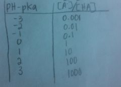
Henderson Hassalbach Trick
Relationship between pH-pKa to [A-]/[HA] |
|
|
|
Isoelectric Point
|
pI=[pKa1+pKa2]/ 2
pH>pI- migrate to anode (+) pH<pI- migrate to cathode (-) |
|
|
Explain a titration curve and which is the best buffering region.
|
pH is 1 unit from the pKa is the best buffer region.
|
|
|
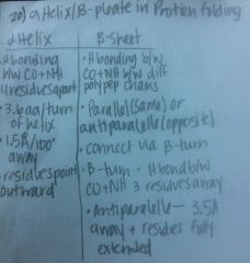
Give characteristics of alpha helix/B pleat of Protein folding.
|
|
|
|
Formula for sterioisomers
|
2^n where n is the number of sterioisomers,
(this typically excludes the first and last carbons, and the carbon that is the keytone, therefore 5 carbon carb has only 3 sterioisomers) |
|
|
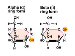
Alpha vs Beta Glucose
|
|
|
|
Give the 2 oxidizing agents used to see the carbohydrate functional groups (aldose, ketose)
|
1. Tollen's Agent (Ag+)- Silver precipitate
2. Benedict's Reagent (Cu2+)- Brick Red precipitate Note: these only detect reducing sugars such as Lactose (hemiacetal and hemiketal, not acetal and ketals) |
|
|
Formula for carbohydrate
|
(CH2O)n
-Aldehyde/Keytone + 2 or more alcohols |
|
|
Define and give the properties for Saturated Fatty Acids
|
Saturated: All carbons have max number of H's
increased Wanderwalls, increased melting point, decreased fluidity, solid at RT. |
|
|
NTP binding and strenght
|
A=T (U)
C≡G |
|
|
Types of RNA
|
mRNA: message to synthesize protiens
tRNA: transfers amino acids in ribosome rRNA: makes up the ribosome |
|
|
Types of Membrane Transport
|
1. Simple Diffusion
2. Facilitated Diffusion 3. Primary Active Transport 4. Secondary Active Transport |
|
|
Simple Diffusion
|
Moves down a concentration gradient- from high concentration to low concentrations
|
|
|
Facilitated Diffusion
|
Moves down a concentration gradient, but needs some help (a membrane protein).
a. Uniport- one thing at a time b. Symport- 2 things at a time in the same direction c. Antiport- 2 things at a time in opposite directions |
|
|
Primary Active Transport
|
Goes against a concentration gradient, from low concentrations to high concentrations. Uses energy directly from the hydrolysis of ATP
Only 2 known 1. Na+-K+ pump: Antiport 2 K+in 3Na+ out 2. Ca2+ ATPase: 2Ca+ out into the cytosol for every ATP |
|
|
Secondary active transport
|
Goes against a concentration gradient, from low to high.
Uses ionic gradient established by primary active transport. ex. Glucose and Na+ in the kidney: increased Na+ in the cell already but once glucose and Na+ bind the symport lets them both in. The Na-K pump only reads the Na+ levels (1 active transport) but the glucose piggybacks in secondary transport |
|
|
Describe the Michaelis-Menten Enzyme Inhibition for competitive and non competitive inhibition
|
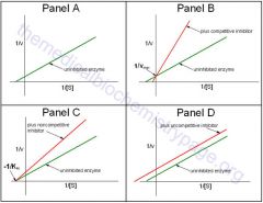
Panel A: Original
Panel B: Competative Inhibitor (same Vmax different Km) Panel C: Non-competative Inhibitor (different Vmax same Km) Panel D: Uncompetative inhibitor (Paralell to original) |
|
|
Relationship between Km (K2/K1) and ES binding
|
As Km increases= decreased ES binding
As Km decreases= increased ES binding Km=K2/K1 |
|
|
Define zero order and first order kinetics
|
First order: V proportional to [S]= As [S] increases V (rate) increases
Zero order: V independent to [S]= As [S] increases V doesn't change |
|
|
Name the 4 check points for regulation of glycolysis
|
1. Increased ATP= Decreased Phosphofructose Kinase (PFK).
2. Increased AMP= increased PFK. 3. Increased Citrate= Decreased PFK 4. Increased H+= decreased PFK functionality. |
|
|
How does pH affect PFK functionality?
|
Increased H+= This decrease in pH augments ATP inhibition of PFK. This assumes lactic acid buildup so decreased PFK functionality.
|
|
|
How does citrate buildup affect PFK functionality?
|
Increased Citrate= This means that there is a lot of citrate buildup from the citrate acid cycle. There is no more need to commit more glucose for degredation. Decreased PFK
|
|
|
How does AMP affect PFK functionality
|
Increased AMP= AMP reverses the inhibition from ATP. Therefore increased PFK. PFK activity increases as ATP/AMP ratio is lowered.
|
|
|
How does ATP affect PFK functionality
|
Increased ATP= Decreased Phosphofructose Kinase (PFK). ATP alosterically inhibits PFK. PFK activity increases as ATP/AMP ratio is lowered.
|
|
|
Starting at Glucose name the essentials thru glycolysis and Krebs
|
|
|
|
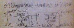
What is gluconeogeneisis
|
Gluconeogenesis is synthesis of glucose. It takes place in the liver.
|
|
|
Explain what happens in the muscle cell and liver cell respectively in terms of glycolysis.
|
|
|
|
What drives Krebs and Gluconeogenesis
|
Increased Acetal CoA= Increased Pyruvate Carboxilate= increased oxaloacetate which drives Krebs and Gluconeogensis
|
|
|
Overall Net ATP of Gycolysis and Gluconeogenesis
|
Glycolysis = 2ATP
Gluconeogensis=6ATP therefore NET -4 ATP |
|
|
True or False: Gluconeogensis is the reverse of Glycolysis
|
False, it is not the exact reversal because there are irreversible steps in glycolysis (where ATP) have been spent, that are bypassed in glyconeogensis to prevent spending ATP to undo it.
|
|
|
How many ATP are the following worth
a. 1 FADH b. 1 NADH |
a. 1FADH=2 ATP
b. 1NADH=3 ATP |
|
|
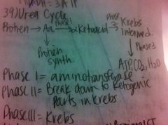
Give a general overview of the urea cycle
|
|
|
|
What are the parts of the Kidney's nepheron and the journey through it.
|

a. Proximal Convoluted Tubule
b. Loop of Henley c. Distal Convoluted Tubule d. Collection Duct |
|

Describe how the Proximal convoluted tubule (a) works
|

Decreased regulation.
100% absorption of Glucose, Protein, and Vitamins. 80% Absorption of H20 and Na/Cl |
|
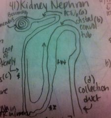
Describe how the distal convoluted tubule (b) works.
|

Close to the loop of Henley.
Impermeable to urea and H20 Lets ions out Increased dilution of the urine |
|

Describe how the loop of henely (c) works.
|

There are three parts to the Loop of Henely.
*= H20 can leave. Urea and NaCl cannot. Increase concentration of urine. **= Urea can leave, but NaCl and H20 can not. Decrease concentration of urine. ***=Actively transports ions (NaCl) out. H20 and urea can not leave. Further dilutes urine. |
|

Describe how the collecting duct (D) works
|
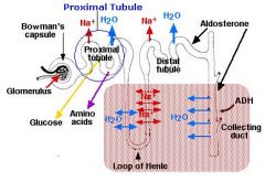
ADH- increased release of ADH causes increased H20 reabsorption which causes increased urine concentration
Aldosterone- increased aldosterone means increased Na in epithelials and increase K+ in lumen. |
|
|
Explain how ADH can be used to counteract a drop in BP due to a hemorrhage for example
|
Premise: Blood pressure drops therefore decreased blood volume
1. ADH- Atria--> Hypothalmus to release vassopressin (ADH) 2. This works in the kidneys to increase H20 resorption into the blood thereby increasing blood volume. 3. This leads to increased blood molarity as well because ADH is circulating in the blood. B. |
|
|
Explain how the renin-angiostensin-aldosterone system can be used to counteract a drop in BP due to a hemorrhage for example
|
1. When BP drops renin is released into the blood stream.
2. Renin floats around and converts inactive forms of angiotensin (produced by liver) into angiotensin I 4. Most angiotensin I is converted to angiotensin II this then a. directly causes vasoconstrition in the arterioles (thereby increasing BP) and b. causes aldosterone to be released from the cortex. Aldosterone increases Na+ reapsorbtion into the blood therefore increasing blood osmolarity and so H20 will flow there and this increases blood volume and thereby increases BP. |