Reading...
![]()
Play button
![]()
Play button
![]()
Use LEFT and RIGHT arrow keys to navigate between flashcards;
Use UP and DOWN arrow keys to flip the card;
H to show hint;
A reads text to speech;
48 Cards in this Set
- Front
- Back
|
Obj.
Describe the development of the meninges |
-develop from 2 primary layers (neural crest & mesoderm)
-primitive single layer forms by 20-35 days -layer separates into ectomeninx & endomeninx/leptomeininges -by end of first trimester adult patterns have formes |
|
|
What does the ectomeninx become?
endomeninx/leptomeininges? |
dura
(pachymeninx) pia & arachnoid (ENDO= inner layers) (arachnoid + pia = leptomeninges) |
|
|
Obj.
Orientation of the continuous primary meningeal layers in the brain and spinal cord (outer to inner) |
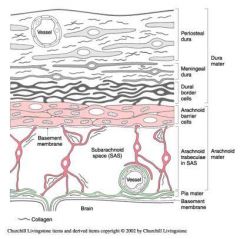
-dura mater
periosteal dura meningeal dura dural border cells -arachnoid mater arachnoid barrier cells arachnoid trabeculae + subarachnoid space -pia mater |
|
|
What is a congenital dermal sinus?
|
developmental defect where ectoderm fails to completely dissociate from neuroectoderm, leaving an epithelium lined channel to the surface of the skin.
*hole in outer dura, spina bifida aperta *associated w/ spina bifida aperta -can result in recurrent meningitis, usually surgery |
|
|
The ectomeninx (future dura) stays attaches to the periosteal CT layer at suture line throughout development. What does this create in the adult?
|
2 dural layers:
periosteal (outer) (connected to bone/skull) & meningeal (inner) |
|
|
In the adult spinal cord the space between the vertebral periosteum dura and the meningeal dura creates what space?
|
epidural space
*no space btwn layers in brain |
|
|
What attaches the dura to the arachnoid?
|
dural border cells
|
|
|
The dural border cell area is considered a 3rd dura layer, what does this area allow for?
|
reduced adherence btwn dura & arachnoid, potential space, "weak spot"
|
|
|
What attaches the arachnoid to the pia?
|
arachnoid trabeculae
|
|
|
What continuous space separates the pia and arachnoid?
|
subarachnoid space
(contains CSF) |
|
|
What is a sub dural hematoma?
|
typically venous bleeding btwn dura and arachnoid
|
|
|
What are dural septa (infoldings)?
|
-portions of meningeal dura that fold inward into the cranial cavity & separates it into compartments
=restrictive barrier for movement of brain w/i cranial cavity |
|
|
What are the 2 primary dural septa?
|
1. falx cerebri
2. tentorium cerebelli |
|
|
Which primary dural septa lies in the longitudinal fissure and attaches anteriorly to the crista gali?
*separates the cerebellar hemispheres* |
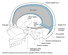
falx cerebri
*attaches posteriorly to the tentorium cerebelli. |
|
|
Which primary dural septa lies axially in the transverse fissure connecting to the anterior clinoid processes?
*separates the cerebrum from cerebellum* |
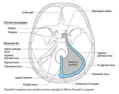
The tentorium cerebelli
|
|
|
What is the falx cerebelli?
|
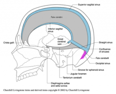
a smaller septum that lies in the midline of the cerebellar hemispheres to varying heights.
|
|
|
What is the diaphragma sella?
|
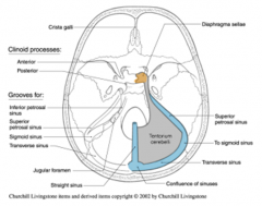
the smallest septum that forms the roof of the hypophyseal fossa , over the sella turica, encircling the penetrating infundibulum.
|
|
|
Describe the dural compartments
|
-lateral supratentorial compartments (separated by falx cerebri)
-infratentorial compartment (bordered by tentorium cerebelli) |
|
|
What effect would an expanding mass have on contents of the dural compartments?
|
can push contents btwn compartments
|
|
|
What are dural sinuses?
|
*large veins btwn periosteal & meningeal layers
formed by two mechanisms: 1. separation of the meningeal and periosteal dura at junctions with the skull. 2. Joining of two layers of meningeal dura at the free edges of dural septa *blood collects in sinuses from connecting veins, drains CSF |
|
|
What is the vascular supply to the dura in the cranium?
|
branches from internal carotid artery
|
|
|
An expanding space occupying hematoma btwn the 2 dural layers, that disrupts the arterial supply btwn the periosteum & periosteal dura is referred to as __________________
|
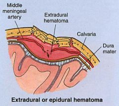
extradural/epidural hematoma
|
|
|
The ________ is insensitive to pain, but the _______ are sensitive
|
brain (insensitive)
dural coverings (sensitive) |
|
|
The anterior and middle cranial fossa are innervated by branches of the _______ nerve.
|
trigeminal nerve
|
|
|
The dura of the posterior fossa receives sensory branches from _________________and may have some sensation through the vagus nerve.
|
C2 and C3 (also C1 when present)
|
|
|
The tentorium is supplied by the __________ nerve, a branch of the opthalmic nerve (V).
|
tentorial nerve
|
|
|
Because the dura above the tentorium is innervated by the trigeminal nerve, a headache from the irritation or infection of the dura in this region will be referred to where?
|
the face
|
|
|
The infratentorial dura is innervated by the cervical nerves, irritation in this area will be referred where?
|
to the back of the head
|
|
|
Q: the difference between an epidural (extradural) hematoma & subdural hematoma is that the subdural hematoma is where?
|
btwn the meningeal dura & arachnoid matter
|
|
|
The _________ mater is avascular and not innervated
|
arachnoid mater
|
|
|
What are the 2 primary parts of the subarachnoid space (btwn arachnoid & pia)?
|
1. arachnoid barrier layer
2. arachnoid trabecule |
|
|
_____________________ has cells with tight junctions that form a barrier to the diffusion of CSF from the subarachoid space.
|
The arachnoid barrier layer
|
|
|
________________are delicate strands of connective tissue spanning across the subarachnoid space to the pia. These act to suspend the brain in the subarachnoid space.
|
Arachnoid trabecule
*allow brain to move in fluid |
|
|
What are arachnoid villi?
|
specializations of the dura arachnoid interface in dural sinuses that allow the drainage of CSF into the venous system
collections of villi= granulations *essential for CSF circulation & normal intracranial pressure |
|
|
The pia mater can be divided into what 2 layers?
|
1. epipial layer - external
2. intima pia layer - close to the glial limitans or glial basement membrane that forms the outermost layer of cerebral cortex *pia is thicker in the spinal cord, very tight around brain |
|
|
Pial cells surround and follow surface blood vessels into the brain of spinal cord substance along w/ a small area of extracellular space known as.......
|
Virchow-Robin space
*allow for exchange of ECF w/ CSF, allow leukemic cells to enter brain |
|
|
The absence of a dural attachment from the spinal cord to the vertebra allows what?
|
allows dura to stretch as the vertebra move
|
|
|
What neurological signs associated w/ stretching of spinal dura & its attachment to nerve roots are used to indicate meningitis?
|
Kernig (inability to straighten leg)
& Brudzinski (passive neck flexion causes involuntary hip flexion) |
|
|
The spinal cord dura is anchored to the spinal column indirectly through the exits of the spinal roots and the___________, that connects the end of the dura to the coccyx.
|
filum terminale externum
(after spinal cord ends, dura continues & is anchored to coccyx) |
|
|
What specialized attachments does the pia mater form in the spinal cord?
|
-filum terminale internum: attaches to the caudal end of the dural sack that makes up the lumbar cistern.
-denticulate ligaments: attach from the spinal cord pia arachnoid to spinal dura at 21 pairs of points between the foramen magnum and the first lumbar spinal nerve |
|
|
What is the primary function of the filum terminale & denticulate ligaments?
|
The filum terminale and denticulate ligaments further help stabilize the spinal cord relative to the dura and spinal column.
|
|
|
What is the primary subarachnoid cistern (enlargement of subarachnoid space) associated with the spinal cord meninges?
|
is the lumbar cistern
-extension of spinal meninges (L1-2) after end of cord, contains cauda equina, space commonly accessed in spinal taps (L3-4) |
|
|
What are meningiomas?
|
tumors of meninges that can for space occupying lesions & compress brain in dural compartments
*usually benign & atypical, located outside brain parenchyma, rarely penetrate brain tissue= slow detection |
|
|
Meningities usually involves virus & bacteria. Bacterial infections typically involve what layers?
|
arachnoid & pia, spread in the subarachnoid space
|
|
|
Infections of meninges (meningitis) typically causes what?
|
thickening of the meninges and obstruction of CSF (cloudy w/ WBCs) return or flow
*acute forms can cause death w/i 2 days |
|
|
Q: from the skull inward the proper order of layers is?
|
dura, arachnoid, subarachnoid, & pia
|
|
|
Q: the dural septum that lies in the longitudinal fissure is.....
|
falx cerebri
|
|
|
Q: bacteria causing meningitis are likely to spread most rapidly where?
|
in subarachnoid space
|