Reading...
![]()
Play button
![]()
Play button
![]()
Use LEFT and RIGHT arrow keys to navigate between flashcards;
Use UP and DOWN arrow keys to flip the card;
H to show hint;
A reads text to speech;
153 Cards in this Set
- Front
- Back
|
|
|
|
Reticulin in space of Disse is made of....
|
type 3 collagen and 4 and 18
McG 397 |
|
|
Kupffer cells are:
a) mobile b) immobile and can express class ... histocompatibility antigens for APC functions (not super efficient though) |
a) mobile
class 2 McG 398 |
|
|
Hepatic stellate cells are also called......
|
lipocytes or ito cells
|
|
|
Hepatic stellate cells.
Functions? |
storing Vit A
hepatic injury → lose Vit A → synthesize collagen → hepatic fibrosis McG 398 |
|
|
gallblader is absent in for example these 2 mammals:.......
|
rat and horse
|
|
|
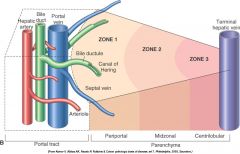
Canals of Hering are also called ..... and are lined by ......
|
cholangioles
low cuboidal epithelium |
|
|
3 functions of bile
|
- excretion (eg cholesterol, bilirubin, xenobiotics → excreted in bile)
- digestion of lipids - neutralize the acid pH of ingesta McG 398 |
|
|
Urobilinogen that is not reabsorped is oxidized to ....
|
stercobilin, which is responsible for the color of the feces
McG 399 |
|
|
1, Normal bilirubin production from heme (0.2 to 0.3 g per day) is derived primarily from the breakdown of senescent circulating erythrocytes, with a minor contribution from degradation of tissue heme-containing proteins. 2, Extrahepatic bilirubin is bound to serum albumin and delivered to the liver. 3, Hepatocellular uptake and 4, glucuronidation in the endoplasmic reticulum generate bilirubin monoglucuronides and diglucuronides, which are water soluble and readily excreted into bile. 5, Gut bacteria deconjugate the bilirubin and degrade it to colorless urobilinogens. The urobilinogens and the residue of intact pigments are excreted in the feces, with some reabsorption and excretion into urine.
|
|
|
bile may be recycled up to ... times per day
|
15
|
|
|
Which liver enzyme in the bilirubin-pathway has the terrible name?
|
bilirubin UDP-glucuronyltransferase
McG 399 |
|
|
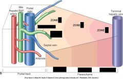
explain: portal streaming
|
refers to the differential flow of portal blood from one segment of the digestive tract to particular lobes of the liver
McG 401 |
|
|
vein from navel to liver (neonates) is called ....
|
umbilical vein
|
|
|
Biliary tree is defended in part by Ig A. Describe how this gets there.
|
gastrointestinal plasma cell → Ig A → lymph vessel → blood stream → hepatocyte → canaliculi
McG 401 |
|
name the morphocologic pattern of necrosis
|
Random hepatocellular injury (Salmonellosis, Su)
|
|
name distribution.
Cause of necrosis? |
- Centrilobular necrosis (periacinar or zone 3)
- least oxigenated, greatest enzymatic activity (activating toxic forms) Causes: right side heart failure, anemia, ... McG 403 |
|
name distribution of necrosis
Describe possible causes |
Paracentral degeneration and necrosis, zonal hepatocellular injury, liver, cow. A wedge-shaped area of hepatocytes is damaged. This wedge is the apex of the diamond-shaped liver acinus (zone 3) and reflects the partitioning of the lobule based on the inflow of blood from each of the individual portal tracts that surround the lobule. This change can be seen as an early manifestation of hepatic hypoxia in animals with anemia or right-sided heart failure and precedes centrilobular necrosis.
|
|
name distribution of necrosis
a cause in Fe? a cause in Su + Eq? |
Midzonal necrosis
Fe: hexachlorophene Su + Eq: aflatoxicoses |
|
|
Latin: inflammation that is centered on the biliary tract
|
Cholangitis
|
|
|
Latin: inflammation of the gallblader
|
Cholecystitis
|
|
|
Latin: inflammation that affects the biliary ducts and hepatic parenchyma
|
Cholangiohepatitis
|
|
|
concentrations of .... above .... can produce icterus (jaundice), especially evident in tissue conatianing ... This concentration is within ref range for ...., however in other Spp hyper.... can occur over ... mg/dl
|
bilirubin above 2 mg/dl → icterus, esp tissues w elastin.
Norm for Eq, in othe Spp hyperbilirubinemia over 0,5 mg/dl (Can) |
|
|
Hepatic problem. How long does it take before bilirubine reaches max in tissues?
|
2 days (so no icterus if animals die shortly after acute hepatic failure)
|
|
|
3 causes of hyperbilirubinemia
|
1) overproduction of bilirubine (hemolyse)
2) Decreased uptake, conjugation or secretion (severe diffuse hepatic disease) 3) Cholestasis (extrahepatic or intrahepatic) McG 407 |
|
|
"physiologic icterus" is common in what Spp?
Deprived of feed they can become icteric because.... |
Eq
uptake of bilirubin from the plasma by hepatocytes is decreased McG 407 |
|
|
Corriedale sheep may have a mutation that leads to deficient conjugated bilirubin excretion. Describe their liver.
|
dark and discoloured due ti polymerized catecholamine metabolites that accumulate in lysosomes
McG 407 |
|
|
Intrahepatic cholestasis can result from.... (X3)
|
1) liver injury (inability to produce/excrete bile)
2) hemolysis (↓oxygen for hepatocytes) 3) inherited abnormalities of bile synthesis McG 407/408 |
|
|
Describe how acholic feces are formed
|
Complete biliary obstruction → no stercobilin (bilirubin-derived pigment produced by bacterial metabolism) → clay-colored stool
|
|
|
Stellate cell + liver injury → change to cell with myofibroblastic appearance → produce collagen types ..., .... and .... They also produces matrix components like ... and ....
|
Collagen types I, III and IV
Matrix components like laminin and chondroitin sulfate proteoglycans. |
|
|
Cirrhosis: definition
It originally meant..... |
Diffuse fibrosis with hyperplastic nodule formation
WHO:"a diffuse process characterized by fibrosis and conversion of normal liver architecture into structurally abnormal lobules" Originally meant: "tawny yellow". So not a strong descriptive term. McG 412 |
|
|
Lobular dissecting hepatitis
Spp? Cause? |
Young dogs
Cause unknown [small and smooth livers, w fine septa w increased fibrosis that dissect the hepatic plates. Often fatal] |
|
|
Chronic passive hepatic congestion eventually leads to fibrosis near ..... which is sometimes termed ... and can progress to ......
|
near central veins
cardiac sclerosis cardiac cirrhosis |
|
|
Abnormal storage of copper may produce an end-stage liver. Name 3 breeds w hereditary problems.
|
Dalmatians, Bedlington and West Highland white terriers.
McG 413 |
|
|
5 potential consequences of hepatic dysfunction and failure
|
1) hepatic encephalopathy
2) hyperbilirubinemia 3) metabolic disturbances 4) vascular and hemodynamic alterations 5) cutaneous (eg. epidermal necrosis in dogs, photosensitization in herbivores) McG 414 |
|
|
Why does liver failure cause CNS signs?
3 theories |
1) if portal blood bypasses the liver, or livercells don't metabolize enough → ↑NH3 in blood →brain.
2) inbalance of excitatory and inhibitory amino acid neurotransmitters (l-glutamate and GABA) 3) ↑ brain concentration of endogenous benzodiazepines McG 414 |
|
|
Latin: bleeding tendencies
|
hemorrhagic diathesis
|
|
|
All clotting factors, except ..., are synthesized in .....
|
VIII
the liver McG 414 |
|
|
Acute liver failure → shortage of clotting factors with short half-life such as: ...... (x4)
Chronic liver disease → factor ..... deficiency |
acute: V, VII, IX, X
chronic: II (prothrombin) McG 414 |
|
|
liver failure → metabolic disturbances → affect platelet function → syntesis of abnormal fibrinogen.
Latin? |
dysfibrinogenemia
McG 414 |
|
|
obstruction of biliary system → ..................... → hemorrhagic diathesis.
Explain |
obstruction of biliary system → impaired fat absorption → limits Vit K uptake → inactivity of factors II, VII, IX, X
McG 414 |
|
|
Box 8-4: Causes of End-Stage Liver (X6)
|
1) Chronic toxicity
2) Chronic cholangitis and/or obstruction 3) Chronic congestion (right side heart failure) 4) Inherited disorders of metal metabolism (copper or iron) 5) Chronic hepatitis 6) Idiopathic McG 413 |
|
|
Hypoalbuminemia: reflect chronic liver disease, not acute, because half-life of albumin is: .....
|
8 days in Can
21 days in Bo |
|
|
Hepatocutaneous syndrome.
- Gross - Histo |
- Dog with severe hepatic disease → crusting, erosions, ulcerations of epidermis → muzzle, mucocutaneous areas of face, footpads and pressure points
- typical: parakeratosis, edema of the superficial epithelium, basal cell hyperplasia McG 415 |
|
|
Primary photosensitization.
Causes? |
St John´s wort (Hypericum perforatum) and buckweat (Fagopyrum esculentum).
Tetracycline, phenothiazine, ... McG 416 |
|
|
Secundary photosensitization.
Pathogenesis? |
loss of 80% of hepatic function or obstruction → impairs excretion of phylloerythrin (made from chlorophyll by gastrointestinal bacteria) in bile → accumulation in blood and cutaneous tissues
McG 416 |
|
|
3 mammals that don´t have a gallblader
|
rat, horse, elephant
McG 416 |
|
|
Which "standard" layer is absent in the gallblader?
|
Muscularis mucosae
McG 416 |
|
|
Exocrine pancreas secretes enzymes to break down lipids: .....
proteins: .... carbohydrates: .... |
lipids: lipase and phospholipase
proteins: trypsin and chymotrypsin carbohydrates: amylase |
|
|
.... is one of the more important hormones involved in pancreatic secretion, and it stimulates water and bicarbonate secretion by duct cells. It is produced by neuroendocrine cells within .....
|
Secretin
duodenal epithelium McG 417 |
|
|
...... , an important hormone, stimulates the release of digestive enzymes from the acinar cells (pancreas).
|
Cholecystokinin
McG 417 |
|
|
Dogs, cats and calves with very little pancreatic tissue develop: ....
While horses develop: ..... |
Exocrine pancreatic insufficiency
Hypoinsulinism McG 417 |
|
|
Congenital cysts in the liver are found in which Spp? (X3)
What is an important DDX? |
Can, Fe, Su
DDx: cysticerci (or other parasitic cysts) McG 418 |
|
|
The typical multiple cysts affecting extensive areas of the liver in Fe, Su and to a lesser extend Can, are believed to be:
....... or ...... |
developmental anomalies of the intrahepatic bile ducts
or benign cystic neoplasia |
|
|
Congenital polycystic liver disease.
Predisposed:...... |
Cairn terriers
Wehst Highland white terriers Fe, esp Persian Fe Capr McG 418 |
|
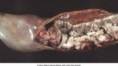
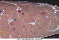
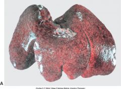
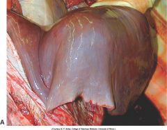
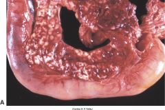
Liver, cut surface, cow. What is wrong with the liver?
|
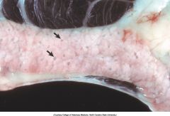
Tension lipidosis, liver, cut surface, cow. Note the area of fatty infiltration (F), the ligamentous attachment adjacent to the affected portion (arrowhead), and the areas of telangiectasis (arrows)
This is common in Bo and Eq. Tension → impede blood supply → accumulation of fat in hepatocytes |
|
|
Chronic passive congestion in old dogs is common. Cause?
|
Endocardiosis (mucoid degeneration) of right atrioventricular valve
McG 419 |
|
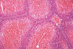
Liver
Pathogenesis + typical name for the lesion |
Chronic passive congestion → persistent hypoxia in centrilobular area → degenerate → sinusses dilate → gross: red
Chronic passive congestion → hypoxia of periportal hepatocytes → fatty degeneration (lipidosis) → Gross: white This is a Nutmeg liver (accentuation of lobular pattern, enhanced reticular pattern) |
|
|
Hepatic veno-occlusive disease.
Characterised by: ...... Not etio-specific, but can follow ....... or ..... induced hepatic injury. Extreme high incidence in ......, possibly because of ingestion of large amounts of ...... |
Intimal thickening + occulsion of central veins
can follow pyrrolyzidine alkaloid or aflatoxin-induced hepatic injury Captive exotic cat → Vit A (?) McG 420 |
|
|
typically, INTRAhepatic portosystemic shunts involve failure of closure of the .....
at birth |
ductus venosus
|
|
|
Intrahepatic shunt are found most often in the ... side of the liver, in .... size dogs. Extrahepatic shunt are found most often in ... size dogs.
|
left side.
Intra: large breeds of dogs Extra: small breeds of dogs |
|
|
Do congenital portosystemic shunt lead to ascites? Why?
|
No
Portal vein pressure is normal. |
|
|
Portal hypertension can be devided into 3 categories:
Prehepatic Intrahepatic Posthepatic Causes per category |
Pr: Portal vein thrombosis, tumor emboli, external compression, portal vein hypoplasia
Intra: chronic liver disease (most common), veno-occlusive disease, amyloidosis, schistosomiasis, arteriovenous fistulae Post: Thrombosis of hepatic veins (= Budd-Chiari syndrome) McG 422 |
|
|
Ammonium biurate crystals, urinary bladder, dog. The bladder contains ammonium biurate crystals. These crystals can result from abnormal ammonia metabolism in dogs with portosystemic vascular anastomoses.
|
|
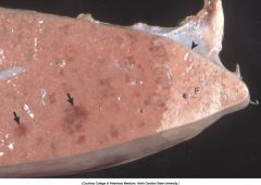
what is this?
Whcih Spp most common? Important DDx? |
Telangiectasia, liver, cut surface, cow. Telangiectasia is a condition in which hepatic sinusoids become dilated and filled with blood. These lesions can be seen as dark red areas on the cut and capsular surface of the liver.
Common: Bo. Also: old Fe. DDx: esp in old Fe → vascular tumors (hemangioma, hemangiosarcoma) McG 423 |
|
|
a free fatty acid (ffa) reaches the liver. What happens?
|
within hepatocyte: ffa → esterified to triglyceride → complexed with apoproteins → Low Density Lipoprotein → released into the plasma.
With the exception of Ru, liver also produces lipids from amino acids and glucose McG 423 |
|
|
Potential mechanisms responsible for excessive accumulation of fat in the liver (X6)
!! |
1) Excessive entry of fatty acids into the liver (eg dietary of fat mobilisation)
2) Abnormal hepatocyte function (eg ↓energy for oxidation of fatty acids) 3) Excessive dietary intake of carbohydrates 4) ↑ esterification of fatty acids to triglycerids in response to glucose and insulin 5) ↓ apoprotein synthesis 6) Impaired secretion of lipoprotein from liver ← hepatotoxins or drugs McG 423/424 |
|
|
Specific causes/syndromes of hepatic lipidosis indomestic animals: (X7)
|
1) dietary a) excess fat/cholesterol b) increased mobilisation from adipose tissue c) Deficiencies: Cobalt and Vit B12 in KRu
2) Toxic and anoxic causes 3) Ketosis 4) Bovine fatty liver syndrome (= ↑ mobilisation + defective hepatocyte function w ↓ export of lipoprotein) 5) Feline fatty liver syndrome. Idiopathic. 6) Ponies, miniature horses, donkeys. Overweight pregnant or lactating mares after stress or anorexia. Predisposed: Shetland ponies. 7) Endocrine disorders such as: diabetes mellitus and hypothyroidism McG 425 |
|
|
Short pathogenesis of ketosis.
Common in which Spp at what time? |
high energy demand → ↑ free fatty acids from fat → esterified to fatty acyl CoA in liver → oxidation in mitochondria to: acetoacetic acid and ß-hydroxybutyric acid → hyperketonemia + hypoglycemia.
Common in Bo during peak lactation Common in Ov during late gestation (particularly ewes w twins) → pregnancy toxemia McG 424/425 |
|
|
Bovine fatty liver syndrome.
Typically at what time? |
Dairy cattle: obese animals, few days before parturition, following an event that causes anorexia (eg retained placenta, metritis, .....)
Beef cattle: obese, few days before parturition. |
|
|
Excessive glucocorticoids = Cushing´s disease in dogs (eg iatrogenic or hyperadrenocorticism).
What do you think happens to the liver? Explain. ND. |
Glucocorticoids → ↑ glycogen synthethase → hepatic storage of glycogen → hepatocytes swell up to 10 times, esp midzonal → liver is enlarged, pale.
"Glucocorticoid-induced hepatocellular degeneration" or " steroid-induced hepatopathy" McG 425 |
|
|
In primary amyloidosis, the fibril is designated: .....
In secondary amyloidosis:.... |
Primary: AL (Amyloid light chain → composed of Ig light chains)
Secondary (or reactive): AA (Amyloid A, precursor: serum amyoild-associated protein = acute phase protein) |
|
|
Inherited amyloidosis occurs in: .......
|
dogs: Shar Pei
cats: Abyssinian, Siamese, other Oriental cats McG 426 |
|
|
Normally, serum copper is bound to .....
and hepatic copper is bound to ... |
serum: ceruloplasmin
liver: metallothionein |
|
|
Copper toxicosis in domestic animals ussually occurs as a consequence of: ..... (X5)
|
1) Dietary excess (esp common in sheep w mineral block for Bo)
2) grazing on pasture w insufficient molybdenum 3) Hepatotoxic phytotoxins, usually pyrrolizidine alkaloids (Heliotropum, Crotalaria, Senecio) → prevent hepatocellular proliferation → ever-increasing copper accumulation in surviving cells. 4) Diseases that produce cholestasis (Copper is excreted in bile) 5) Hereditary: Bedlington, Dalmatian, West higland white. Bedlington: autosomal recessive, impaired biliary excretion of copper. McG 427 |
|
|
Describe a typical pathogenesis of chronic copper intoxication in Ru
|
Excess intake or too little molybdenum → Cu accumulates in liver → event triggers release → acute severe intravascular hemolysis → acute anemia → hepatocellular necrosis, midzonal and centrilobular
McG 427 |
|
|
Describe pathogenesis of copper intoxication in Bedlington terrier
|
mutation(s) in MURR1 gene → encodes chaperone protein involved in copper excretion by hepatocytes → Cu accumulates in centrilobular region of liver → necrosis, inflammation, fibrosis → end-stage liver
McG 427 |
|
|
Hemosiderin is an ....-containing, golden-brown, granular pigment derived from ......
|
iron-containing
derived from ferritin (the initial iron-storage protein) McG 427 |
|
|
Name 2 species in which congenital melanosis of the liver occurs.
|
Su and Ru
McG 428 |
|
|
Live flukes → very dark excreta, contain mixture of ... and ... → discoloration in migratory tracts, esp those produced by ..... in Bo livers. This "pirgment" is called parasite ....
|
mixture of iron and porphyrin
Fascioloides magna Parasite hematin McG 428 |
|
|
Lesions in liver of:
puppy with infectious canine hepatitis adult dog with canine hepatitis |
puppy: snly scattered foci of hepatocellular necrosis ( they rappidly succumb)
adult: both foci of hepatocellular necrosis and widespread centrilobular necrosis (fulminant disease) McG 429 |
|
|
canine adenovirus 1, has a predilection for:..... (X3)
|
- hepatocytes
- vascular endothelium (→ widespread petechia and ecchymoses) - renal endothelium |
|
|
What is a really characteristic lesion of canine adenovirus 1 infection?
|
wall of gallbladder is thickened by edema
McG 429 |
|

|
The liver from a dog infected with infectious canine hepatitis (ICH) can be slightly enlarged and friable with a blotchy yellow discoloration. Sometimes fibrin is evident on the capsular surface. Note the petechiae on the serosal surface of the intestines caused by vascular damage from canine adenovirus type I infection
|
|
|
Describe the pathogenesis of " blue eye" in infectious canine hepatitis
|
Some dogs recover from the disease and develop an immune-complex uveitis (type III hypersensitivity) → degeneration and necrosis of corneal endothelium → corneal edema → blue colour
McG 429 |
|
|
Abortigenic herpesviruses.
1) often also affect neonates up to 2w of age. Why not older animals? 2) Characteristic lesions (MD):.... 3) Name a few examples of these viruses in different Spp |
1) Onze animals develop the ability to maintain normal body temperature, they are less likely to be affected (hypothermia is pivotal → also explains why these herpesviruses like to xxx in nose first)
2) multifocal, randomly distributed, small (<1mm) areas of necrosis in several fetal organs 3) abortigenic equine herpesvirus (EHV-1), infectious bovine rhinotracheitis virus (BHV-1), Caprine herpesvirus, canine herpesvirus (CHV-1), Feline viral rhinotracheitis virus (FeHV-1), Pseudorabies (SHV-1) |
|
|
Abortigenic herpesviruses als affect neonates, and reach their organs as follows:
xxx in oronasal epithelium → enter bloodstream via ........... cells → viremia → organs |
mononuclear phagocytic cells
McG 429 |
|
|
Rift valley fever
- Transmitted by ..... - Zoonotic? - principally affects which Spp? - Causing:.... - virus is member of family ..... genus ..... - Especially ↑ prevelance after periods of .... - Gross? |
- arthtropod (mosquito)-transmitted
- Yes, zoonotic - Ru - Abortion and extensive mortility in young animals - family Bunyaviridae, genus Phlebovirus (spp: rift valley fever virus) - after periods of unusually high rainfall - Gross: Liver → enlargement, yellow, areas of congestion, adult animal: pale foci, sometimes centrilobular hepatic necrosis. Systemic → diffuse petechiae&ecchymoses, DIC (potential hemorrhagic diathesis). Gallblader → edema and hemorrhages of wall In histo: sometimes eosinophilic INIB´s McG 430 |
|
|
Wesselsbron disease.
- transmitted by ...... - Zoonotic? - Clinical signs - virus is member of the .... |
- arthropod (mosquito)-transmitted
- yes, zoonotic - occasionally: multifocal areas of hepatocellular necrosis + icterus in lambs. Adult sheep usually survive, focal areas of hepatocellular necrosis, abortion. - Member of the Flaviviridae McG430 |
|
|
Bacterial infections of the liver, with formation of hepatic abscesses, are particularly common in feedlot cattle. Usually as a sequal to......
|
a sequal to toxic ruminitis → damage mucosa → ruminal microflora (esp Fusobact necroph) → portal circulation
McG 431 |
|
|
Bacillary hemoglobinuria.
- Et - Pathogenesis |
- Clostridium haemolyticum
- spores of Cl haemol → p.o. → inside Kupffer cells → once ↓ oxygen tension: xxx. Nidus is necrotic hepatic tissue created by: liver flukes or biopsy → release toxins. Most important: Phospholipase C → hepatocellular necrosis, intravascular hemolysis → acute and higly fatal disease of Ru. McG 433 |
|
|
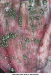
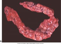
|
Focal hepatic necrosis, Clostridium haemolyticum (bacillary hemoglobinuria), liver, cut surface, cow. These large areas of necrosis are sharply delineated from the adjacent parenchyma, are usually pale, and are surrounded by an intensely hyperemic zone of acute inflammation
|
|
|
Focal hepatic necrosis, Clostridium haemolyticum (bacillary hemoglobinuria), liver, cut surface, cow. These large areas of necrosis are sharply delineated from the adjacent parenchyma, are usually pale, and are surrounded by an intensely hyperemic zone of acute inflammation
|
|
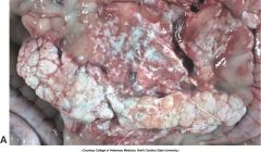
|
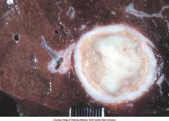
Chronic hepatic abscesses, Corynebacterium pseudotuberculosis, liver, sheep. Note the thick fibrous capsule and the pale caseous exudate characteristic of pus produced by Corynebacterium pseudotuberculosis in sheep.
|
|
|
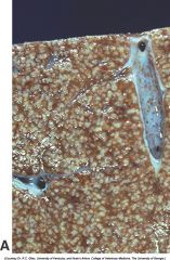
Tyzzer´s disease.
- ET - Spp - Disease is characterised by - Which stain? |
- Cl piliforme
- Lab animals (bacteria live in rodent intestines), sporadical in foals, described in Bo, Fe, Can, and others - Abdominal lnn: enlarged, edematous, hemorrhagic. Liver: enlarged, random pale foci of necrosis surrounded by inflam infiltrate. - Warthin-Starry or Gomori´s silver stain McG 433/434 |
|
|
yzzer’s disease (Clostridium piliforme). A, Liver, horse. Disseminated gray-white 1- to 2-mm foci of necrosis surrounded by suppurative inflammation. B, Foal. Clostridium piliforme can be identified by the haphazard distribution of filamentous bacteria in the cytoplasm of hepatocytes. Giemsa stain. C, Foal. Clostridium piliforme can be readily seen with special stains such as Giemsa and Warthin-Starry stains. Warthin-Starry stain
|
|
|
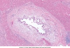
which adult nematodes can be found in the hepatic parenchyma of Can and Fe?
|
Capillaria hepatica
McG 435 |
|
|
Hepatosis dietetica is characterized by ......... to .... hepatic necrosis
|
hemorrhagic centrilobular to massive
McG 440 |
|
|
White liver disease
Spp? Cause? |
fatty liver in Ov caused by insufficient cobalt intake (a necessary cofactor in synthesis of Vit B12 and other enzymes → anemia → liver lesions
McG 440 |
|
Can
|
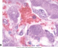
Note the yeast form of Histoplasma in the cytoplasm of Kupffer cells and macrophages. H&E stain.
|
|
|
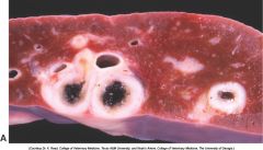
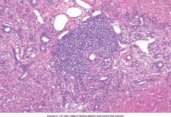
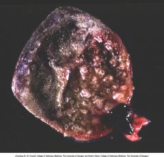
Cysticercosis, liver, cut surface, sheep. The thick fibrous capsule usually indicates the death of the larva.
|
|
|
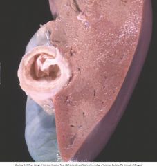
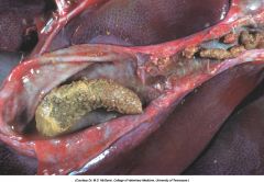
Fasciola hepatica infection. Chronic intrahepatic cholangitis (Fasciola hepatica), liver, cow. When Fasciola hepatica metacercariae are ingested, they migrate to the liver and then take up residence within the bile ducts. Mature flukes reside in the larger extrahepatic and intrahepatic bile ducts and cause chronic cholangitis and bile duct obstruction that lead to ectasia and stenosis of the ducts and periductular fibrosis that thickens the walls so that the ducts become increasingly prominent, as shown here.
|
|
|
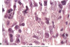
Chronic intrahepatic cholangitis, liver, cat. Fluke infections of the biliary tree of cats produce a characteristically pronounced periductular fibrosis, dilated bile duct, and papillary projections of biliary epithelium, although flukes may be difficult to find. H&E stain.
|
|
|
Feline lymphocytic cholangitis, liver, cat. Large numbers of lymphocytes and plasma cells surrounding bile ducts and biliary hyperplasia in portal areas are the hallmarks of this disease. The inflammation most often affects the periphery of the bile ducts and could be termed a pericholangitis, but the syndrome is referred to as cholangitis. H&E stain.
|
|
|
Massive necrosis, Hepatosis dietetica, liver, pig. A, Areas of hemorrhagic centrilobular necrosis and massive necrosis appear as dark regions of different size scattered throughout the liver.
|
|
|
Massive necrosis, Hepatosis dietetica, liver, pig. Acute centrilobular necrosis is the principal lesion of this disorder.
|
|
|
What si: idiosyncratic drug reaction
|
responses only seen in small minority of exposed animals
|
|
|
Not just the hepatocytes are affected by toxic drugs.
Biliary epithelium:.... (x2) Kupffer cells: ..... Endothelial cells: .... Stellate cells: .... (activation, not damage) |
biliary epithelium: trimethoprim sulfa. sporidesmin
Kupffer: edotoxin endothelial: arsenicals stellate: Vit A McG 442 |
|
|
Hepatotoxic injury can be classified into 6 categories based on the cellular target: ....
|
1) Production of injurous metabolites or adducts with essential cellular enzymes by Cyt P450 (SER). (Example: carbon tetrachloride)
2) Adducts between drugs and cellular material → neoantigens 3) stimulation of proapoptotic pathways (direct or immune mediated) 4) Damage cell membrane → influx of calcium → a.o. activation of proteases → damage actin filaments → blebbing and lysis of cell membrane. (example: carbon tetrachloride) 5) Cholestasis because of: - disrupt pumps that secrete bile constituents (eg estrogen, erythromycin) - disrupt actin filaments → no normal pulsating contraction to move bile 6) Mitochandrial damage (Example: IV tetracycline) → 1. microvesicular steatosis 2. release of reactive oxygen spp 3. ↓ production of ATP 4. release of Cytochrome-c → apoptosis McG 443 |
|
|
Describe the "biotransformation" by Cyt P450 to metabolize lipid-soluble chemicals into water-soluble compounds
|
Phase I. Chemicals are bioactivated to high energy reactive intermediate molecule
Phase II. formation of covalent bonds with polar molecules, such as glucuronic acid Phase III transport across cell membrane into bile lumen McG 443 |
|
|
Describe toxic effect of Carbon tetrachloride
|
metabolized by Cyt P450 to CCL3• → damage cell membrane → disrupt calcium homeostasis. Effect most severe in centrilobular (periacinar) areas.
McG 443 |
|
|
Describe toxic effect of Carbon tetrachloride
|
metabolized by Cyt P450 to CCL3• → damage cell membrane → disrupt calcium homeostasis. Effect most severe in centrilobular (periacinar) areas.
McG 443 |
|
|
Name the (3?) most important genera
Toxins? Gross Histo |
Toxins: Mycrocystins (eg mycrocystin LR)
Gross: hemorrhagic gastroenteritis + red, swollen hemorrhagic liver Histo: zonal to massive hepatic necrosis and hemorrhage McG 444 |
|
|
Describe toxic effect of Carbon tetrachloride
|
metabolized by Cyt P450 to CCL3• → damage cell membrane → disrupt calcium homeostasis. Effect most severe in centrilobular (periacinar) areas.
McG 443 |
|

3 genera
toxin Gross Histo |
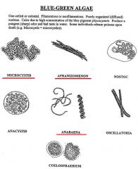
Toxins: Mycrocystins (eg mycrocystin LR)
Gross: hemorrhagic gastroenteritis + red, swollen hemorrhagic liver Histo: zonal to massive hepatic necrosis and hemorrhage McG 444 |
|
|
Pyrrolizidine alkaloids.
foudn in a.o. which genera? |
Senecio
Cynoglossum Amsinckia Crotalaria Echium Trichodesma Heliotropium McG 444 |
|
|
Pyrrolizidine alkaloids.
How do they induce toxic effects? |
Cyt P450 → converted to pyrrolic esters → are alkylating agents: react w cytosolic and nuclear proteins and nucleic acids (antimitotic effect)
McG 444 |
|
|
Pyrrolizidine alkaloids.
Characteristic injury |
Megalocytosis (antimitotic effects), fibrosis, biliary hyperplasia
(If exposure is not constant: nodular regeneration of parenchyma) McG 444 |
|
describe toxic effect
|
Cycads: contain cycasin (=nontoxic glycoside) → ingestion → deconjugated by intestinal bacteria → methylazoxymethanol (= toxic metabolite) → absorption → hepatic metabolism → alkylating agents →
a) chronic in Bo: hepatocellular megalocytosis, nuclear hyperchromasia, hepatic fibrosis + "dying back" of axons in dorsal funiculus → proprioceptive deficits in hind legs b) acute in Ov: acute gastrointestinal dysfunction + centrilobular hepatic necrosis c) also toxic to Can McG 445 |
|
|
At least 14 different types of aflatoxin are produced in nature. Aflatoxin .... is considered the most toxic and is produced by both ...... and ........ Aflatoxin G1 and G2 are produced exclusively by ..........
|
At least 14 different types of aflatoxin are produced in nature. Aflatoxin B1 is considered the most toxic and is produced by both Aspergillus flavus and Aspergillus parasiticus. Aflatoxin G1 and G2 are produced exclusively by A. parasiticus.
|
|
|
Aflatoxin.
Describe pathogenesis |
Aflatoxin → Cyt P450 → toxic intermediate → binding to DNA, RNA or proteins → Carcinogenic, toxic and teratogenic effects
McG 445 |
|
|
Aflatoxicosis.
Acute and chronic lesions |
Acute → hemorrhagic central to massive necrosis, lipidosis and biliary proliferation
Chronic → ill-thrift. immune suppression, occasionally hepatic failure. Liver is firm, pale: lipidosis, necrosis, biliary hyperplasia, centrilobular to bridging necrosis McG 445 |
|
|
Sporidesmin
Produced by.... Pathogenesis + common name Most affected organ-part |
Produced by Pithomyces chartarum, a fungus that grows well in dead rye grass
Pathog: necrosis epithelium of biliary ducts (esp Ov, less in Bo) → cholestasis → failure to excrete phylloerythrin → photosensitization → common name: facial eczema. Particularly affects left liver lobe (= ventral in ruminant liver) which has increased proportion of blood draining from dd. McG 445/446 |
|
|
Phomopsins
toxin produced by..... Spp affected:...... Gross Histo |
Produced by Phomopsis leptostromiformis, a fungus that growns on lupine.
Affects Bo, Ov and occasionally Eq. Gross: Atrophic and fibrotic liver. Histo: diffuse scattered hepatocyte necrosis, diffuse fibrosis, biliary hyperplasia. McG 446 |
|

Which mushroom?
Which 2 toxins? Gross Histo |
Amanita phalloides (death cap)
Amatoxin (inhibition of RNA polymerase II function) Phalloidin (disruption of intracellular actin filaments) Gross: hepatic hemorrhage and shrinkage Histo: Hepatic lipidosis, hemorrhage, centrilobular to massive necrosis. McG 446 |
|
|
Phosphorus.
which 2 forms? Histo? |
red phosphorus (unimportant) and white phosphorus → lipidosis and periportal necrosis
McG 446 |
|
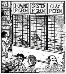
If a pig eats a clay pigeon, and then has centrilobular to massive hepatic hemorrhage and necrosis, what is the diagnose?
And the DDx? |
Cresol intoxication
DDx: hepatosis dietetica and ingestion of cotton seed meal McG 446 |
|
|
Why are Fe more susceptible then Can to many toxic chemicals?
|
Fe are relatively deficient in hepatic glucuronyltransferase activity (phase II enzyme).
McG 447 |
|
|
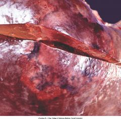
Livers from horses affected with equine serum hepatitis can be small, flabby and pale or discolored by bile pigment.
|
|
|
Is hepatocellular nodular hyperplasia more common in male dogs?
|
No.
No breed or sex predilection. McG 449 |
|
|
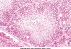
Hepatic nodular hyperplasia, dog
|
|

|
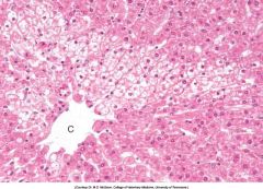
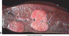
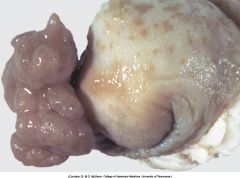
Hepatocellular adenomas, liver, dog. Hepatocellular adenomas form discrete masses of hepatocytes that compress adjacent normal parenchyma.
|
|
|
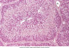
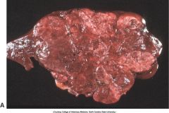
Hepatocellular carcinoma, liver, dog. A multi lobular carcinoma has replaced much of the normal liver.
|
|
|
Hepatocellular carcinomas are uncommon in all domestic spp, but occur more freq in ... particularly ...
|
Ru particularly Ov
|
|
|
Most common primary hepatic neoplasm in cats
|
cholangiocellular (bile duct) adenoma
|
|
|
a cystic variant of a cholangioma (typically w a nonencapsulated, multilocular cystic structure)
|
biliary cystadenoma
|
|
|
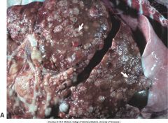
Cholangiocellular (bile duct) carcinoma, liver. Dog. Multiple nodules of tumor, some of which are umbilicated (arrows).
Usually arise from intrahepatic ducts, all species, single mass or multiple. Usually firm, raised, umbilicated, gray, unencapsulated. Often scirrhous response, metastasis in lnn, lung, abdominal cavity. McG 451/452 |
|
|
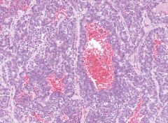
Neuroendocrine tumors, carcinoid, liver, dog. Carcinoids are malignant neoplasms of neuroendocrine cells of the liver or bile ducts. Histologically the tumor is composed of small elongated or spindle-shaped basophilic cells that form ribbons or rosettes and contain numerous vascular spaces. H&E stain.
|
|
|
latin for gallstone
|
cholelith
|
|
|
Important viral cause for acute cholecystitis in:
- a Ru - a Can |
- Rift Valley fever (RFF virus → phlebovirus → bunyaviridae)
- infectious canine hepatitis (CAV-1) McG 453 |
|
|
Choleliths, gallbladder, pig. Choleliths are concretions formed from the constituents of bile.
|
|
|
Cystic mucinous hyperplasia, gallbladder, dog.
Only reported in Can and Ov. Only appreciated by opening gallblader Mucosa gray-white, spongelike. Occasionally polypoid masses. Numerous 1-3mm cysts. Copious amount of mucus. No significance to host. Cause not known. |
|
Dog, Gallblader.
What kind of tumor? These are rare neoplpasms, but most common in ........ |
Adenoma of gallblader.
Most common in young cattle. McG 454 |
|
|
Exocrine pancreatic atrophy
- Breed predelection - Usual age - DDx (X1) |
- German Shepherd, rough-coated collies (autosomal recessive)
- 6 to 12 M of age - Hypoplasia of exocrine pancreas in calves (distinction between atrophy and hypoplasia can be difficult (atrophic cells can more often contain lipofuscin) |
|
|
Pacinian corpuscles (arrows), pancreas, cat. The feline pancreas contains numerous pacinian corpuscles, which may be visible grossly as about 1-mm clear foci.
|
|
|
fluid-filled nonepithelialized fibrous sacs that form within pancreas or adjacent tissue are described in Can and Fe.What are they called?
|
Pancreatic pseudocysts
McG 456 |
|
|
What is the best name in most instances:
- acute pancreatitis - acute pancreatic necrosis |
- acute pancreatic necrosis
(obese sedentary bitches are especially predisposed) McG 456 |
|
|
3 major proposed mechanisms of pancreatitis
|
1) Obstruction of the ducts.
2) Direct injury to acinar cells (T-2, zinc-toxicosis, sulfonamides, trauma,..) 3) Disturbances of enzyme trafficking within the cytoplasm of acinar cells →inappropriate activation of proenzymes |
|
|
.... is believed to be a key player in pancreatitis.
Onze activated it can activate ... and ... into ... and ... |
Trypsin:
Proelastase →elastase Prophospholipase → phospholipase A |
|
|
Acute pancreatic necrosis, acute pancreatitis, pancreas, dog. Note the expansion of the pancreas by areas of hemorrhage and edema.
|
|

|
Chronic pancreatitis, pancreas, dog. Lobules are more prominent due to fibrosis, and the pancreas is paler than normal. The white, raised, granular areas in the pancreas and mesentery are foci of fat necrosis that result from enzymatic digestion of lipids that then become mineralized.
|
|
|
Pancreatic nodular exocrine hyperplasia, pancreas, dog.
Common, especially in older Can and Fe. |
|
|
Pancreatic carcinoma. Stomach and pancreas (center), ventral-dorsal view, dog. Pancreatic carcinoma has invaded the mesentery, wall of the stomach, and gastrosplenic ligament. Note the lobulated appearance of the mass, which is formed by neoplastic exocrine pancreatic epithelial cells and scirrhous connective tissue. Proximal duodenum (bottom), liver (top), and spleen (right [left anatomically]).
|