Reading...
![]()
Play button
![]()
Play button
![]()
Use LEFT and RIGHT arrow keys to navigate between flashcards;
Use UP and DOWN arrow keys to flip the card;
H to show hint;
A reads text to speech;
49 Cards in this Set
- Front
- Back
|
What is the lower respiratory tract?
|
Begins with the trachea and includes the main stem bronchi, bronchial tubes and lungs (alveoli).
|
|
|
What lines the trachea, bronchi and bronchial tubes?
|
Ciliated pseudostratified columnar epithelium, contained in a mucous membrane.
|
|
|
What do the cilia do?
|
Move a layer of mucous toward the laryngopharynx where it can be swallowed or coughed up.
|
|
|
What produce the mucus in the lower respiratory tract? What does the mucus do?
|
Goblet cells. Traps debris.
|
|
|
Where is the trachea?
|
(C6-T4) Continuous with the larynx, a midline structure that extends from C6 vertebra to the sternal angle at the level of T4.
|
|
|
Where does the trachea bifurcate?
|
The transverse thoracic plane passes through the sternal angle b/t T4-T5, this is where it bifurcates into two bronchi.
|
|
|
What is the size of the trachea?
|
3/4" diameter
3 & 1/2" to 5" long in adults |
|
|
What is the shape of the trachea?
|
A cylinder with a cross-sectional profile shaped like a horseshoe.
|
|
|
What is the trachea composed of?
|
-20 cartilage rings
-smooth muscle -mucous glands |
|
|
What do the cartilage rings in the trachea look like?
|
U-shaped, made of hyaline cartilages
|
|
|
What does the smooth muscle of the trachea do?
|
Functions to open the trachea while breathing.
|
|
|
What is the blood supply to the trachea?
|
Borrows from thryroid;
-superior and inferior thyroid arteries -superior, middle, and inferior thyroid veins |
|
|
What are the primary (main stem) bronchi differences?
|
Right main bronchus - larger diameter, more vertical, shorter length
Left main brochus - smaller diameter, less vertical, longer length More lung on R side b/c of heart on L side. |
|
|
What branch off the primary main bronchus?
|
Secondary lobar bronchi
|
|
|
What are the secondary lobar bronchi?
|
Go to the lobes of the lungs.
Right lung - 3 lobes and 3 lobar bronchi Left lung - 2 lobes and 2 lobar bronchi |
|
|
What branches from the secondary lobar bronchi?
|
Tertiary (segmental) bronchi.
|
|
|
What are the tertiary (segmental) bronchi?
|
Go to bronchopulmonary segments.
Right lung - 10 segments and 10 segmental bronchi Left lung - 8 segments and 8 segmental bronchi |
|
|
What are the terminal bronchioles?
|
Branches of segmental bronchi.
|
|
|
What are the respiratory bronchioles?
|
Contain the alveoli.
|
|
|
What are the alveoli?
|
Air sacs of the lungs. Single cell in thickness, organized in clusters, surrounded by dense capillary network.
|
|
|
What is the arterial blood supply to the bronchi?
|
3 bronchial arteries (2 L and 1 R) from descending aorta, supply bronchial tubes NOT alveoli
NOT part of pulmonary circulation |
|
|
What is the venous blood supply to the bronchi?
|
Bronchial veins drain to the azygos veins which are tributaries to the superior vena cava.
|
|
|
What is the surface anatomy of the lung?
|
-fissures and lobes
-cardiac notch -lingula -apex and base -hilum -visceral pleura |
|
|
What is the oblique fissure of the lung?
|
L lung - divides upper and lower lobes
R lung - divides middle lobe from the lower lobe, and upper lobe from the middle lobe |
|
|
What is the horizontal fissure of the lung?
|
Only in R lung, divides the upper lobe from the middle lobe.
|
|
|
What is the cardiac notch of the lung?
|
In upper lobe of L lung, adjacent to heart.
|
|
|
What is the lingula of the lung?
|
In upper lobe of L lung, adjacent to the cardiac notch. Shaped like a tongue.
|
|
|
What are the base and apex of the lung?
|
Apex - above first rib, AKA cupola
Base - adjacent to respiratory diaphragm |
|
|
What is the hilum of the lung?
|
Contains structures entering and exiting the lungs, i.e. bronchi, pulmonary vessels and nerves, lymphatic vessels and bronchial vessels.
|
|
|
What is the visceral pleura of the lung?
|
Serous membrane covering the external surface of lungs.
|
|
|
What structures are involved in pulmonary circulation?
|
-pulmonary trunk
-pulmonary arteries (L and R) -alveolar capillary network -pulmonary veins |
|
|
Where does the pulmonary trunk come from?
|
Emerges from the R ventricle
|
|
|
What are the pulmonary arteries?
|
L and R, have branching patterns similar to bronchi. Carry deoxygenated blood.
|
|
|
What is the alveolar capillary network?
|
Functional part of the pulmonary circulation, provides for exchange of blood gases with air in the alveoli.
|
|
|
What is the total surface area of the capillary network?
|
Must be equal in size to all of the combined capillaries in all the organs in the body.
|
|
|
What are the pulmonary veins?
|
Collect oxygenated blood from alveoli and return it to the L atrium.
|
|
|
What are the sensory neurons (GVA) that innervate the lower respiratory tract?
|
Vagus nerve CN X
|
|
|
What kind of sensory receptors are in the lower respiratory tract and where are they located?
|
stretch receptors - in alveoli and smooth muscle
irritant receptors - respiratory epithelium tactile receptors - cough reflex baroreceptors - pulmonary arteries chemoreceptors - pulmonary veins (detect blood gases) |
|
|
What supplies motor (GVE neurons) fibers to the lower respiratory tract?
|
Autonomic fibers contained in pulmonary nerve plexus.
-sympathetic -parasympathetic |
|
|
What do the sympathetic nerve fibers that innervate the lower respiratory tract do?
|
Go to smooth muscle and glands to inhibit glandular secretion, induce vasoconstriction of bronchial vessels, and dilate bronchial tubes.
|
|
|
What do the parasympathetic nerve fibers that innervate the lower respiratory tract do?
|
Go to smooth muscle and glands to increase glandular secretion, induce vasodilation of bronchial vessels, and constrict bronchial tubes.
|
|
|
What contributes to the parasympathetic innervation to the lower respiratory tract?
|
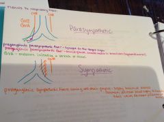
Pulmonary nerve plexus by the vagal nerves (CN X), which synapse with post-ganglionic parasym. fibers on the surfaces of bronchial tubes and vessels.
|
|
|
What are the lymphatic plexuses of the lower respiratory tract?
|
-superficial plexus
-deep plexus |
|
|
What is the superficial plexus?
|

Lies deep to the visceral pleura, drains lymph from the visceral pleura and lung parenchyma to the bronchopulmonary lymph nodes in the hilum.
|
|
|
What is the deep plexus?
|
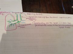
In the walls of bronchi, drains lymph from the bronchi to the pulmonary lymph nodes near the hilum and then goes to the bronchopulmonary nodes in the hilum.
|
|
|
What are the lymph nodes in the lower respiratory tract and where are they located?
|
In sequence of lymphatic drainage
1. pulmonary nodes-in lungs at the bifurcations of larger bronchi 2. bronchopulmonary (hilar) nodes)-in hilum 3. tracheobronchial nodes-at the bifurcation of the trachea 4. tracheal nodes-lateral surfaces of the trachea |
|
|
What are the lymph trunks of the lower respiratory tract?
|
-right bronchomediastinal lymph trunk
-left bronchomediastinal lymph trunk |
|
|
What does the right bronchomediastinal lymph trunk drain?
|
Drains lymph from the right side of the trachea to the right lymphatic duct (behind subclavian)
|
|
|
What does the left bronchomediastinal lymph trunk drain?
|
Drains lymph from the left side of the trachea to the thoracic duct.
|