Reading...
![]()
Play button
![]()
Play button
![]()
Use LEFT and RIGHT arrow keys to navigate between flashcards;
Use UP and DOWN arrow keys to flip the card;
H to show hint;
A reads text to speech;
53 Cards in this Set
- Front
- Back
|
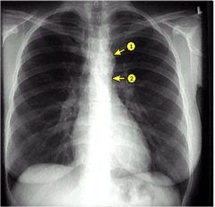
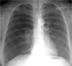
1. aortic knob
2.aortopulmonic window |
|
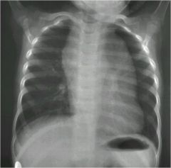
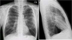
what is going on in this child?
|
thymic hyperplasia
|
|
|
if you had a CXR of a child and you thought they may have a thymic hyperplasia, what should be your next move?
|
CT of the chest
|
|
|
the most common thing seen in CT as a superior or anterior mediastinal tumor is what?
** |
substernal thyroid (goiter)
|
|
what is the pathology?
|
teratoma
|
|
|
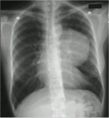
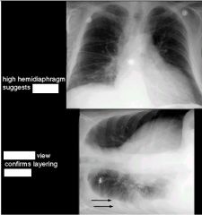
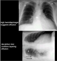
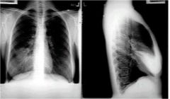
which diaphragm is higher?
|
right
|
|
|
what is the sniff test? (normal finding, and abnormal
|
normally both diaphragms go down
but in diaphragmatic paralysis there is paradoxical motion, where the diaphragms go up (due to negative pressure) |
|
|
Herniation of Bachdelek: 3 traits
(probably not on test) |
Big
in Babies in the Back (more commonly on left side) |
|
|
Herniation of Morgagni
|
right sided anterior herniation of the diaphragm
|
|
|
Hiatal hernia... 2 types (describe both, and which is most common)
*** |
Paraesophageal: fundus of stomach is herniated into the chest, esophagus in the normal area
Sliding: most common, whole stomach and esophagus are in the chest |
|
this person was given a sniff test...what is going on?
|
sniff test with paradoxical movement of left hemidiaphragm
|
|
|
as you go down the spinal cord in a lateral view what should happen to its appearance?
|
it gets darker
|
|
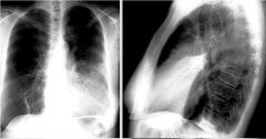
what is going on here
|
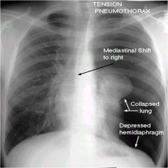
right diaphragmatic leaflets
tension pneumothorax (hole in visceral pleura) left lung starts to look 'dirty' because it is getting all the blood |
|
|
best view to see a tension pneumothorax?
|
PA
|
|
|
if you suspect a pneumothorax on a normal CXR what should be your next step?
** |
upright expiratory chest film
|
|

|
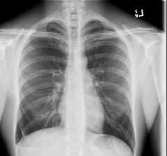
just wanted to show this
|
|
|
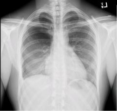
again just look at this
|
|
|
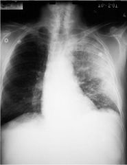
effusion
|
|
|
|
|
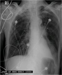
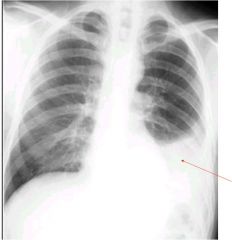
2 important diagnoses?
|
mediastinal shift left
pleural effusion |
|
TEST
|
pneumothorax that has collected inferiorlly
|
|
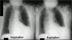
This PA chest was performed
In expiration….Why? *** |
suspected pneumothorax
this is an expiratory chest film |
|
Pneumothorax
COPD CHF Consolidation |
COPD
|
|
|
bacterial will cause a alveolar or interstitial CXR?
|
predominantly alveolar
|
|
|
viral/mycoplasma will cause a alveolar or interstitial CXR?
|
interstitial
|
|
|
fungal will cause a alveolar or interstitial CXR?
|
actually it is combination
TRICK QUESTION! |
|
|
Apical-posterior segments are filled up in what type of pneumonia?
|
reactivation TB
|
|
|
posterior segment of upper lobes that are lighting up on CXR is normally caused by what?
|
aspiration in supine position
|
|
|
Expansion or bowing of fissures is caused by?
|
Klebsiella
|
|
|
if you see rapid spread or abcess on CXR what is the likely cause?
|
staph
|
|
|
in a neutropenic pt, what type of findings might you see in a pt who has pneumonia on CXR?
|
may have no findings in a neutropenic patient (limited inflam. response)
|
|
|
what can make pneumonia look worse?
|
hydration
|
|
|
when a pt has pneumonia, and you take an Xray right at the beginning, what might you find?
|
X-rays may lag the onset and the resolution of pneumonia
so you might not see much |
|
what sign
|
silhouette sign
|
|
sign and where?
|
silhouette sign of lingula
|
|
cause of this pneumonia?
|
anthrax
see widened mediastinum |
|
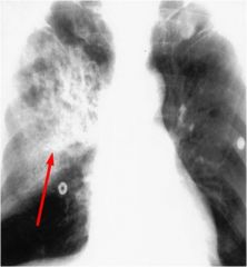
if this person had TB, what would you call this?
|
Renkie Complex (Ghon lesion)
|
|
|
lung cancer LEAST commonly associated with cigs?
in what sex? |
adenocarcinoma
women |
|
|
what percent of primary lung tumors are malignant?
|
90
|
|
|
when you see a CT of someone's chest it goes down to the adrenal glands...Why?
***TEST |
hypervascular
one of the first places to see a metastatic deposit |
|
|
what type of cancer may have air bronchograms?
|
adenocarcinoma
|
|
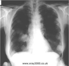
what can be seen on this CXR?
|
metastatic lesions
|
|
|
Birth control pills
Pregnancy (during and after) Surgery Trauma Malignancy Immobilization Cross country car or airplane trips are all what? *** |
risk factors for PE
|
|
|
where do PEs most often occur?
*** |
bilateral and in lower lungs
|
|
|
what will cause tumor emboli?
|
renal cancer
|
|
|
what is the method of choice to find a PE?
*** |
CT
|
|
|
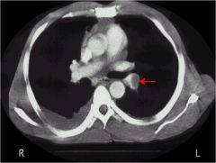
Westermark sign
|
clot so big it blocks the blood going to a lung segment
refers to dilation of the pulmonary artery proximal to an embolism with collapse of distal vessels |
|
|
you give a VQ scan. It shows a normal ventilation, but the perfusion is lacking...what does this show?
|
PE
|
|
|
If you see V/Q Mismatch, what is going on?
***TEST |
PE
(note: asthma and COPD do that too...so make sure they don't have a history of either or your V/Q mismatch isn't as helpful) |
|
|
d-dimer does what?
|
measures the clotting in the blood
shows PE |
|
|
THERE WILL BE A CT FROM LECTURE ON THERE
|
look at them
|
|
what is the triple rule out? what does this show?
|
Triple Rule out:
Aoritc dissection PE Coronary Artery occlusion this is PE (i think) |
|
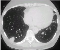
what does this show? what causes it?
|
Wedge shaped peripheral defect, infarct due to PE
|