Reading...
![]()
Play button
![]()
Play button
![]()
Use LEFT and RIGHT arrow keys to navigate between flashcards;
Use UP and DOWN arrow keys to flip the card;
H to show hint;
A reads text to speech;
22 Cards in this Set
- Front
- Back
|
Hereditary papillary renal cell carcinoma is a syndrome characterized by predisposition to develop multiple, bilateral papillary renal tumors. Linkage analysis mapped the gene to chromosome:
a. 7q31 b. 3p25 c. 10q11 d. 11q13 |
a. 7q31
Hereditary papillary renal cell carcinoma is a cancer syndrome mapped to chromosome 7q31.1-q34, a region containing the MET oncogene, which is mutated in hereditary papillary renal cell carcinoma families. Although trisomy 7 is commonly associated with sporadic papillary renal cell carcinomas, only 13% of sporadic papillary tumors have MET mutations, suggesting that sporadic and hereditary disease may develop by different mechanisms. Cytogenetic features of most common renal neoplasm are as follows: WT1 gene on chromosome 11p13 in nephroblastoma, p57/kip2 gene on chromosome 11p15 in nephroblastoma associated with Beckwith-Wiedemann syndrome, VHL gene on chromosome 3p25 in clear cell renal cell carcinoma associated with von Hippel-Lindau syndrome, MET gene on chromosome 7q31 in papillary renal cell carcinoma associated with hereditary papillary renal cell carcinoma. |
|
|
Simple cysts are the most common cystic lesion of the kidneys. Their incidence rate increases with:
a. Age b. Cholesterol level c. Blood pressure d. Hormone level e. None of the above |
a.
The incidence rate of simple renal cysts increases with age. In general, they are asymptomatic. They can be solitary to multiple and are usually located in the cortex. The size varies but is usually <5 cm. |
|
|
Several classifications of renal cystic diseases have been proposed based on microscopic findings, clinical presentation, or radiologic appearance.
P.148 The primary distinctions are genetic and nongenetic. Which of the following is NOT a genetic cystic disease? a. Adult polycystic kidney disease b. Infantile polycystic kidney disease c. Juvenile nephronophthisis d. Medullary sponge kidney e. None of the above |
d.
Genetic renal cystic diseases include adult (autosomal dominant) polycystic kidney disease, infantile (autosomal recessive) polycystic kidney disease, juvenile nephronophthisis (autosomal recessive), medullary cystic disease (autosomal dominant), and autosomal recessive and rare multicystic disorders (von Hippel Lindau, tuberous sclerosis, etc.). Nongenetic renal cystic diseases include multicystic dysplastic kidney, benign multilocular cyst, simple cyst, medullary sponge kidney, sporadic glomerulocystic kidney disease, acquired renal cystic disease, and calyceal diverticulum. |
|
|
What is the correct association between polycystic kidney disease and liver pathology?
a. Autosomal dominant polycystic kidney - hepatic cysts b. Autosomal recessive polycystic kidney - hepatic cysts c. Autosomal dominant polycystic kidney - congenital hepatic fibrosis d. Medullary sponge kidney - congenital hepatic fibrosis e. None of the above |
a.
Autosomal dominant polycystic kidney disease is associated with hepatic cysts that become evident with increased age. Autosomal recessive polycystic kidney disease is associated with congenital hepatic fibrosis in all affected surviving individuals. |
|
|
Which are the most common sites of metastases from renal cell carcinoma?
a. Lung, bone, and liver b. Bladder, colon, and prostate c. Esophagus, stomach, and duodenum d. Anal canal, liver, and esophagus e. None of the above |
a.
The common sites are lung, liver, bone, lymph nodes, adrenal, brain, opposite kidney, heart, spleen, and skin. Liver metastasis is associated with an ominous prognosis. Adrenal metastasis occurs in <19% in ipsilateral and 10% to 11% in contralateral adrenal. Adrenal metastasis is more common with large upper pole tumors in patients with metastatic disease. The most common sites and approximate frequency of metastasis of renal cell carcinoma are lung in 50% to 60%, lymph nodes, liver, and bone in 30% to 40% each, opposite kidney in 10% to 25%, and brain in 5%. |
|
|
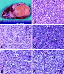
CD10+, Vimentin+
del (3p) Aggressive |
Clear cell renal cell carcinoma
|
|
|
CD10+, Vimentin+, AMACR+
Loss of Y, gains of 7 & 17 Indolent |
Papillary renal cell carcinoma
|
|
|
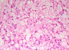
CD10–/+, Vimentin –, CD117+
Loss of Y, 1, 2, 6, 10, 13, 17, and 21 Indolent |
Chromophobe renal cell carcinoma
|
|
|
CD10–, Vimentin –, high MW keratin+ del (1q)
Aggressive |
Collecting (Bellini) duct carcinoma
|
|
|
CD10–, Vimentin –, CEA+
Unknown chromosomes Aggressive |
Renal medullary carcinoma
|
|
|
CD10–/+, Vimentin +/–, CD117+ Highly variable: loss of 1, 14. t(5;11). Benign
|
Oncocytoma
|
|
|
HMB45+
LOH at TSC2 on 16p Benign |
Angiomyolipoma
|
|
|
TFE3+ (nuclear)CD10+
Xp11.2 translocation Indolent |
Xp11.2 translocation carcinoma
RCC |
|
|
|
monotonous proliferation of uniform small round cells with evenly distributed fine chromatin, separately by uniform vascular network
Also called bone metastasizing renal tumor Clear cell sarcoma |
|
|
Clear cell sarcoma
monotonous proliferation of uniform small round cells with evenly distributed fine chromatin, separately by uniform vascular network Also called bone metastasizing renal tumor |
|
|
congenital tumor resembling infantile fibromatosis or leiomyoma, with fascicles and whorls of bland myofibroblasts and thin collagen fibers; cellular variant resembles infantile fibrosarcoma
Also called fetal, mesenchymal or leiomyomatous hamartoma Mesoblastic nephroma |
|

|
pure epithelial tumor composed of embryonal-type epithelium with bland features
Rare, 0.2% of adult renal epithelial neoplasms; < 100 cases described Also called metanephroid renal tumor, nephroblastoma-like adenoma of kidney Metanephric adenoma |
|
|
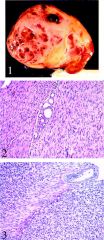
Chromophobe type, renal cell carcinoma
Differential diagnosis: · oncocytoma - no hard cell membranes, no mitotic figures, no loss of chromosomes 2, 6, 10 or 17 (Mod Pathol 2005;18:161) · eosinophilic papillary or clear cell carcinomas - Hales colloidal iron negative |
|

|
Juxtaglomerular Cell Tumor is a rare renal tumor presenting in young adulthood with severe hypertension due to excessive renin secretion, that is corrected by surgical removal of the tumor. Histologically, it resembles hemangiopericytoma and glomus tumors.
|
|

|
characteristic rhomboid-shaped renin protogranules
Juxtaglomerular cell tumor Cytogenetics: -9 and -11 |
|

|
Nephrogenic metaplasia is a metaplastic process seen after surgical manipulation of the urinary tract, or in association with trauma, stones, and inflammation. Papillary, polypoid, or cystic structures lined by cuboidal epithelium
|
|

|
Endocervicosis is an uncommon mullerian lesion consisting of irregularly-shaped glands proliferating deep in the bladder wall. Patients often give history of having undergone caesarean section.
|