Reading...
![]()
Play button
![]()
Play button
![]()
Use LEFT and RIGHT arrow keys to navigate between flashcards;
Use UP and DOWN arrow keys to flip the card;
H to show hint;
A reads text to speech;
24 Cards in this Set
- Front
- Back
|
Acanthocyte/Spur Cell
|
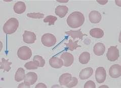
Irregular, spiculated red cells with few unevenly distributed surface projections of various length and diameter
|
|
|
Agglutination
|
An antibody coats the erythrocyte causing bridging and clumping
|
|
|
Anisocytosis
|

A variation in the size of RBC, may be due to the presence of macrocytes, microcytes or both
|
|
|
Basophilic Stippling
|

Presence of small, dark-blue bodies within the erythrocyte; observed in cells containing residual RNA
|
|
|
Crenation
|
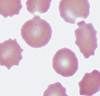
RBC changes from a disc shape to spheres with projections; Result of pH change due to slow drying of blood films
|
|
|
Darocyte
|

Tear-shaped RBC that disrupts the bone marrow architecture
|
|
|
Discocyte
|

Discoid-shaped RBC, seen normally in dogs
|
|
|
Echinocyte/Burr Cells
|

Spiculated cells with numerous short, evenly spaced, blunt to sharp surface projections with uniform size and shape
|
|
|
Ghost Cells
|

A dead cell where the outside remains visible but the nucleus and cytoplasmic structures are not stainable
|
|
|
Heinz Bodies
|

Round structures representing denatured hemoglobin caused by certain oxidant drugs/chemicals; up to 5% normal in cats
|
|
|
Howell Jolly Bodies
|

Basophilic nuclear remnants seen in young erythrocytes in response to anemia; removed by spleen
|
|
|
Hypochromic
|

Decreased staining intensity due to insufficient hemoglobin; usually seen with iron deficiency
|
|
|
Keratocytes
|

Spiculated red cells with two or more pointed projections
|
|
|
Macrocyte
|

RBC's that are larger than normal with an increased MCV; usually reticulocytes
|
|
|
Microcytes
|

RBC's with a smaller than normal diameter and decreased MCV; seen in iron deficiency
|
|
|
Nucleated RBC
|

Cells released into circulation early during anemia; normal in non-mammalian animals
|
|
|
Poikilocyte
|

Abnormally shaped RBC
|
|
|
Polychromasia/Polychromatophilic
|

RBC that exhibit a bluish tint when stained with Wright's stain; organelles remain in cytoplasm indicating young cells
|
|
|
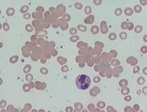
Rouleaux
|

A grouping of erythrocytes in stacks; seen with increased fibrinogen or globulin
|
|
|
Schistocyte/Shizocyte
|

RBC fragments; usually from shearing of the red cell by intravascular trauma; DIC
|
|
|
Spiculated Cells
|

Erythrocytes with one or more surface projections
|
|
|
Spherocyte
|

Darkly staining red cells with reduced or no central pallor; not easily detected except in dogs; suggest IMHA
|
|
|
Stomatocyte
|

Erythrocyte with an oval-shaped central pallor
|
|
|
Target Cell/Leptocyte/Codocyte
|

Thin erythrocytes with increased membrane or decreased volume
|