![]()
![]()
![]()
Use LEFT and RIGHT arrow keys to navigate between flashcards;
Use UP and DOWN arrow keys to flip the card;
H to show hint;
A reads text to speech;
67 Cards in this Set
- Front
- Back
|
Membrane Potential Vm |
electrical voltage across cell membrane remember with a cell that has no sodium pump and is only permeable to K, K would not leak out because of electroneutrality - this is not true some DOES leak out- establishing Membrane Potential |
|
|
Diffusion driven by |
- Concentration Gradients - Electrical potential differences + -> - - -> + |
|
|
Measure Membrane PD _____ with respect to _____ |
inside , outside |
|
|
FORMULA Nernst Equation |
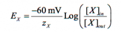
z = valance charge FOR A NEGATIVE NUMBER |
|
|
Membrane vs Equilibrium Potential |
Membrane - true voltage PD across membrane Equilibrium - voltage (determined by the Nernst equation) that perfectly balancesthe chemical diffusional driving force determined by a concentration difference across the cellmembrane |
|
|
Typical Equilibrium Potentials |
EK = - 100 mV ENa = + 50 mV ECl = - 70 mV |
|
|
Resting Membrane Potential |
ionic permeability of cell membranes is unchanging with time |
|
|
Membrane Ionic Conductances (gNa, gk, gCl) |
easier way to measure rmp directly proportional to permeability i = gV iK = gK (Vm - Ek) iCl = gCl (Vm - ECl) iNa = gNa (Vm - ENa) |
|
|
Vm is stable then total net current iT = |
iT = 0 (t = sum of all i's) |
|
|
Positive current Negative current |
Positive current (ik) = outward current, makes Vm more negative Negative current (iCl) = inward current, makes Vm more positive |
|
|
Hyper polarizing Depolarizing |
H - More negative D - Less negative/more positive |
|
|
Most cells resting potential |
~ -70 mV |
|
|
Vm FORMULA |
Vm = gkEk + gNaENa + gClECl / gT |
|
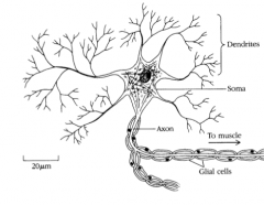
Motor Neuron Anatomy |
Motor neuron - innervates muscle In Spinal Cord Soma - cell body (nucleus, receives connections) Dendrites - branching, receives connections Outside Spinal Cord nerve - bundle of axons with many neurons |
|
|
Action Potential |
Short in duration (3msec) - neuron impulse |
|
|
Phases of AP |
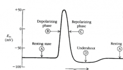
A - resting potential (-70 mV) B - rapid depolarization (peak at 40 mV) C - rapid depolarization (back to resting) D - after hyperpolarization undershoot (-90) A - return to resting |
|
|
What causes AP? |
NA and K channels |
|
|
Voltage-Gated Channels of AP |
Na+ Channel - m gate (activation) - h gate (inactivation) K+ Channel - n gate (activation) |
|
|
AP Channels - Na+ |
At Rest: Na+ closed Depolarization: activates/open rapidly - inc in GNa more depolarization, + feedback inc GNa more Repolarization: when Vm gets more positive, channel will inactivate/close (dec GNa and iNa) |
|
|
AP Channels - K+ |
At Rest: K+ open - causes negative rp Depolarization: more activate/open and remain open after inactivation of Na channels Repolarization: an inc of gK causes Vm to get more negative - produces after shoot |
|
|
Ratio of Na:K channels |
4:1 - reason for the depolarization |
|
|
Gates + Phases
|
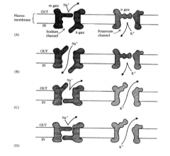
A: M + N closed, H open B: M + H open, N closed C: M + N open, H closed D: M + H closed, N open |
|
|
Hyperkalemia |
Higher than normal extracellular K+ levels Less negative EK Vm becomes less negative, closure of H gates Na+ channels won't open -> NO AP |
|
|
Ionic Concentration Gradient |
Even though AP result in an inc in Na in and an dec of K out, this is countered by the sodium pump -> pumps out extra Na and restores lost K |
|
|
Ouabain |
No immediate effect on AP, the neuron could still fire 1000s of AP before the gradient would be dissipated enough to cause an effect |
|
|
Initiation of AP |
Vm must be depolarized to threshold AP are all are none - same amplitude and duration |
|
|
Refractory Periods |
Absolute - 2nd AP can't occur no matter what Relative - 2nd AP can occur if stimulus is large enough, must be after relative refractory |
|
|
Tetraethyl ammonium cation (TEA+) |
Blocks K+ channels, doesn't prevent AP - prolongs duration and removes afterhyperpolarization |
|
|
Tetrodotoxin (TTX) |
Blocks Na+ irreversibly - not used clinically Blocks AP LETHAL |
|
|
Local anesthetics (lidocaine) |
Blocks Na+ reversibly, used clinically |
|
|
Action Potential Propagation |
Action potentials travel along neurons axon AP occurs at distance (x) = 0 |
|
|
λ |
Length constant -> each λ traveled = decrease in potential by 63% |
|
|
AP conduction Antidromic Orthodromic |
Can conduct in 2 directions - rarely happens
Anti - abnormal direction of conduction Ortho - normal conduction |
|
|
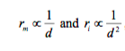
λ FORMULA |

rm = membrane resistance (1/gT) ri = intracellular resistance |
|
|
rm and ri |
|
|
|
Myelin |
Evolutionary advantage - packing of millions of high speed axons - increases λ by increasing rm Current must flow axially down the axon to find gaps (nodes of Ranvier) - also where the Na channels are solely located - nerve conduction jumps from node to node (saltatory conduction) - highest conduction in alpha motor neurons |
|
|
Multiple Sclerosis (MS) |
caused by demyelination, decreases λ |
|
|
Unmeylinated vs myelinated axon |
Unmeylinated: grey, velocity proportional to square root of diameter, can be fast at very small diameters Myelinated: white, long axons rapidly conducting |
|
|
Local potentials |
- Initiate AP - Graded potentials - result from ion-permeable channels - amplitude and duration vary - can sum in time (temporal) and distance (spatial) |
|
|
Three types of local potentials |
- Synaptic potentials - Endplate potentials - Generator potentials |
|
|
Synaptic potentials |
2 types of postsynaptic potentials - EPSP - elicit AP - depolarize - IPSP - inhibit AP - hyper polarize Typically small events, just one can't affect AP - can be summed |
|
|
EPSP |
- equally permeable to Na and K - inc gK normally hyperpolarizes, but with gNa inc depolarizes |
|
|
IPSP |
- permeable to K or Cl - inc in gK hyper polarizes, gCl no change - cancels effects of EPSP - typically hyperpolarizing, but can also be depolarizing if ECl is less negative than Vrest |
|
|
Summation of EPSP / IPSP |
Can occur, because there is no rapidly activating Na channel - can't fire AP, summation leads to AP at hillock Does NOT sum algebraically |
|
|
Silent IPSP |
No change in Vm but can still counter EPSP |
|
|
Neural inhibition |
Half of all neural synapses are inhibitory by increasing IPSP, dec activity of postsynaptic neurons |
|
|
ADHD |
Inc brain activity, (normal function is to inhibits brain centers) this decreased hyperkinesis |
|
|
Tetanus |
causes from injection via puncture wound blocks inhibitory synapses - loss of inhibition, muscle contraction - asphyxiation and death |
|
|
End Plate Potential EPP |
Endplate is in the skeletal muscle - motor neuron innervates NMJ - inc gNa and gK - Depolarizing, always elicits AP in muscle fiber (because much larger area) - no inhibitor EPPS |
|
|
Generator / Receptor Potentials |
- detection of physical stimuli - sensory receptors - touch/pressure receptor - carries info to CNS - frequency of AP varies with pressure |
|
|
Pacinian corpuscle |
touch receptor in palms, fingers 2 regions - depressed naked region - Ap propagates along axon, can elicit AP - unmyelinated - fast acting Na channels, non excitable no AP |
|
|
Thin Filament is made up of |
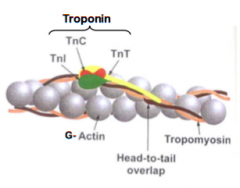
Actin - troponin 3 subunits G-actin - globular tropomyosin - thin lines |
|
|
Thick Filament is made up of |
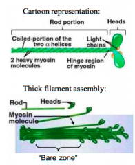
myosin, heads line up head to head - bare zone in the middle |
|
|
Which band remains constant? |
A BAND |
|
|
Sliding Filament theory |
1. Rigor state - tight binding 45 angle, NO ATP 2. ATP binds, myosin can dissociate from actin 3. ATPase activity of myosin hydrolyzes ATP, have ADP and Pi bound to myosin 4. REGULATED STEP Myosin head swings and wings to a new actin - 90 degree 5. Pi released POWER STROKE, actin pushed 6. ADP released back to rigor |
|
|
3 Subunits of Troponin |
TnC - Ca++ binding site TnT - bound to tropomyosin TnI - glue, holds it all together |
|
|
What covers the myosin binding sites on actin |
Tropomyosin, ca++ causes a conformational change of troponin-tropomyosin |
|
|
Ca++ comes from? |
SR through EC coupling |
|
|
EC Coupling Process |
AP - DHP opens ryanodine receptor (plug + cork) |
|
|
Termination of contraction caused by? |
Lower levels of Ca++ - calsequestrin |
|
|
Isometric |
Same length, doesn't shorten but generates force |
|
|
Isotonic |
same tension, muscle shortens, shortens more with lighter loads |
|
|
Single twitch Summation Summation -> unfused tetanus Summation -> complete tetanus |
- muscle relates after stimuli - muscle can't relax fully b/w 2 stimuli - multiple stimuli far enough allow muscle to relax slightly - muscle reaches steady tension - stops with fatigue |
|
|
Why is tetanus tension 3-5x more than twitch? |
because of the time needed to stretch |
|
|
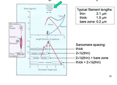
Length tension relationship |
|
|
|
Strenuous vs. endurance exercise |
- sprinting - high energy expenditure, long lag recovery - long distance - lower energy expenditure, maintained longer |
|
|

|