![]()
![]()
![]()
Use LEFT and RIGHT arrow keys to navigate between flashcards;
Use UP and DOWN arrow keys to flip the card;
H to show hint;
A reads text to speech;
39 Cards in this Set
- Front
- Back
|
Geniculo-striate Pathway |
Each eye receives inout from the left and right visual field. Temporal visual field projects to nasal retinal. Nasal visual field projects to temporal retina. Nasal retinal fibres cross over at optic chiasm and project to lateral geniculate nucleus. |
|
|
Lateran Geniculate Nucleus (LGN) |
Has 6 layers 1,4,6: Contralateral eye. 2,3,5: Ipsilateral eye. |
|
|
M channel |
Motion contrast |
|
|
P channel |
Colour, fine details. |
|
|
K channel |
Blue, Motion |
|
|
Neural Processing within the LGN |
It receives approximately 90% of the fibres from the retina. 10% goes to the superior colliculus. LGN neurons have centre surround receptive fields. LGN regulates neural signals en-route to the visual cortex. |
|
|
How the LGN regulates neural signals en-route to the visual cortex |
It combines inputs from various sources to regulate neural firing. Feedback projections from V1. 10 Action potentials from the retina is equal to 4 action potentials to the V1 |
|
|
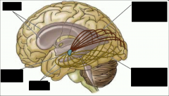
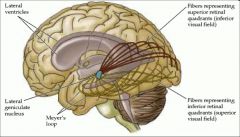
Retinotopic organization in the visual system |
The visual system is organized retinotopically. Each portion of the retina is represented on a topographic map at multiple levels of the geniculo-striate system. Damage to a piece in the map will result in blindness for the corresponding area. |
|
|
|
|
|
Cellular Organization of visual cortex |
The primary visual cortex (V1) is the first cortical region that receives visual information from the retina. Often referred to as the Striate cortex because of distinct stripe pattern (in V2). Cortex has 6 layers. Layer 4 receives sensory inputs. |
|
|
Receptive fields of V1 neurons |
Simple cells. Neurons respond maximally when the stimulus is in the preferred orientation. Orientations selectivity. Different neurons respond to different orientations. V1 Neurons as feature detectors. |
|
|
Simple cells |
Have side by side opponent receptive fields. Monocular receptive fields. |
|
|
Complex cells |
Respond best to bars at a particular orientation that move in a specific direction. |
|
|
End Stopped Cells |
Fire when lines of a specific length move in a particular direction. Also respond to moving edges or angles. |
|
|
Cortical Magnification |
Fovea accounts for approximately 0.01% of the area of the retina. However 10% of the visual cortex is devoted to the fovea. |
|
|
The role of feature detectors in vision |
Linking firing rates with perception. Selective adaptation and the contrast threshold. |
|
|
Linking firing rates with perception |
Selective adaptation. Neurons that respond to a particular property will fatigue if they are continuously exposed to the same stimulus. |
|
|
Selective adaptation and the contrast threshold |
Difference in intensity needed so that the bars are barely visible. |
|
|
Selective rearing experiments |
Cats were raised in environments with only horizontal or vertical stripes for 5 hours a day for 5 months. V1 neurons responded only to line orientations that were consistent with the environment that the cat was raised in. |
|
|
Columnar organization of V1 |
Location columns, Orientations columns, Ocular dominance columns, Hypercolumns. |
|
|
Location Columns |
Columns whose receptive fields are concentrates in the same part of the retina. |
|
|
Orientation Columns |
Columns whose receptive fields prefer a specific orientation (tied to location columns) |
|
|
Ocular dominance columns |
Columns whose receptive fields are predominately from one eye. |
|
|
Hypercolumns |
Columns that combine location specificity, orientation selectivity, and ocular dominance. Ice cube model. |
|
|
Hypercolumns in V1 |
Each region of visual space is represented by a V1 module. Blobs are sensitive to colour but not orientation or form. Colour information sent to V4/V8. Neurons outside of blobs respond to orientation and form but not colour. |
|
|
Representations of Objects in V1 |
Objects activate numerous hypercoloumns in V1 that correspond to stimulated portions of the retina. Features of the objects are coded by simple, complex, and end-stop cells in each of the hypercolumns. |
|
|
|
|
|
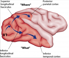
Object discrimination |
Monkey has to remember under which object the food is placed. Inferior temporal lesions cause a deficit. 'what'. |
|
|
Landmark Discrimination |
Monkey has to remember that the food is in hidden under the object that is near the cylinder. Parietal lesions cause a deficit. 'where'. |
|
|
Patient DF: Visual form agnosia |
Bilateral lesions to the occipital-temporal cortex. She could not recognize objects or copy simple drawings. She could draw items from memory. |
|
|
Dorsal |
Fast response. M-channel. Absolute metrics. Moment to moment. Visually guided action. 'non-conscious'. |
|
|
Ventral |
Slow response. M+P channels. Scene based metrics. Long term memory. Object recognition. 'Conscious'. |
|
|
Patient D.B. and 'blindsight' |
34 yr old male, began experiencing migraine headaches at 14. Visual auras were present prior to headaches which developed into a scotoma on the patients left side. D.B. was still able to localize targets in his blind field. |
|
|
Alternate visual pathways |
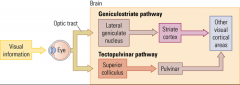
Visual information can reach the cortex outside of the primary visual pathway. |
|
|
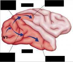
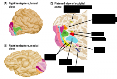
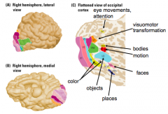
Organization of the ventral stream |
There are regions outside occipital cortex that are specialized for processing different kinds of visual information. |
|
|
|
|
|
Prosopagnosia |
The inability to recognize faces despite normal perceptual abilities. Cannot even recognize their own family members. Recognize people using voices, hair, posture. Caused by lesions to the right fusiform gyrus (FFA) |
|
|
Congenital Prosopagnosia |
Ventral stram areas must develop over time in order to correctly recognize objects and faces. |
|
|
Category learning and the ventral stream |
Cells in area TE respond to complex combinations of object features. Cells with similar selectivity are clustered together in columns. Object identification requires activation of many neurons in a column. These neurons are shaped by learning. |