![]()
![]()
![]()
Use LEFT and RIGHT arrow keys to navigate between flashcards;
Use UP and DOWN arrow keys to flip the card;
H to show hint;
A reads text to speech;
37 Cards in this Set
- Front
- Back
- 3rd side (hint)
|
History |
Gorter & Grendel 1920s: bilayer of phospholipids Davson & Denielli 30s: sandwich model, hydrophilic proteins coating bilayer Singer & Nicholson current: fluid mosaic model |
|
|
|
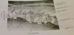
Structure of Plasma Membrane |
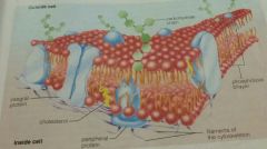
Composed of phospholipids, proteins, cholesterol and carbohydrates (glycolipids and glycoproteins) |
|
|
|
Phospholipids |
Amphipatic, with a hydrophilic phosphate head and 2 hydrophobic fatty acid tails. Self assemble into a bilayer in aqueous environment for stability. |
|
|
|
Proteins in plasma membrane |
Integral Proteins: embedded in membrane, hydrophilic and hydrophobic regions. Peripheral Proteins: attached to the layer, loosely bound to the surface |
|
|
|
Membrane Protein Functions |
Transport: substances in/out of cell, Na-K pump. Enzymatic Activity: Protein may be and enzyme, catalizes a reaction. ATP in cellular respiration. Signal Transduction: Allows cell to communicate. Receptor (protein) have a binding site, allows chemical messanger (hormones, neurotransmitters) to bind. Cell-Cell Recognition: Glycoproteins serve as identification tags. Blood type, antigens. Intercellular Joining: of adjacent cells Attachment to Cytoskeleton and Extracellular Matrix: maintain cell and/or stabilize location of membrane proteins. |
|
|
|
Carbohydrates |
Bonded, glycolipids or glycoproteins. Distinguishes cells, involved in cell-cell recognition. Type and distribution vary among species, individuals and cell type. |
|
|
|
Cholesterol |
Wedged between phospholipids. High temp decreases fluidity of membrane. Low temp increases fluidity of membrane. |
|
|
|
Fluid Mosaic Model |
Membrane components always in motion. Mosaic cause embedded proteins. Freeze fracture, fusion of mouse and human cell. |
|
|
|
Plasma Membrane Functions |
Shape & Strength: to cell Biological Barrier: separate and protect cell from surroundings Regulates: movement in/out of cell, selectively permeable allow some through but not other. |
|
|
|
Selective Permeability Of Membrane |
Depends on lipid bilayer and transport proteins. |
|
|
|
Lipid Bilayer (in transport) |
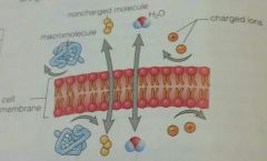
Nonpolar molecules are hydrophobic, pass easily. Polar molecules are hydrophilic, have difficulty passing. Charged particles are sorrounded by water molecules and can't pass. Large molecules don't fit through the membrane pores. |
|
|
|
Transport proteins |

Are integral proteins, help polar and charged molecules pass. Channel Proteins: have a hydrophilic channel. Carrier Proteins: hold on to the molecule and change shape, making the molecule pass. |
|
|
|
Diffusion |

Particles are in constant motion (Brownian Motion), move from areas of high to low concentration. Reach dynamic equilibrium. Each type of molecule diffuses along its own concentration gradient. |
|
|
|
Rate of Diffusion Factors |
Concentration Gradient: the difference in concentration. Drives diffusion cause it represents potential energy. Temperature: particle movement Distance: closer distance = more bumping = faster diffusion or smaller distance to cover Area of Membrane: increased SA = increased rate of diffusion Permeability of Membrane: membrane more permeable to some cell than others |
|
|
|
Passive Transport |
Diffusion of substances across the membrane with no energy investment. Facilitated Diffusion = with the help of transport proteins. For small, polar molecules and ions. |
|
|
|
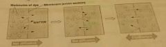
Osmosis |
Diffusion of water across a selectively permeable membrane. Changes volume of solution for same concentration. If solute can't diffuse. Aquaporis: channel proteins that allow movement of water. |
|
|
|
Potential Energy |
The concentration gradient drives diffusion because it represents potential energy. Potential is the potential movement (kinetic enrgy) of the particle. Since the particles are close together (at high concentration, on the gradient) they bump into each other more, which drives diffusion (cause of particle movement). |
|
|
|
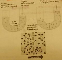
Osmotic Pressure |
Force pulling solvent (H2O) from the hypotonic solution to the hypertonic one. |
|
|
|
Tonicity |
The ability of a solution sorrounding the cell to cause it to gain or lose water. Always a comparison. |
|
|
|
Hypotonic |
Solute concentration of solution less than of cell. Water enters the cell |
Hypothermia |
|
|
Isotonic |
Solute concentration of solution same as cell. No net movement of water = dynamic equilibrium |
|
|
|
Hypertonic |
Solute concentration of solution more than of cell. Water exits the cell |
Hyperactive |
|
|
Effect of Tonicity in Cells |
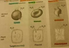
Animals: sensitive cause no cell wall - Hypotonic = swell and lyse (burst). - Hypertonic = cause shriveling. - Isotonic = best for animal cell. Plants: depend on it for mechanical support, cell wall can resist pressure - Isotonic = become flaccid cause no internal pressure. - Hypertonic = cause cell yo undergo plasmolysis, membrane pulls away from cell wall. - Hypotonic = water intake creates tug or pressure, keeps plant cells firm. |
|
|
|
Active Transport |

Uses energy (ATP) to move solutes against their concentration gradient. From low to high concentration. Protein pumps = carrier proteins involved. Na+/K+ pump, proton pump |
|
|
|
Bulk Transport |

For large molecules like proteins and polysaccharides. Uses ATP. Endocytosis: taking in material, with new vesicles from the membrane Exocytosis: moving out |
|
|
|
Types of Endocytosis |
P |
|
|
|
Types of Endocytosis |

Phagocytosis: Eating. Engulfment of solid materials = formation of vacuoles. Pinocytosis: Drinking. Uptake of liquids and dissolved solutes = tiny vesicles. Receptor-Mediated: to aquire specific substances. Proteins on membrane with specific receptors bind to substances, then brought in with a vesicle. Lock/Key. |
|
|
|
Exocytosis |

Exports materials by fusing vesicles with plasma membrane, releasing contents to outside of cell. Used to export wastes and by secretory cells. Endocrine glands release hormones, neurons release neurotransmitters |
|
|
|
Cell Size and SA/V Ratio |
Cell metabolism sets limits on its size. Must be able to efficiently bring in nutrients and and get rid of wastes to survive. Maximize surface area and minimize volume. = surface area-to-volume ratio As diffusion inside the cell is slower than through the membrane cause of distance and the concentration gradient. |
|
|
|
SA/V Ratio |
Used to compare efficiency (of diffusion) If object is small SA/V ratio is large= effective If object is large SA/V ratio is small= ineffective |
|
|
|
Cell Shape |
Can modify the SA/V. Important to cells that exchange a lot with their surroundings. Plant roots: root hairs. Intestines: villi and microvilli to take in nutrients Lungs: alveoli Blood vessels: capillaries |
|
|
|
Multicellular Organism |
Consist of more than one cell. Often specialized. |
|
|
|
Specialized Cells |
For different functions, in multicellular organisms. Have different sizes, shapes and number of different organelles. Become more effective and efficient at performing one task. Over 200 in the human body. |
|
|
|
Principle of Complementarity |
Function reflects structure and structure determines function. |
|
|
|
Cell Differentiation |
Process by which cells become specialized in structure and function. |
|
|
|
Stem Cells |
Unspecialized cells, can divide inroads specialized cells and self-renew. Embryonic: totipotent, into all cell types Adult: multipotent, into multiple cells |
|
|
|
Structiral Hierarchy of Organism |
Organism: complex functioning whole, sum of all it's components. Organ Systems: association of organs with a common function Organs: structure, two or more tissues Tissues: groups of cells of similar structure that perform a related function Specialized Cells: different structure and function |
|