![]()
![]()
![]()
Use LEFT and RIGHT arrow keys to navigate between flashcards;
Use UP and DOWN arrow keys to flip the card;
H to show hint;
A reads text to speech;
116 Cards in this Set
- Front
- Back
|
What is the charge of toluidine blue? |
Positive (cationic). |
|
|
What color is toluidine blue when it binds to carboxylated polysaccharides? |
Pinkish-purple. |
|
|
What color is toluidine blue when it binds to the free phosphate of nucleic acids? |
Greenish-blue to purple. |
|
|
What color is toluidine blue when it binds to polyphenolic compounds such as lignin and tannins? |
Green to green-blue to bright blue. |
|
|
What information should be included when drawing a specimen? |
Magnification, stain, species, and cut. |
|
|
Describe IKI solution. |
Iodine dissolved in an aqueous solution that binds with starch to produce an intense blue/purple color. |
|
|
Describe Sudan IV. |
Used for staining lipids. It has the appearance of reddish brown crystals |
|
|
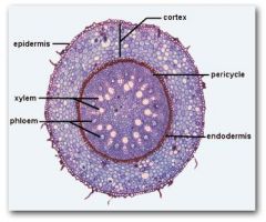
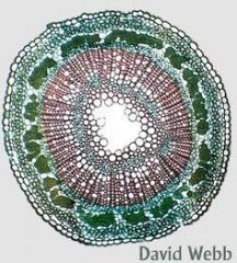
Cross section of a eudicot root. |
|
|
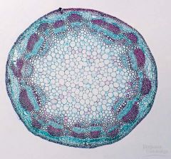
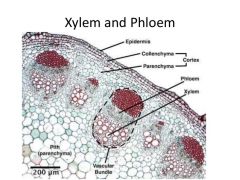
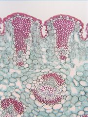
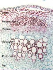
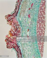
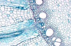
Cross section of a eudicot stem. |
|
|
What are sclerenchyma fiber cells located? |
|
|
|
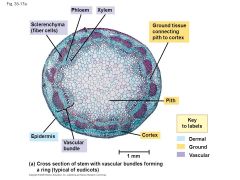
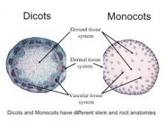
Where are the ground, vascular, and dermal tissues located in monocots and eudicot stems. |
|
|
|
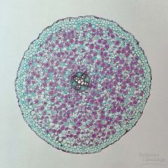
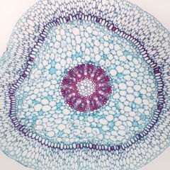
Cross section of a monocot root. |
|
|
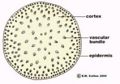
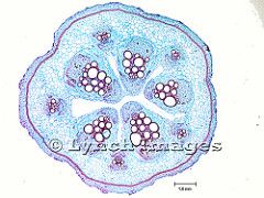
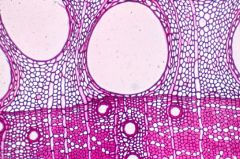
Cross section of a monocot stem. |
|
|
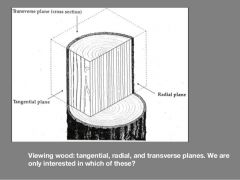
Describe the major planes we use for view plant anatomy. |
|
|
|
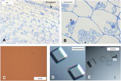
Calcium carbonate cystolith. |
|
|
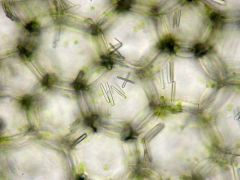
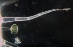
Calcium oxalate raphide. |
|
|
Multicystatin crystal |
|
|
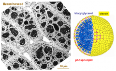
Oil droplet. |
|
|
Starch grain. |
|
|
Where are collenchyma cells? |
|
|
|
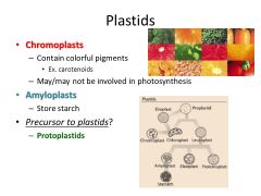
Describe plastids. |
|
|
|
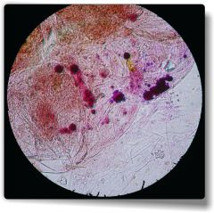
Anthocyanin. |
|
|
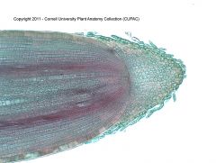
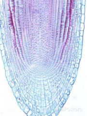
Zea mays; closed calyptrogen. Root cap not connected to root. Cell division pattern: cortex, epidermis. |
|
|
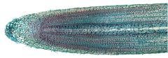
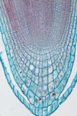
Raphanus closed dermatocalyptrogen. Root cap connected to root. Cell division pattern: epidermis, root cap. |
|
|
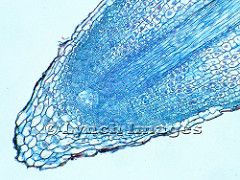
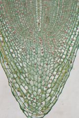
Allium open initial cell organization Root cap connected to root. |
|
|
Botrychium; 1 initial cell. Root cap connected to root. |
|
|
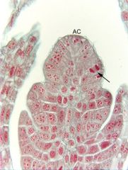
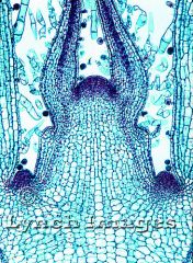
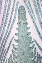
Equisetum shoot apical meristem. Single initial cell. Radial, anticlinal, periclinal cell division. |
|

|
Pinus shoot apical meristem. Single cell layer. Anticlinal and periclinal cell division. |
|
|
Coleus shoot apical meristem. Tunica corpus cell organization. Tunica arises from anticlinal cell division; corpus arises from periclinal cell devision. |
|
|
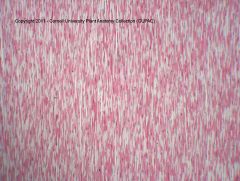
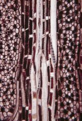
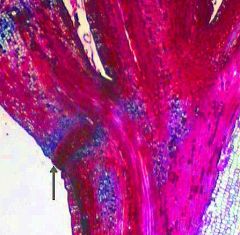
Xylary fibers. |
|
|
Xylary fibers again. |
|
|
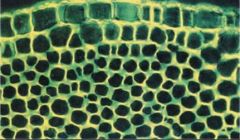
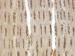
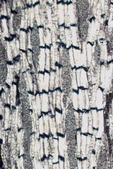
Extraxylary fibers (dark green). |
|
|
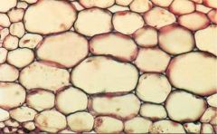
Parenchyma |
|
|
Collenchyma |
|
|
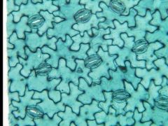
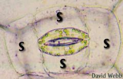
Eudicot guard cells. |
|
|
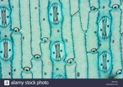
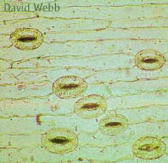
Monocot guard cells. |
|
|
Sunken stomata. |
|
|
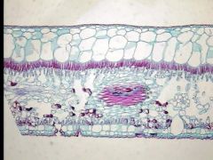
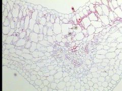
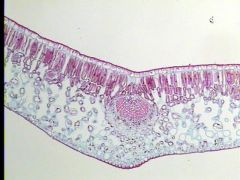
Pelargonium leaf. |
|
|
Ligustrum leaf. |
|
|
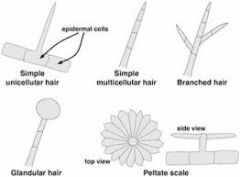
Describe the different types of trichomes. |
|
|
|
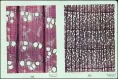
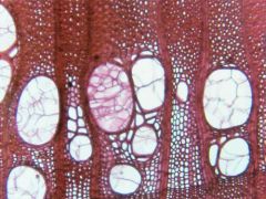
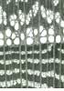
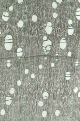
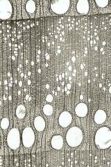
Ring porous and diffuse porous. |
|
|
Guard cell with subsidiary cells. |
|
|
Guard cells with no subsidiary cells. |
|
|
Bulliform cells. |
|
|
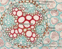
Where are the metaxylem, protoxylem, pericycle, endodermis, and phloem located in a eudicot root. |
|
|
|
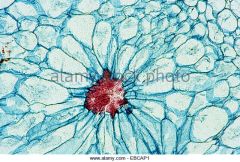
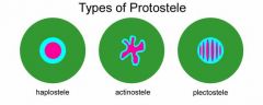
What are the three main types of protostele? |
|
|
|
What are the types of xylem development? |

xylem in brown; arrows show direction of development from protoxylem to metaxylem.protoxylem is usually distinguished by narrower vessels formed of smaller cells. |
|
|
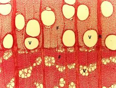
Bi-collateral vascular bundles in cucurbita. |
|
|
What is the difference between protoxylem and metaxylem. |
|
|
|
Angular collenchyma. |
|
|
Lamellar collenchyma. |
|
|
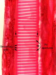
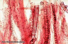
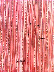
Scalariform vessel perforation in longitudinal cut. |
|
|
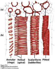
What are the different kinds of perforation plates (secondary wall patterns) in metaxylem? |
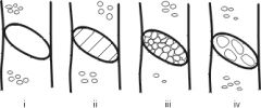
simple, scalariform, reticulate, foraminate |
|
|
Annual and helical secondary wall patterns in protoxylem. |
|
|
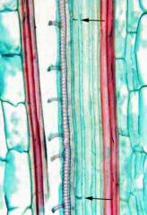
Two sieve plates (arrows) can be seen as "blue bars" which demonstrate the length of a single sieve tube element. |
|
|
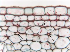
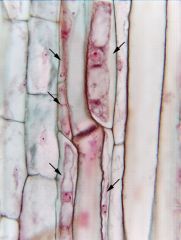
Notice that the cells on the left are all short, block-shaped and have protoplasts (not sieve tube members). The arrows point to several narrow, cytoplasmic cells that are in contact with sieve tube members. They appear to be companion cells. |
|
|
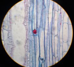
P-protein plug. |
|
|
Non storied cambium seen in temperate species. |
|
|
Storied cambium seen in tropical species. |
|
|
In what cut would we see storied (or non storied) cambium or uniseriate & multiseriate rays? |
Tangential. |
|

|
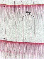
Tangential section of rays in pine wood. |
|
|
Transverse section of rays in pine wood. |
|
|
Radial section of rays in pine wood. |
|
|
What is the fence analogy? |
The radial section allows you to see the fences from the side. Thus you can see the arrangement of the different fence posts as well as the height of the fence. However, you can’t see its thickness.The tangential view is like looking at the fence from one of its ends. You can see its height and thickness but you can’t see the sides of the fence or is length. The transverse you can the fence posts from the top. |
|
|
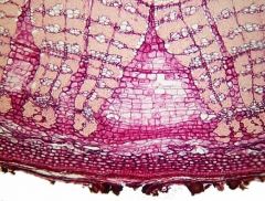
The expanded v-section of ray parenchyma phloem cells ( #3) fill the gapwhich arises as the earlier laid phloem deriving from a then smaller circumferenced vascular cambium are pushed out with the expanding layers of xylem. |
|
|
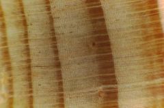
Early wood (lighter) and late wood. |
|
|
Lenticels. |
|
|
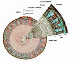
Where is the periderm? |
|
|
|
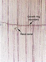
Resin canal. |
|
|
Vitis secondary phloem in longitudinal section. Note sieve tube members, compound sieve plates and lateral sieve areas. |
|
|
Robinia secondary phloemshowing dormancy callose on sieve plates as seen in radial section. Produced by storied cambium. |
|
|
Wood of Quercus borealis. |
|

|
Wood of Acer. |
|
|
Wood of Pinus. |
|
|
Wood of Robinia. |
|
|
Wood of Thuja occidentalis. |
|
|
Wood of Celtis occidentalis. |
|
|
What do uniseriate and multiseriate rays look like? |
|
|
|
Wood of Ulmus. |
|
|
Wood of Juglans. |
|
|
Wood of Castanea. |
|
|
Leaf abscission zone. |
|
|
Orchid root cross section (velamen, exodermis, cortex, endodermis, stele). |
|
|
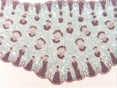
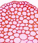
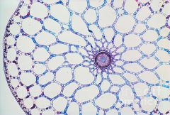
Aerenchyma cells. |
|
|
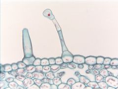
Glandular trichomes. |
|
|
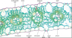
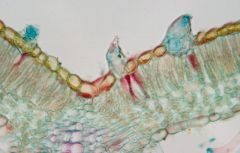
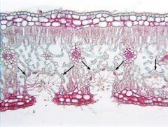
Shade (A) vs sun (B) leaf. |
|
|
What do bundle sheath cells look like? |
|
|
|
Cannabis trichomes. |
|
|
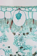
Cytolith of Ficus (rubber plant). Note multiseriate epidermis. The cystolith is a cacium carbonate inclusion. |
|
|
Uniseriate and glandular trichomes of Lycopersicon. |
|
|
Lateral root placement in Zea alternates with primary xylem (opposite the primary phloem). Note its origin is beneath the endodermis in the pericycle. |
|
|
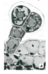
Root apical meristem of Brassica showing three tier organization, type 1: epidermis shares initial with cortex (open). |
|
|
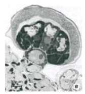
Root apical meristem of Linum(flax, Linaceae) showing three tier organization, type 2: epidermis shares initial with root cap (closed dermatocalyptrogen). |
|
|
Shoot apex of Elodea, a common submerged aquatic plant. Note the number of tunica layers, thickness of leaves, irregularity of axillary buds. |
|
|
What is merocrine secretion? |
Excretion via exocytosis or transport(passive/active) |
|
|
What is holocrine secretion? |
Secretory compound released afterdegeneration/lysis of secretory structure |
|
|
Capitate trichome. |
|
|
Peltate trichome. |
|
|
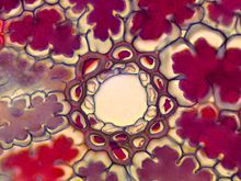
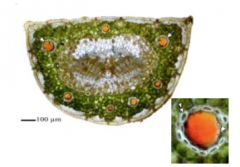
Resin duct in pine needle. |
|
|
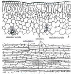
Lactifers in Allium leaves. |
|
|
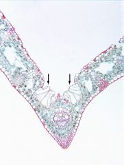
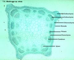
Where are the metaxylem and metaphloem in the Medicago stem? |
|
|
|
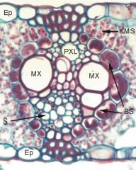
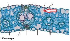
Where are the metaxylem and metaphloem in the Zea leaf? |
|
|
|
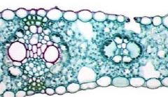
Bundle sheath extensions (Zea mays). |
|
|
Wood of juniper. |
|
|
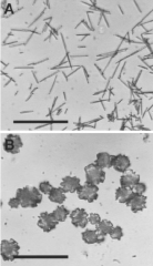
A (raphides) and B(druses). |
|
|
secondary cell walls in xylem. |
|
|
What are the pores in hardwood? |
Vessels; gymnosperms only have (tracheids). |
|
|
What kind of secondary cell wall thickening does protoxylem have? |
Annular and spiral. (Smaller in diameter than metaxylem). |
|
|
What kind of secondary cell wall thickening does metaxylem have? |
Pitted. (Larger in diameter than protoxylem). |
|
|
What is the difference between protophloem and metaphloem? |
Protophloem is thinner and the sieve areas are inconspicuous. Metaphloem is wider, has distinct sieve areas, and has companion cells. |
|
|
What is phloroglucinol used for? |
A stain specific for lignin and suberin used to stain secondary walls red. |
|
|
Stomatal crypts. |
|
|
Substomatal chamber. |
|
|
Where are the dorsal and ventral parts of the guard cell? |
|
|
|
How many vascular bundles are in each needle of a pine species with five needles per fasicle? |
1 vascular bundle. |
|
|
How many vascular bundles are in each needle of a pine species with two-three needles per fasicle? |
2 vascular bundles. |