Reading...
![]()
Play button
![]()
Play button
![]()
Use LEFT and RIGHT arrow keys to navigate between flashcards;
Use UP and DOWN arrow keys to flip the card;
H to show hint;
A reads text to speech;
65 Cards in this Set
- Front
- Back
|
What resulted from the invention of image intensifiers
|
replaced flourescent screens
increased image brightness |
|
|
What is the primary function of the flouroscopy
|
The primary function of fluoroscopy is real-time
imaging to provide visualization of dynamic pro cesses as they occur. |
|
|
What has a higher radiation exposure rate; flouro or plain film
|
flouro
|
|
|
What is the exposure rate of typical flouro
|
45mGy/min
|
|
|
What is the exposure rate of a typical plain film
|
900mGy/min
|
|
|
What is higher the TOTAL exposure time for plain film or flouro
|
flouro (x rays occur for a much shorter period of time
|
|
|
What is the total exposure for a 10 minute flouro study of the abdomen
|
450mGy
|
|
|
what is the exposure total for an abdominal film
|
3mGy
|
|
|
What is done to compensate for the increased radiation dose
|
the exposure rate (as mentioned before) is less.
|
|
|
What is a flourescent screen
|
theis is s a matererial that immediates visible light in response to a stimuli such as an x ray
|
|
|
Does red light have a short or long wavelength
|
long
|
|
|
What was the purpose of red adaption googles
|
to allow the flouroscopist to remain adjust to dark room and peform activity outside to retain dark room adaptation
|
|
|
Why were image intensifiers developed
|
To overcome the deficiencies of viewing the
dim fluorescent screen image, image intensifier devices were developed and introduced in 1953. |
|
|
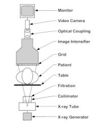
What are the components of a flouroscopy machine
|
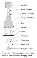
|
|
|
How does the X ray generator work
|
it allows selection of kVp and mA that is delivered to the x ray tube
|
|
|
What are 3 additional controls added to a flouroscopy machine that are different than what is seen in a typical x ray machine
|
low continuous tube current
rapid pulsed exposure automatic brightness control |
|
|
What are 3 types of X-ray generators used for flouro
|
single phase
three phase constant potential high frequency |
|
|
What 2 methods are used to energize the x ray tube for flouroscopy
|
continous exposure
pulsed exposure |
|
|
What is the current in continous flouroscopy
|
For continuous fluoroscopy, the genera-
tor provides a steady tube current while the fluo- roscope is activated. |
|
|
How fast are the images acquired in continous exposure
|
. Images are acquired at a rate
of 30 frames per second, resulting in an acquisi- tion time of 33 msec per image |
|
|
How is the exposure during a pulsed flouroscopy
|
For pulsed fluo-
roscopy, the exposure is delivered in short pulses, 3–10 msec in length. |
|
|
How fast are the images acquired in pulsed exposure
|
ypically, a pulse rate of 30
pulses per second is used, with some units allow- ing the selection of lower pulse rates (15 or 7.5 pulses per second) |
|
|
What is an advantage of pulsed exposure
|
improved temporal resolution...motion artifact is reduced by shorter acquisition time
|
|
|
What are pulsed exposures particularly useful for
|
pulsed fluoroscopy useful for examining
rapidly moving structures such as those seen in cardiovascular applications. |
|
|
Can pulsed exposure reduce radiation dose
|
yes, especially when it is set lower
|
|
|
Why is reproducibility so important
|
Good exposure reproduc-
ibility is critical for fluoroscopic systems equip- ped with digital subtraction angiography (DSA), because differences in tube voltage between maskand contrast images can cause incomplete subtraction. |
|
|
What type of generator produces the best reproducibility
|
high frequency generators
|
|
|
What type of generators are capable of producing the shortest exposure pulse
|
constant potential
|
|
|
Do high frequency generators and three phase generators produce as good a short pulse as constant potential
|
no, slighly longer
|
|
|
What is the function of the automatic brightness control (ABC)
|
this acts to keep the overall image brightness seen on the monitor at a
constant level as the image intensifier is panned over body parts of differing thickness and attenuation |
|
|
How does the ABC work
|
by automatically adjustion the kVp and the mA settings
|
|
|
What does the X-ray tube do
|
The x-ray tube converts electrical energy pro-
vided by the generator into an x-ray beam |
|
|
How does the X-ray tube work
|
Within the x-ray tube, electrons are produced by
a heated filament and accelerated toward a posi- tively charged tungsten anode. The interaction of the electrons with the anode results in the emis- sion of x rays. |
|
|
Is the anode postively charged
|
yes
|
|
|
What is the anode made out of
|
tungsten
|
|
|
What is the focal spot
|
the area of the anode struck by electrons
|
|
|
What is desired; large or small focal spot
|
small (sharper image)
|
|
|
How is the focal spot size adusted
|
by changing the anode angle
|
|
|
How is the anode angle changed
|

|
|
|
What is the typical range of an anode angle
|
7-20 degrees
|
|
|
What is a problem of inherent fluoroscopy (especially with interventional procedures where there is a long exposure time)
|
heating of the anode
|
|
|
Does the anode need a large heat capacity
|
yes
|
|
|
What is done to improve the heat capacity of the anode
|
To improve heat dissi-
pation, high-speed anode rotation may be used (over 10,000 rpm). In addition, a circulating water or oil heat exchanger with cooling fans is commonly installed |
|
|
What is the type of exposure control that can be used for interventional or angiographic procedures
|
grid-con-
trolled pulsing to produce very short (millisec- ond) exposures for cine image recording or pulsed fluoroscopy. In a grid-controlled tube, the cathode is at a variable negative potential, ca- pable of pinching the electron flow off and on for short exposure pulses. |
|
|
How does the grid controlled tube stop the flow of electrons
|
In a grid-controlled tube, the
cathode is at a variable negative potential, ca- pable of pinching the electron flow off and on for short exposure pulses. |
|
|
What is a problem when using maximum FOV in interventional or angiogrphic procedures
2 |
limits the heat capacity
When a large FOV is needed to image with a large image intensifier or film changer, the anode angle must be large enough to allow adequate coverage without cut- off of the beam intensity. |
|
|
How does the filament size and anode angle effect the focal spot size
|

Diagram on the left (a)
shows a large anode angle, which provides large radiation field cov- erage and a small effective focal spot size. However, the actual focal spot track on the anode is narrow, resulting in low heat capacity. The center diagram (b) illustrates a configuration with the same ef- fective focal spot size and a small anode angle. This configuration results in greater heat capacity but small field coverage. To satisfy the requirements of both large field coverage and large heat capac- ity, the filament size must be increased, resulting in a larger effec- tive focal spot size, as shown in c |
|
|
What is the result of a large anode angle, small filament size on radiation field and effective focal spot
|

LEFT IMAGE: shows a large anode angle, which provides large radiation field cov-
erage and a small effective focal spot size. However, the actual focal spot track on the anode is narrow, resulting in low heat capacity |
|
|
What does a smaller anode angle do to the effective focal spot size
|
decrease it
|
|
|
What does a large anode angle do to the effective focal spot size
|
increase is
|
|
|
What does a LARGE anode angle and a large filament size do to the effective focal spot and radiation field size IMAGE ON RIGHT
|
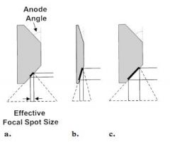
INCREASE
|
|
|
What happens to heat capacity with a small focal spot
|
decreases the heat capacity of the tube
|
|
|
What is the function of a collimator
|
define the shape of the X-ray beam
|
|
|
What are the 2 sets of bades that are present within the collimator
|
round and rectangular
|
|
|
What is the function of a round collimator
|
conforms the X-ray beam to a circular FOV
|
|
|
What is the function of the rectangular collimator
|
this can be used manually during the examination to reduce the size of the X-ray beam
|
|
|
What is the benefit of manual collimation
|
Collimation reduces
the exposed volume of tissue, resulting in re- duced scatter production and improved image contrast. |
|
|
How does manual collimation reduce radiation to the patient
|
coning the x-ray
beam to the area of clinical interest will reduce overall patient dose by minimizing direct expo- sure and scatter exposure to sensitive organs that may be adjacent to the beam |
|
|
After the Xray is generator and moves through the collimator where does it go next
|
|
|
|
What are the filters of a flouroscopy machine called
|
countour or wedge filters
|
|
|
What is the function of filters
|
to provide further beam shaping and collimation
|
|
|
What is another type of filter besides wedge or contour
|
equilization filters
|
|
|
What is the function of an equilzation filter
|
Equalization filters re-
duce glare from unattenuated radiation near the edge of the patient and equalize light exposure to the video camera. |
|
|
What do filters look like
|
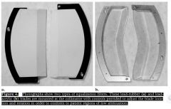
|
|
|
it operates in the radiographic mode. Tube current is measured in hundreds of mA instead of less than 5 mA, as in image-intensifying fluoroscopy.
|
it operates in the radiographic mode. Tube current is measured in hundreds of mA instead of less than 5 mA, as in image-intensifying fluoroscopy.
|