Reading...
![]()
Play button
![]()
Play button
![]()
Use LEFT and RIGHT arrow keys to navigate between flashcards;
Use UP and DOWN arrow keys to flip the card;
H to show hint;
A reads text to speech;
33 Cards in this Set
- Front
- Back
|
What is the Periodontal Ligament?
|
• Soft connective tissue (and associated cells/structures) situated between and connecting cementum to alveolar bone.
• It ranges in thickness (when in a state of health) from about 0.1 – 0.4 mm. |
|
|
Formation of the PDL -
Where does the PDL develop? |
• From the outer mesenchymal cells of the dental follicle.
|
|
|
Formation of the PDL -
When does differentiation into fibroblasts begin? |
• Soon after the beginning of root formation, although there is considerable variation from tooth to tooth.
|
|
|
What are the 4 roles of the PDL?
|
1. Homeostasis
2. Supportive 3. Sensory 4. Nutrition |
|
|
What is Homeostasis?
(In relation to the ongoing remodelling of the periodontium) |
Homeostasis refers to the dual function of formation and resorption.
Note - The turnover of alveolar bone and collagen within the PDL is higher within the periodontium than anywhere else within the body. |
|
|
How is the PDL supportive?
|
The PDL attaches the tooth to the jaw and has a cushioning effect via an hydraulic mechanism which
incorporates the blood vessels right through to the hydrophilic nature and water absorptive properties of the ground substance. |
|
|
How is the PDL Sensory?
|
The PDL has an extraordinary proprioceptive
capacity with up to 50% of the brain’s proprioceptive capacity devoted to oral functions. A healthy dentition can detect thicknesses of as little as 5 microns between the teeth. |
|
|
How does the PDL provide Nutrition?
|
Blood vessels carry anabolites etc as required by the cells within the PDL
(includes the more superficial osteocytes and cementocytes given that cementum is avascular). |
|
|
What is the Periodontal Ligament made of?
|
• Cells
• Fibres • Ground Substance • Blood vessels • Nerve supply • Lymphatics • Cementicles |
|
|
The 6 cell types?
|
• Synthetic Cells.
• Resorptive Cells. • Progenitor Cells. • Epithelial Rests of Malassez • Immunological Cells. • Endothelial and Neural Cells. |
|
|
What makes up Synthetic Cells?
|
- Osteoblasts
- Fibroblasts - Cementoblasts |
|
|
What makes up Resorptive Cells?
|
- Osteoclasts
- Fibroclasts - Cementoclasts |
|
|
What are Progenitor Cells?
|
Undifferentiated mesenchymal cells with the ability to differentiate into their predetermined cell type.
|
|
|
What is Epithelial Rests of Malassez?
|
Remnants of Hertwig’s root sheath.
|
|
|
What makes up Immunological Cells?
|
• Macrophages – responsible for phagocytosis. Make up 4% of total cell population within the PDL.
• Mast cells – small numbers only in healthy periodontium. Release histamine in inflammatory responses. |
|
|
Where are Endothelial and Neural Cells found?
|
Blood vessels and nervous system.
|
|
|
Principal Fibres of the PDL
How do the fibres of the Alveolar Crest Group run? |

Fibres from the crest of the alveolar bone run coronally/obliquely into the cementum (just below CEJ).
|
|
|
Principal Fibres of the PDL
How do the fibres of the Horizontal group run? |

The Horizontal fibres are the next group apical to the alveolar crest group.
They run from cementum to bone in a horizontal direction. |
|
|
Principal Fibres of the PDL
How do the fibres of the Oblique group run? |

Oblique Group run at an angle from the alveolar bone apically towards the tooth, anchoring into the cementum.
Note - the most prevalent of all of the fibres. |
|
|
Principal Fibres of the PDL
How do the fibres of the Apical group run? |

These fibres radiate from the apical cementum to the alveolar bone at the base of the socket.
|
|
|
Principal Fibres of the PDL
How do the fibres of the Interradicular group run? |
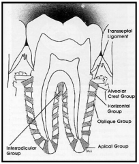
Found in multi-rooted teeth.
These fan out from the alveolar crest of the interradicular bone and insert into the cementum of the furcations of teeth. |
|
|
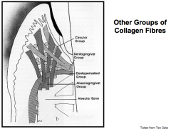
Other Collagen Fibres of the PDL
How do the fibres of the Dentogingival group run? |

Run from the cervical cementum immediately below the CEJ and extend into the lamina propria of the free and attached gingivae.
Note - They are the most numerous of the fibres outside of the strict PDL. |
|
|
Other Collagen Fibres of the PDL
How do the fibres of the Alveologingival group run? |

Alveologingival Group run from the bone of the alveolar crest to the lamina propria of the free and attached gingiva.
|
|
|
Other Collagen Fibres of the PDL
How do the fibres of the Circular group run? |

Circular Group form a circular ring around the tooth within the region of the free gingiva which interlocks with the other fibres, helping bind the free gingiva to the tooth.
|
|
|
Other Collagen Fibres of the PDL
How do the fibres of the Dentoperiosteal group run? |

These fibres leave the cementum between the dentogingival fibres and the first true fibres of the PDL, the alveolar crest group.
They then run over the alveolar crest and insert into the periosteum covering the outer cortical plates. |
|
|
Other Collagen Fibres of the PDL
How do the Transseptal fibres run? |
Transseptal Fibres run from cementum of one tooth to cementum of another tooth, traversing the interseptal bone.
|
|
|
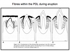
Fibres of PDL during Eruption
|
|
|
|
Ground Substance is a major component of the PDL.
Components of Ground Substance - |
• Estimated to be 70% water which significantly
aids with the protective function of the PDL. • Other components include proteoglycans and glycoproteins (i.e. combinations of various proteins and polysaccharide units). |
|
|
Blood Vessels of the Periodontal Ligament
The very rich blood supply is derived from: |
• The appropriate superior and inferior alveolar arteries along with other supplies from the lingual and palatine arteries.
|
|
|
Blood Vessels of the Periodontal Ligament
The Alveolar arteries run through bone and into the PDL through what? |
• Special canals termed Volkmann’s canals.
|
|
|
Blood Vessels of the Periodontal Ligament
Apical vessels - |
• Apical vessels also branch before entering the tooth
and supply some of the apical regions Of the PDL. |
|
|
Nerves of the Periodontal Ligament
The PDL is very richly innervated with nerves, they enter through two main sources, these are? |
• Those which branch from the nerves entering the root apex.
• Those which enter into the middle and cervical thirds through canals within the alveolar bone. |
|
|
Nerves of the Periodontal Ligament
The two types: |
• Sensory – associated with nociception and mechanoreception.
• Autonomic – regulate the blood vessels of the PDL. |