Reading...
![]()
Play button
![]()
Play button
![]()
Use LEFT and RIGHT arrow keys to navigate between flashcards;
Use UP and DOWN arrow keys to flip the card;
H to show hint;
A reads text to speech;
34 Cards in this Set
- Front
- Back
|
True or false.
Acute pericarditis is a syndrome. |
True
|
|
|
Acute pericarditis is most commonly ____________, self-limited to _________ weeks with what two symptoms and signs?
|
Idiopathic (viral)
1-3 weeks Sharp substernal pleuritic positional pain Pericardial friction rub |
|
|
What does the ECG of acute pericarditis look like?
|
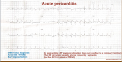
ST elevation does not confine to coronary territory, has upward concavity (seems to mimic STEMI sometimes)
|
|
|
What are the two types of pericarditis?
|
Serous and fibrinous
|
|
|
What are some differences between the two types?
|
Serous has smoother surface, few neutrophils/lymps/macros usually with effusion of 50-200mL of thin fluid (protein <50% serum level)
Fibrinous has dry, roughened, shaggy, bread ad butter surface, many more neutrophils/lymphs/macros, Is called serofibrinous if it is with effusion. |
|
|
What are some causes of serous pericarditis?
|
Heart failure, lymphatic obstruction by tumor, hypoalbuminemia
|
|
|
What are some causes of fibrinous pericarditis?
|
Viral myopericarditis, uremia, acute MI, metastatic malignancy, autoimmune
|
|
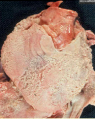
What is shown here?
|
Fibrinous pericarditis
|
|
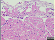
What is shown here?
|
Fibrinous pericarditis = lower layer of more condensed fibrin a few lymphocytes and macrophages
|
|
|
What are three more pathologic types of pericarditis?
|
Hemorrhagic
Purulent Constrictive |
|
|
What is seen with hemorrhagic pericarditis?
|
Serous, fibrinous or purulent plus hemorrhage, +/- effusion or exudate with blood added
|
|
|
What is seen with purulent (suppurative) pericarditis?
|
Red granular surface coated with pus, lots of subsurface neutrophils, up to 500 mL exudate in the pericardium
|
|
|
True or false. These three types (hemorrhagic, purulent, and constrictive) are very rare.
|
True
|
|
|
What are the two forms that post MI pericarditis can take?
|
1. Extension of visceral pericarditis to parietal over large transmural infarct (uncommon, <5% of infarctions)
2. Dressler syndrome 2-12 weeks after infarction (probably autoimmune but has become rare). |
|
|
Patients with what two autoimmune diseases get pericarditis? Percentages?
|
Lupus (as part of polyserositis with simultaneous pleuritis and peritonitis) and rheumatoid arthritis (30%)
|
|
|
Drug-induced pericarditis occurs with what two drugs?
|
Procainamide (sometimes as part of polyserositis) and hydralazine
|
|
|
What three diseases is hemorrhagic pericarditis associated with?
|
1. Metastatic carcinoma
2. Leukemia (thrombocytopenia) 3. Tuberculosis |
|
|
What are important tests for unexplained pericarditis?
|
Skin test for tuberculosis and chest x-ray
|
|
|
Describe constrictive pericarditis.
Is it rare? What is it commonly due to? What is the pathology similar to? |
Encasement of the heart in a dense fibrinous or fibrocalcific scar which prevents cardiac hypertrophy or dilatation.
Rare, commonly due to previous purulent or tuberculous pericarditis. |
|
|
What else is on the differential for constrictive pericarditis?
How do you test for it? What will the test show? How do you treat it? |
Restrictive cardiomyopathy
Echocardiogram, computerized tomography, or MRI --> thickened pericardium Strip it surgically! |
|
|
What are the normal pericardial effusion values?
What is the purpose of the effusion? |
15-50 mL of thin serous fluid in pericardium to lubricate between pericardial sac and heart
|
|
|
Describe the outcomes of the pericardial effusion changes:
Sudden increase up to 250 mL Between 250 and 300 mL Slow increase up to 1 L |
OK
Can be fatal OK |
|
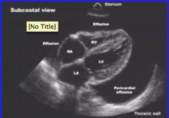
What is shown here?
|
Pericardial effusion on ultrasound examination
|
|
|
What are common causes of pericardial effusion?
|
Viral myopericarditis
Metastatic malignancy Autoimmune disease Drug-induced Renal failure Bleeding (hemopericardium) |
|
|
True or false.
Cardiac tamponade is a syndrome. |
True
|
|
|
What are some signs of cardiac tamponde?
What will the echocardiogram show? |
Jugular venous distention, muffled heart sounds, hypotension, pulses paradoxus
Diastolic collapse of right atrium and right ventricle |
|
|
What is Swan-Ganz?
|
Equalization of pressures
|
|
|
What is the treatment of cardiac tamponade?
|
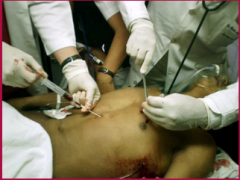
Pericardiocentisis
|
|
|
What are cardiac myxomas?
Are they rare? What sex are they more common in? What part of the heart are they most common in? |
Benign gelatinous mesenchymal neoplasms of the endocardium
Rare More common in females Most in left atrium |
|
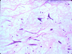
What is shown here?
|
Scattered stellate cells with scant cytoplasm in a bluish myxoid ground substance.
|
|
|
What are some symptoms of cardiac myxoma?
|
Dyspnea +/- orthopnea, cough +/- hemoptysis, fatigue, fever, transience neurological symptoms +/- syncope
|
|
|
What are some signs of cardiac myxoma?
|
Loud first heart sound, diastolic rumble, diastolic tumor plop, holosystolic murmur
|
|
|
What are some complications of cardiac myxoma?
|
Intermittent mitral obstruction, embolization (50% of patients, to brain in 50%), MI, sudden death
|
|
|
What is the diagnosis and treatment of cardiac myxoma?
|
Diagnosis = echocardiogram
Treatment = surgical excision (curative) |