Reading...
![]()
Play button
![]()
Play button
![]()
Use LEFT and RIGHT arrow keys to navigate between flashcards;
Use UP and DOWN arrow keys to flip the card;
H to show hint;
A reads text to speech;
94 Cards in this Set
- Front
- Back
|
What is the origin of the iliofemoral ligament (Y ligament of bigelow)?
|
Anterior Inferior Iliac Spine (AIIS)
|
|
|
What structure of the pelvis is an anteriorly raised region that represents the union of the ilium and pubis?
|
The iliopectineal eminence
|
|
|
The acetabulum is anteverted by what degree and obliquely oriented by what degree?
|
Anteverted by 15 degrees
Obliquely oriented by 45 degrees caudally |
|
|
Is the femoral neck anteverted or retroverted and by what degree in relation to the femoral condyles?
|
Anteverted approximately 14 degrees in relation to the femoral condyles
|
|
|
What is the average femoral neck shaft angle?
|
127 degrees
|
|
|
What do the trabecular patterns of the hip help determine?
|
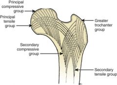
Trabecular patterns help determine the presence of osteopenia and displacement of femoral neck fractures
|
|
|
The hip joint capsule extends anteriorly across the femoral neck to the trochanteric crest. However, it extends posteriorly only partially across the femoral neck, so what regions of the hip are actually left extracapsular because of this?
|
The basocervical and intertrochanteric crest regions are extracapsular
|
|
|
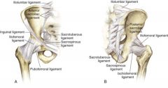
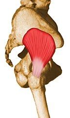
What 3 ligaments form the hip capsule?
|
The iliofemoral ligament (Y ligament of Bigelow)
The ischiofemoral ligament The pubofemoral ligament |
|
|
What is the strongest ligament in the body?
|
The iliofemoral ligament (Y ligament of Bigelow) is the strongest ligament in the body and attaches the anterior-inferior iliac spine to the intertrochanteric line in an inverted Y manner.
|
|
|
What is the purpose of the ligamentum teres?
|
Transmits an arterial branch of the posterior division of the obturator artery to the femoral head which is more important in pediatrics than adults
|
|
|
Why is the piriformis not used as a starting point for a nail on a pediatric patient?
|
It would damage the posterosuperior retinacular vessels, causing avascular necrosis of the femoral head
|
|
|
From birth to 4 years old, what is the primary blood supply to the femoral head?
|
Primary medial and lateral circumflex arteries (from deep femoral artery)
Ligamentum teres with posterior division of obturator artery |
|
|
From 4 years old to adulthood, what is the primary blood supply to the femoral head?
|
Negligible amount from lateral circumflex artery
Minimal amount from ligamentum teres Posterosuperior and posteroinferior retinacular from medial femoral circumflex artery |
|
|
In adulthood, what is the primary blood supply to the femoral head?
|
Medial femoral circumflex to lateral epiphyseal artery
|
|
|
When performing a posterior acetabular approach in an adult, why should you not completely transect the quadratus femoris muscle?
|
In order to avert damage to the main blood supply to the femoral head– the medial femoral circumflex artery
|
|
|
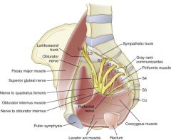
What structures outline the boundaries for the greater and lesser sciatic foramina?
|

The greater and lesser sciatic foramina
|
|
|
What structure constitutes the inferior border of the greater sciatic foramen and the superior border of the lesser sciatic foramen, effectively separating the greater and lesser sciatic foramina
|
The sacrospinous ligament (anterior sacrum to the ischial spine)
|
|
|
What forms the inferior border of the lesser sciatic foramen?
|
The sacrotuberous ligament (posterolateral sacrum to the ischial tuerosity)
|
|
|
What exits the greater and lesser sciatic foramina?
|
The piriformis, sciatic nerve, and other important structures exit the greater sciatic foramen.
The short external rotators of the hip exit the lesser sciatic foramen. |
|
|
What 3 muscles are hip flexors?
|
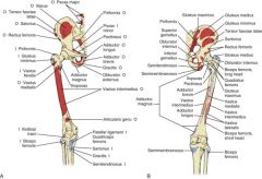
Hip flexor muscles are the iliopsoas, rectus femoris, and sartorius.
|
|
|
What are the 4 hip extensor muscles?
|
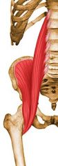
Hip extensor muscles are the gluteus maximus and hamstrings (semitendinosus, semimembranosus, and long head of biceps femoris)
|
|
|
What are the 2 main hip abductors? What muscle provides abduct in a flexed hip?
|

Hip abduction results primarily from the actions of the gluteus medius and minimus. The tensor fasciae latae also helps with abduction in a flexed hip.
|
|
|
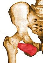
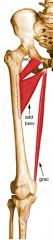
Hip adduction is primarily performed by what 5 muscles?
|
Hip adduction results primarily from the actions of the adductor brevis, adductor longus, adductor magnus, pectineus, and gracilis
|
|
|
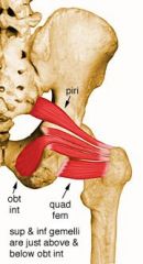
What 6 muscles provide hip external rotation?
|

Hip external rotation results from the action of the
P-GO-GO-Q: piriformis, gemellus superior, obturator externus, gemellus inferior, obturator internus, quadratus femoris |
|
|
Hip internal rotation is provided by what 7 muscles?
|

Hip internal rotation is provided by secondary actions of the anterior fibers of the gluteus medius and gluteus minimus and by the tensor fasciae latae, semimembranosus, semitendinosus, pectineus, and posterior part of the adductor magnus.
|
|
|
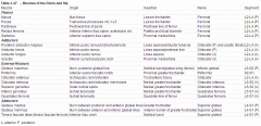
What is the origin, insertion, nerve, and spinal segment of the following hip FLEXOR?
Iliacus |

Origin: Iliac fossa
Insertion: Lesser trochanter Innervation: Femoral Spinal Segment L2-L4 (P) - Common to all flexors |
|
|
What is the origin, insertion, nerve, and spinal segment of the following hip FLEXOR?
Psoas Major |
Origin: Transverse processes of L1-L5
Insertion: Lesser trochanter Innervation: Ventral Ramus L1-L3 Spinal Segment L2-L4 (P) - Common to all flexors |
|
|
What is the origin, insertion, nerve, and spinal segment of the following hip FLEXOR?
Psoas Minor |
Origin: Transverse processes T12-L1
Insertion: Pectineal line of femur Innervation: Ventral Ramus of L1 Runs superficial to psoas major, sometimes indistinguishable |
|
|
What is the origin, insertion, nerve, and spinal segment of the following?
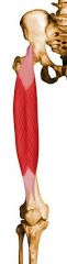
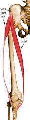
Tensor Fascia Latae |

Origin: ASIS
Insertion: Iliotibial band Action: Helps stabilize and steady the hip and knee joints by putting tension on the iliotibial band of fascia (in extension) Innervation: Superior gluteal - Common to abductors Spinal Segment: L4-S1 (P) - Common to all abductors |
|
|
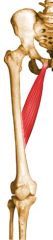
What is the origin, insertion, nerve, and spinal segment of the following hip FLEXOR?
Rectus Femoris |
Origin: Anterior inferior iliac spine, acetabular rim
Insertion: Patella and tibial tubercle Innervation: Femoral Action: Flexes hip and extends knee Spinal Segment L2-L4 (P) - Common to all flexors |
|
|
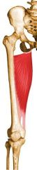
What is the origin, insertion, nerve, and spinal segment of the following hip FLEXOR?
Sartorius |
Origin: Anterior superior iliac spine
Insertion: Proximal medial tibia @ pes anserinus Innervation: Femoral Action: Flex, abduct, externally rotate hip Spinal Segment L2-L4 (P) - Common to all flexors |
|
|
What is the origin, insertion, nerve, and spinal segment of the following hip EXTERNAL ROTATOR?
Obturator Externus |
Origin: External surface of obturator membrane and anterior bony margins of obturator foramen
Insertion: Posteromedial surface of greater trochanter of femur Innervation: Obturator Spinal Segment: L2-L4 (A) |
|
|
What is the origin, insertion, nerve, and spinal segment of the following hip FLEXOR?
Pectineus |
Origin: Pectineal line of pubis
Insertion: Pectineal line of femur Innervation: Femoral nerve usually, although it may sometimes receive additional innervation from the obturator nerve as well (L2, L3, L4) Spinal Segment L2-L4 (P) - Common to all flexors |
|
|
What is the origin, insertion, nerve, and spinal segment of the following hip ADDUCTOR?
Adductor brevis |

Origin: Inferior pubic ramus
Insertion: Linea aspera/pectineal line Innervation: Obturator (P) Spinal Segment: L2-L4 (A) - Common to all adductors |
|
|
What is the origin, insertion, nerve, and spinal segment of the following hip ADDUCTOR?
Adductor Longus |
Origin: Anterior pubic ramus
Insertion: Middle 1/3 Linea aspera between adductor magnus and brevis medially and vastus medialis laterally Action: Adducts & flexes thigh Innervation: Obturator (Anterior division ) Spinal Segment: L2-L4 (A) - Common to all adductors |
|
|
What is the origin, insertion, nerve, and spinal segment of the following hip ADDUCTOR?
Posterior Adductor Magnus |
Origin: Inferior pubic ramus, ischial ramus, and inferolateral area of ischial tuberosity
Insertion: Linea aspera/adductor tubercle Action: Powerful thigh adductor; superior horizontal fibers also help flex the thigh, while vertical fibers help extend the thigh Innervation: Posterior division of obturator nerve innervates most of the adductor magnus; vertical or hamstring portion innervated by tibial nerve (L2, L3, L4) Spinal Segment: L2-L4 (A) - Common to all adductors |
|
|
What is the origin, insertion, nerve, and spinal segment of the following hip ADDUCTOR?
Gracilis |

Origin: Inferior symphysis/pubic arch
Insertion: Proximal medial tibia, just posterior to sartorius Innervation: Obturator (A) Spinal Segment: L2-L4 (A) - Common to all adductors |
|
|
What is the origin, insertion, nerve, and spinal segment of the following hip EXTERNAL ROTATOR?
Gluteus Maximus |
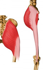
Origin:Ilium & sacrum
Insertion: Primarily in fascia lata at the iliotibial band; also into the gluteal tuberosity on posterior femoral surface Innervation: Inferior gluteal Action: Major extensor of hip joint, assists in laterally rotating the thigh; upper and middle third section of the muscle are abductors Spinal Segment: L5-S2 (P) |
|
|
What is the origin, insertion, nerve, and spinal segment of the following hip ABDUCTOR?
Gluteus Medius |
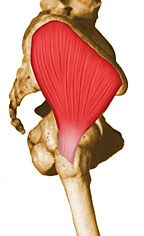
Origin: Ilium between posterior and anterior gluteal lines
Insertion: Greater trochanter lateral & superior Action: Major abductor of thigh; anterior fibers help to rotate hip medially; posterior fibers help to rotate hip laterally Innervation: Superior gluteal - Common to abductors Spinal Segment: L4-S1 (P) - Common to all abductors |
|
|
What is the origin, insertion, nerve, and spinal segment of the following hip ABDUCTOR?
Gluteus Minimus |
Origin: Ilium between anterior and inferior gluteal lines
Insertion: Anterior border of greater trochanter Innervation: Superior gluteal - Common to abductors Spinal Segment: L4-S1 (P) - Common to all abductors |
|
|
What is the origin, insertion, nerve, and spinal segment of the following hip EXTERNAL ROTATOR?
Piriformis |

Origin: Anterior sacrum
Insertion: Superior border greater trochanter Action: Lateral rotator of the hip joint; also helps abduct the hip if it is flexed Innervation: Nerve to Piriformis (L5-S2) |
|
|
What is the origin, insertion, nerve, and spinal segment of the following hip EXTERNAL ROTATOR?
Obturator Internus |
Origin: Ischiopubic rami/obturator membrane
Insertion: Medial greater trochanter, in common with superior and inferior gemelli Innervation: Nerve to the obturator internus and superior gemellus -- a branch of the sacral plexus (L5, S1) (L5, S1) Spinal Segment: L5-S2 (A) |
|
|
What is the origin, insertion, nerve, and spinal segment of the following hip EXTERNAL ROTATOR?
Superior Gemellus |

Origin: Outer ischial spine
Insertion: Medial greater trochanter, in common with obturator internus Rotates the thigh laterally; also helps abduct the flexed thigh Innervation: Nerve to the obturator internus and superior gemellus -- a branch of the sacral plexus (L5, S1) (L5, S1) Spinal Segment: L5-S2 (A) |
|
|
What is the origin, insertion, nerve, and spinal segment of the following hip EXTERNAL ROTATOR?
Inferior Gemellus |

Origin: Posterior Ischial tuberosity
Insertion: Medial greater trochanter, in common with obturator internus Innervation: Nerve to Quadratus femoris Spinal Segment: L5-S1 (A) |
|
|
What is the origin, insertion, nerve, and spinal segment of the following hip EXTERNAL ROTATOR?
Quadratus Femoris |

Origin: Ischial tuberosity - Lateral margin of obturator ring above ischial tuberosity
Insertion: Quadrate line of femur Innervation: Nerve to quadratus femoris Spinal Segment: L5-S1 (A) |
|
|

|
|
|
What nerves make up the lumbosacral plexus?
|
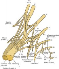
T12-S3.
|
|
|
The sciatic nerve is made up of L4-S3 and has a preaxial and postaxial division, what are these divisions?
|
The tibial nerve is preaxial
The peroneal nerve is postaxial. The peroneal division is more lateral |
|
|
What is the most common nerve injury with primary total hip arthroplasty?
|
The peroneal division of the sciatic nerve
|
|
|
What is the only muscle innervated by the peroneal nerve above the fibular neck?
|
The short head of the biceps femoris
|
|
|
On what muscle does the peroneal nerve run?
|
The deep surface of the long head of the biceps femoris
|
|
|
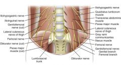
The lumbar plexus is found on the anterior surface of the ___________ muscle under (and within) the substance of the ____________ muscle
|
The lumbar plexus is found on the anterior surface of the QUADRATUS LUMBORUM under (and within) the substance of the PSOAS MAJOR muscle
|
|
|
Which nerve pierces the psoas and then lies on the anteromedial surface of the psoas
|
The genitofemoral nerve
|
|
|
What 2 muscles does the femoral nerve lie between at its origin?
|
The iliacus and psoas
|
|
|
Describe the path of the lateral femoral cutaneous nerve as it exits the pelvis
|
Lies on the surface of the iliacus muscle and exits the pelvis under the lateral attachment of the inguinal ligament
|
|
|
What muscle is the "key" for naming in the pelvis, ie, why is the superior gluteal nerve and artery named "superior"?
|
The piriformis
|
|
|
From lateral to medial, what are the nerves that exit the sciatic foramen?
|
Mnemonic: POP'S IQ.
Pudendal, Obturator internus, Postfemoral cutaneous, Sciatic, Inferior gluteal, Quadratus femoris |
|
|
What nerves leave via the greater sciatic foramen and then reenter the pelvis via the lesser foramen?
|
Pudendal and Nerve to the Obturator Internus
|
|
|
What are the borders of the femoral triangle?
|
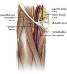
The sartorius laterally, the pectineus medially, and the inguinal ligament superiorly
|
|
|
What are the structures found within the femoral triangle?
|
NAVEL (from lateral to medial...venous near the penis)
Nerve (femoral), Artery, Vein, Empty Space, Lymphatics |
|
|
What are the muscles that make up the floor to the femoral triangle, lateral to medial?
|
Iliacus, Psoas, Pectineus, Adductor Longus
|
|
|
Why might Pott's disease of the spine manifest as hip pain?
|
Because of the attachment of the iliopsoas to the lumbar spine
|
|
|
At the apex of the femoral triangle, a cutaneous nerve branches off and travels to the foot medially. What is the nerve and what is the muscle it runs under proximally?
|
The saphenous nerve, runs under the sartorious
|
|
|
The obturator nerve exits the pelvis via the obturator canal then splits into anterior and posterior divisions within the canal. What does the anterior division and posterior division innervate?
|
Anterior: Proceeds anteriorly to the obturator externus and posteriorly to the pectineus, supplying the adductor longus, adductor brevis, and gracilis; it then delivers cutaneous branches to the medial thigh
Posterior: The posterior division supplies the obturator externus, adductor brevis upper part of the adductor magnus, and then delivers other branches to the knee joint |
|
|
Retractors placed behind what structure could injure the obturator nerve and artery?
|
The transverse acetabular ligament
|
|
|
Is the following nerve from the anterior or posterior division of the lumbosacral plexus? What spinal levels make up the nerve? What muscles/sensation is provided by the nerve?
Tibial |
Division: Anterior
Spinal Levels: L4-S3 Innervations: Semimembranosus, semitendinosus, biceps brachii (long head), adductor magnus, superior gemellus, soleus, plantaris, popliteus, tibialis posterior, flexor digitorum longus, flexor hallucis longus |
|
|
Is the following nerve from the anterior or posterior division of the lumbosacral plexus? What spinal levels make up the nerve? What muscles/sensation is provided by the nerve?
Quadratus femoris |
Division: Anterior
Spinal Levels: L4-S1 Innervations: Quadratus femoris, inferior gemellus |
|
|
Is the following nerve from the anterior or posterior division of the lumbosacral plexus? What spinal levels make up the nerve? What muscles/sensation is provided by the nerve?
Obturator internus |
Division: Anterior
Spinal Levels: L5-S2 Innervations: Obturator internus, superior gemellus |
|
|
Is the following nerve from the anterior or posterior division of the lumbosacral plexus? What spinal levels make up the nerve? What muscles/sensation is provided by the nerve?
Pudendal |
Division: Anterior
Spinal Levels: S2-S4 Innervation - Sensory: perineal Motor: Bulbocavernosus, urethra, urogenital diaphragm |
|
|
Is the following nerve from the anterior or posterior division of the lumbosacral plexus? What spinal levels make up the nerve? What muscles/sensation is provided by the nerve?
Coccygeus |
Division: Anterior
Spinal Levels: S4 Innervation: Coccygeus |
|
|
Is the following nerve from the anterior or posterior division of the lumbosacral plexus? What spinal levels make up the nerve? What muscles/sensation is provided by the nerve?
Levator ani |
Division: Anterior
Spinal Levels: S3-S4 Innervation: Levator ani |
|
|
Is the following nerve from the anterior or posterior division of the lumbosacral plexus? What spinal levels make up the nerve? What muscles/sensation is provided by the nerve?
Peroneal |
Division: Posterior (Peroneal Posterior)
Spinal Levels: L4-S2 Innervations: Biceps (short head), tibialis anterior, extensor digitorum longus, peroneus tertius, extensor hallucis longus, Peroneus longus and brevis, extensor hallucis brevis, extensor digitorum brevis |
|
|
Is the following nerve from the anterior or posterior division of the lumbosacral plexus? What spinal levels make up the nerve? What muscles/sensation is provided by the nerve?
Superior Gluteal |
Division: Posterior
Spinal Levels: L4-S1 Innervation: Gluteus medius and minimus, tensor fascia lata |
|
|
Is the following nerve from the anterior or posterior division of the lumbosacral plexus? What spinal levels make up the nerve? What muscles/sensation is provided by the nerve?
Inferior Gluteal |
Division: Posterior
Spinal Levels: L5-S2 Innervation: Gluteus Maximus |
|
|
Is the following nerve from the anterior or posterior division of the lumbosacral plexus? What spinal levels make up the nerve? What muscles/sensation is provided by the nerve?
Piriformis |
Division: Posterior
Spinal Level: S2 Innervation: Piriformis |
|
|
Is the following nerve from the anterior or posterior division of the lumbosacral plexus? What spinal levels make up the nerve? What muscles/sensation is provided by the nerve?
Posterior femoral cutaneous nerve |
Division: Posterior
Spinal Level: S1-S3 Innervation: Sensor - Posterior Thigh |
|
|
What is the interval for the anterior (Smith-Peterson) approach to the hip?
|
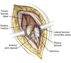
Sartorius (femoral nerve) and Tensor Fasciae Latae (superior gluteal nerve)
|
|
|
During a Smith-Peterson approach to the hip, where can the lateral femoral cutaneous nerve be injured?
|
Either anterior or medial to the sartorious, about 6-8 cm below the ASIS
|
|
|
What artery penetrates the TFL just anterior to the lateral femoral cutaneous nerve is at risk for injury with the Smith-Peterson approach?
|
The superficial circumflex artery
|
|
|
What is the interval for the anterolateral (Watson-Jones) approach to the hip?
|
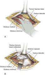
No true interneural plane. The intermuscular plane is the TFL and Gluteus Medius (bother superior gluteal nerve)
|
|
|
In the Watson-Jones (anterolateral) approach to the hip, TFL denervation can occur if dissected too far superiorly because the superior gluteal nerve lies where in relation to what structure?
|
About 5 cm above the acetabular rim
|
|
|
What is the interval for the lateral (Hardinge's) approach to the hip?
|
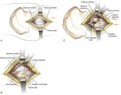
Interval: Splitting of the gluteus medius and vastus lateralis in tandem
Dissection: □ Incise the skin and the fascia lata to expose the gluteus medius and the vastus lateralis. □ Incise the gluteus medius from the greater trochanter, leaving a cuff of tissue and the posterior half to two thirds attached. □ Extend this incision to split the gluteus medius proximally. □ Split the vastus lateralis distally along its anterior fourth down to the femoral shaft. □ Detach the gluteus minimus from its insertion. □ The hip capsule is now exposed for further dissection |
|
|
What is at risk in the lateral (Hardinge's) approach to the hip if the split is taken too proximal?
|
Injury to the femoral nerve and possible denervation of the gluteus medius (superior gluteal nerve) if the split is too proximal (more than 5 cm proximal to the greater trochanter)
|
|
|
What is the interval for the posterior (Moore's or Southern's) approach to the hip?
|
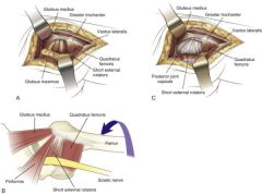
Splitting the fibers of the gluteus maximus
Dissection: □ Incise the skin and the fascia lata along the posterior border of the femur, and then split the fibers of the gluteus maximus bluntly. □ Expose the short external rotators close to their insertions into the greater trochanter, and reflect them laterally to protect the sciatic nerve and expose the posterior hip capsule. □ A portion of the quadratus femoris may be taken down with the short external rotators, but be aware of the significant bleeding that can come from the inferior portion of this muscle (ascending branches of the medial femoral circumflex artery). |
|
|
How could sciatic neurapraxia occur with the posterior approach to the hip? What artery is at risk with this approach?
|
Sciatic neurapraxia if the nerve is not properly protected by the short external rotators.
Damage to the inferior gluteal artery during the splitting of the gluteus maximus can occur |
|
|
What are the superficial and deep intervals for the medial approach (Ludloff's) to the hip?
|
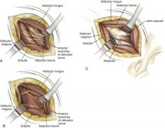
Superficial: Between adductor longus and gracilis
Deep: Adductor brevis and adductor magnus |
|
|
What structures are at risk with the medial approach to the hip?
|
Anterior division of the obturator nerve and medial femoral circumflex artery (between the adductor brevis and the adductor magnus/pectineus)
|
|
|
What approach to the pelvis provides access to the posterior column?
|
Kocher-Langenbach (posterolateral approach)
|
|
|
What approach to the pelvis exposes the anterior column?
|
Ilioinguinal approach, it relies on mobilization of the rectus abdominis and iliacus
|
|
|
There are 3 windows available with the ilioinguinal approach to the pelvis. What is provided with each window?
|
▪ The first window gives access to the internal iliac fossa, the anterior sacroiliac joint, and the upper portion of the anterior column.
▪ The second window (between the iliopectineal fascia and the external iliac vessels) provides access to the pelvic brim from the anterior sacroiliac joint to the lateral portion of the superior pubic ramus. ▪ The third window (below the vessels and the spermatic cord) gives access to the symphysis pubis |
|
|
What approach to the pelvis provides access to both the anterior and posterior columns with one approach?
|
The extended iliofemoral incision allows access to both columns by reflecting the gluteal muscles and tensor posteriorly and dividing the obturator internus and piriformis
|
|

|
|
|
|

|
|

|
|