![]()
![]()
![]()
Use LEFT and RIGHT arrow keys to navigate between flashcards;
Use UP and DOWN arrow keys to flip the card;
H to show hint;
A reads text to speech;
147 Cards in this Set
- Front
- Back
- 3rd side (hint)
|
Developmental anomalies |
▫Epitheliogenesis imperfecta of tongue ▫Cleft palate ▫Megacolon ▫Duplication of colon ▫Atresia coli ▫Atresia ani |
|
|
|
Abnormal smooth surface of tongue due to small filiform papillae. |
Epitheliogenesis imperfecta of tongue |
|
|
|
This is also known as smooth tongue. |
Epitheliogenesis imperfecta of tongue |
|
|
|
Epitheliogenesis imperfecta of tongue occurs in what breed of cattle? |
Holstein-Friesian |
|
|
|
It occurs as a defect in autosomal recessive gene |
Epitheliogenesis imperfecta of tongue |
|
|
|
This is most common congenital abnormality that occurs due to failure of oral-nasal cavity to divide leaving cleft. |
Cleft palate |
|
|
|
It may also extend towards lips producing 'harelip' condition. |
Cleft palate |
|
|
|
There is distension of colon which abruptly terminates in rectum due to mutant gene in dogs. |
Megacolon |
|
|
|
Some alleles of ______ are associated with more severe defects, such as deafness and aganglionic megacolon. |
Alleles of Ednrb |
|
|
|
Mutations in this gene are also responsible for Hirshsprung disease in humans. |
Endothelin B receptor gene |
|
|
|
This disease is characterized by intestinal aganglionosis, which is occasionally associated with hypopigmentation and/or deafness. |
Hirshsprung disease |
|
|
|
This syndrome in horses is due to a mutation in the horse endothelin B receptor gene |
lethal white foal syndrome |
|
|
|
It is a defect that is associated with malformation in the body of vertebrae T4 and T5 in the dog |
Duplication of colon |
|
|
|
In dog, the colon is duplicated from _______ to _______. |
caecum to rectum |
|
|
|
It is the absence of colon |
Atresia coli |
|
|
|
In calf, the absence of colon occurs and the intestine terminates in blind caecum |
Atresia coli |
|
|
|
This is absence of anal opening. |
Atresia ani |
|
|
|
Types of atresia ani |
▫Type I – Congenital anal stenosis ▫Type II – Imperforate anus alone ▫Type III – Combined with more cranial termination of the rectum as a blind pouch ▫Type IV – Discontinuity of the proximal rectum with normal anal and terminal rectal development |
|
|
|
Gender affected with increased incidence of atresia ani |
Females |
|
|
|
Breeds in dogs affected with increased incidence of atresia ani |
✔miniature or toy poodles ✔Boston terriers |
|
|
|
Postoperative complications that occur (atresia ani) |
▫fecal incontinence ▫Colonic atony secondary to prolonged preoperative distension |
|
|
|
It is the inflammation of mucosa of oral cavity. |
Stomatitis |
|
|
|
Condtions included in stomatitis |
✔Gingivitis ✔Glossitis ✔Cheilitis ✔Tonsilitis ✔Palatitis/Lampas |
|
|
|
Inflammation of gums |
Gingivitis |
|
|
|
Inflammation of tongue. |
Glossitis |
|
|
|
Inflammation of lips. |
Cheilitis |
|
|
|
Inflammation of tonsil. |
Tonsilitis |
|
|
|
Inflammation of palates. |
Palatitis/Lampas |
|
|
|
Etiology of stomatitis |
Trauma due to nails, wire, or any sharp object like needle. Physical - due to hot milk, medicines etc. Chemical - Alkali / acids. Microorganisms - Bacteria, virus, fungi. |
|
|
|
Macroscopic features of stomatitis |
▫ Catarrhal stomatitis: Mucous exudation in oral cavity. ▫ Vesicular stomatitis: Vesicles in oral mucosal e.g. FMD. ▫ Erosive stomatitis: Erosions in oral mucosa e.g. Rinderpest. ▫ Fibrinous stomatitis: False membrane in oral mucosa. ▫ Ulcerative stomatitis: Presence of ulcers in oral mucosa e.g. mucosal disease. |
|
|
|
Microscopic features of stomatitis |
▫Congestion of oral mucosa. ▫Presence of erosions, vesicles or ulcers. ▫Infiltration of neutrophils, lymphocytes and macrophages. ▫Presence of fibrinous exudate in the form of diphtheritic membrane. |
|
|
|
Inflammation of esophagus |
Esophagitis |
|
|
|
Characterized by catarrhal inflammation, ulceration or stenosis due to fibrosis. |
Esophagitis |
|
|
|
Etiology of esophagitis |
✔Trauma ✔Parasitis |
|
|
|
Macroscopic features of esophagitis |
Congestion Ulcer formation Red streaks of catarrhal inflammation. Stenosis due to fibrous nodules or inflammatory exudate. |
|
|
|
Microscopic features of esophagitis |
Congestion, haemorrhage. Ulceration. Infiltration of neutrophils, lymphocytes. Sub-epithelial fibrosis/nodules by Spirocerea lupi. |
|
|
|
Inflammation of crop caused by fungi. |
Ingluvitis |
|
|
|
Characterized by ulcerative or diphtheritic lesions |
Ingluvitis |
|
|
|
Etiology of ingluvitis |
Candida albicans (formerly Monilia albicans) |
|
|
|
Macroscopic features of ingluvitis |
✅Turkish towel-like appearance in crop mucosa. ✅Round and raised ulcers. ✅In moniliasis, formation of diphtheritic membrane. |
|
|
|
Microscopic features of ingluvitis |
✅Necrotic and ulcerative lesions. ✅Fibrinous inflammation with infiltration of mononuclear cells. |
|
|
|
Other term for Moniliasis |
Crop mycosis |
|
|
|
It is accumulation of fermentation gases in rumen due to failure of eructation as a result of obstruction or due to excessive production of gases characterized by distended rumen and dyspnea. |
Tympany |
|
|
|
Other term for tympany |
Bloat |
|
|
|
Etiology of tympany |
▫Choke of oesophagus. ▫Sudden change in animal feed with high content of legumes. ▫Excessive lush green fodder. |
|
|
|
Categories of bloat |
◾Frothy bloat (primary tympany) ◾Free gas bloat (secondary tympany) |
|
|
|
Type of bloat which is caused by the formation of a stable foam in the rumen. |
Frothy bloat (primary tympany) |
|
|
|
Example of frothy bloat |
Legume bloat |
|
|
|
Type of bloat which is due to excessive production of gaseous compounds from fermentation or as a result of an obstruction preventing the escape of gas compounds. |
Free gas bloat (secondary tympany) |
|
|
|
Primary tympany occurs most commonly in two settings: |
✅Pasture bloat – Animals on pasture, particularly those containing alfalfa or clover ✅Feedlot bloat – Animals feed high levels of grain, especially when it is finely ground |
|
|
|
Factors that reflect the forage’s potential for causing bloat. |
◾Protein content ◾rates of digestion and ruminal passage |
|
|
|
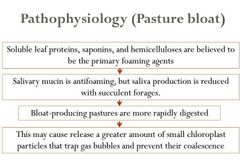
Pathophysiology of pasture bloat |
|
|
|
|
Possible causes of feedlot bloat |
◾production of insoluble slime by certain species of rumen bacteria in cattle fed high-carbohydrate diets ◾the entrapment of the gases of fermentation by the fine particle size of ground feed. |
|
|
|
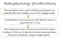
Pathophysiology of feedlot bloat |
|
|
|
|
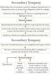
Pathophysiology of secondary tympany |
|
|
|
|
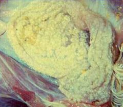
Macroscopic features of tympany |
▫Rumen is distended due to excessive accumulation of gases (C02, H2S, CO). ▫Distended rumen compresses diaphragm to hinder respiration. ▫Tarry colour blood, pale liver and rupture of diaphragm. ▫On rupture of rumen gas comes out (dry tympany). |
|
|
|
The gas is trapped in small bubbles in the ruminal fluid forming foams and is not easily removed, and this type of bloat is produced by saponin and water soluble proteins and due to reduction in surface tension in the absence of fatty acids that favours froth formation. |
Frothy bloat |
|
|
|
Microscopic features of tympany |
▫Haemorrhage in lungs, pericardium, trachea and lymph nodes. ▫Atelectasis in lungs. |
|
|
|
Tx for bloat |
▫Cannulation of rumen |
|
|
|
Inflammation of rumen in ruminant animals caused by change in diet, chemicals or drugs. |
Rumenitis |
|
|
|
Characterized by seropurulent exudate or ulcer formation with or without parakeratosis. |
Rumenitis |
|
|
|
Etiology of rumenitis |
✅Change in diet, corn or alfalfa hay. ✅Chemicals/drugs e.g. potassium antimony tartrate. ✅Fusobacterium necrophorum infection |
|
|
|
Macroscopic features of rumenitis |
✔Ulcers ✔Spherical white nodules of 1-2 cm diameter size ✔Sloughing of mucosa |
|
|
|
Microscopic features of rumenitis |
✔Seropurulent exudate ✔Ulcers Infiltration of lymphocytes and neutrophils. ✔Fibrous nodules due to hyperplasia of fibroblasts. ✔Parakeratosis |
|
|
|
Inflammation of reticulum in ruminant animals caused by trauma/perforation by foreign body including sharp object like needles, wires, etc. |
Reticulitis |
|
|
|
It is characterized by abscess formation, adhesions, peritonitis and pericarditis. |
Reticulitis |
|
|
|
Etiology of reticulitis |
Foreign body - sharp objects like needles, wires etc. |
|
|
|
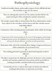
Pathophysiology of reticulitis |
|
|
|
|
Macroscopic features of reticulitis |
✔Perforation of reticulum by foreign body. ✔Abscessation/suppuration. ✔Peritonitis, adhesions of reticulum with diaphragm. ✔Pericarditis due to foreign body (traumatic reticulo pericarditis). |
|
|
|
Other term for traumatic reticulitis |
Hardware disease |
|
|
|
Microscopic features of reticulitis |
✔Infiltration of neutrophils, macrophages, lymphocytes. ✔Proliferation of fibroblasts producing adhesions. ✔Liquefactive necrosis |
|
|
|
Inflammation of abomasum in ruminants caused by chemicals/drugs, bacteria, virus or parasites. |
Abomasitis |
|
|
|
Characterized by congestion, edema and/or haemorrhagic ulcers. |
Abomasitis |
|
|
|
Etiology of abomasitis |
✅Chemicals/drugs. ✅Bacteria e.g. Clostridium septicum cause of braxy. ✅Parasites e.g. Theileria sp. |
|
|
|
Macroscopic features of abomasitis |
▫Congestion ▫oedema of abomasal folds ▫haemorrhage in braxy. |
|
|
|
Microscopic features of abomasitis |
▫Catarrhal, haemorrhagic abomasitis. ▫Presence of Gram positive rods in case of braxy. ▫Neutrophilic and lymphocytic infiltration. ▫Congestion and haemorrhages. ▫Ulceration with lymphocytic infiltration. |
|
|
|
This condition is common in cattle and buffaloes. |
Impaction of rumen and reticulum |
|
|
|
It is caused by heavy carbohydrate diet and characterized by atony of rumen, indigestion, acidosis and haemorrhage on serous membranes. |
Impaction of rumen and reticulum |
|
|
|
Etiology of rumen and reticular impaction |
✅Overfeeding of carbohydrate feed. ✅Lack of water. ✅Defective teeth or damaged tongue. ✅Paralysis of rumen. ✅Simple indigestion can result from suddenly changing the feed, using: ◾spoiled or frozen feeds introducing urea to a ration ◾turning cattle onto a lush cereal grain pasture ◾introducing feedlot cattle to a high-level grain ration |
|
|
|
Simple indigestion is usually associated with __________. |
sudden change in the pH of the ruminal contents |
such as a decrease in ruminal pH due to rapid fermentation of ingested carbohydrates or an increase in ruminal pH due to forestomach hypomotility and putrefaction of ingested feed |
|
|
TRUE OR FALSE. Simple indigestion can also result from accumulation of excessive quantities of relatively indigestible feed that may physically impair rumen function. |
True |
|
|
|
Macroscopic features of rumen & reticulum impaction |
✅Atony of rumen due to lactic acid production. ✅Rumen is filled with hard, caked undigested food with foul odour. ✅Hemoconcentration, anuria, blood becomes dark in colour. |
|
|
|
Microscopic features of rumen & reticulum impaction |
✅Haemorrhage in lungs. ✅Desquamation of ruminal epithelium. ✅Lesions of acidosis/toxicosis. |
|
|
|
Inflammation of stomach in non-ruminant animals having simple stomach caused by chemicals/drugs, bacteria, virus, parasite. |
Gastritis |
|
|
|
Characterized by congestion, edema, haemorrhage and ulceration. |
Gastritis |
|
|
|
Inflammation of proventriculus in poultry. |
proventriculitis |
|
|
|
Etiology of gastritis |
✅Physical - overfeeding, trauma. ✅Chemicals - Acid/alkali. ✅Microorganisms such as bacteria, virus, fungi. ✅Parasites e.g. Trichostrongyles sp., Hemonchus sp. ✅Uremia |
|
|
|
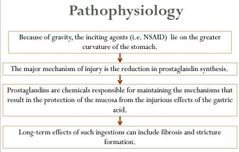
Pathophysiology of gastritis |
|
|
|
|
Macroscopic features of gastritis |
✅Congestion, oedema and haemorrhage of mucosal surface. ✅Thick mucous exudate in stomach. ✅Presence of vesicles/ulcers on gastric mucosa |
|
|
|
Microscopic features of gastritis |
✅Congestion and haemorrhage of gastric mucosa. ✅Presence of ulcer and necrosis. ✅Infiltration of mononuclear cells. ✅Lymphoid hyperplasia. |
|
|
|
Parasitic nematode found in the mucosa of the stomach in cats |
Ollulanus tricuspis |
|
|
|
Characterized by increased number of goblet cells, congestion and infiltration of neutrophils and mononuclear cells in mucosa of intestine. |
Catarrhal enteritis |
|
|
|
Etiology of catarrhal enteritis |
✅Foreign bodies and coarse feed ✅Chemical - drugs ✅Microorganisms - E.coli, Salmonella sp., viruses ✅Parasites - Coccidia |
|
|
|
Macroscopic features of catarrhal enteritis |
✔Presence of catarrhal exudate in lumen of intestine and congestion. ✔Thickening of the wall of intestine. ✔Diarrhea. ✔Presence of parasites in lumen of intestine |
|
|
|
Microscopic features of catarrhal enteritis |
✔Increased number of goblet cells in intestinal villi, reduced length of villi. ✔Congestion ✔Infiltration of polymorphonuclear and mononuclear cells. |
|
|
|
Characterized by inflammation of the intestines along with haemorrhagic exudate. |
Hemorrhagic enteritis |
|
|
|
Etiology of hemorrhagic enteritis |
✅Bacteria - E. coli, Bacillus anthracis, Salmonella sp. ✅Virus - Coronavirus, BVD ✅Parasites - Coccidia |
|
|
|
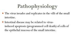
Pathophysiology of hemorrhagic enteritis |
|
|
|
|
Macroscopic features of hemorrhagic enteritis |
✅Haemorrhagic exudate in intestines; blood mixed intestinal contents. ✅Petechial or echymotic haemorrhage in mucosa and submucosa of intestine. ✅Presence of erosions/ulcers in mucosa. |
|
|
|
Microscopic features of hemorrhagic enteritis |
✅Haemorrhage in the mucosa of intestine. ✅Infiltration of neutrophils and mononuclear cells. ✅Erosion or ulcers in intestinal mucosa. ✅Presence of coccidia in the mucosa. |
|
|
|
It is the chronic inflammation of intestine characterized by proliferative changes like proliferation of fibrous tissue, infiltration of mononuclear cells and plasma cells in lamina propria leading to hardening of intestinal wall. |
Chronic enteritis |
|
|
|
Etiology of chronic enteritis |
✅Mycobacterium paratuberculosis in bovines ✅Intestinal helminths ✅E. coli in poultry |
|
|
|
This microorganism is excreted in large numbers in feces of infected animals and in lower numbers in their colostrum and milk. |
Mycobacterium paratuberculosis |
It is resistant to environmental factors and can survive on pasture for >1 yr; survival in water is longer than in soil. |
|
|
Infection of Mycobacterium paratuberculosis |
fecal-oral route |
|
|
|
Introduction of the disease into a noninfected herd is usually through _______. |
herd expansion or replacement purchases |
|
|
|
Factors affecting resistance to infection of M. paratuberculosis |
increases with age, and cattle exposed as adults are much less likely to become infected |
|
|
|
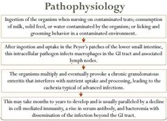
Pathophysiology of chronic enteritis |
|
|
|
|
Macroscopic features of chronic enteritis |
✔Thickening of the wall of intestine (corrugations in Johne's disease) ✔Thick mucous cover over mucosa of intestine ✔Transverse corrugations in the large intestine. ✔Granulomatous nodules in duodenum. ✔Small, round, raised necrotic foci on serosal surface of intestine covering whole length of intestine. |
|
|
|
The granulomatous inflammation leads to _____________ as evidenced by diarrhea and weight loss. |
intestinal thickening and malabsorption |
|
|
|
Microscopic features of chronic enteritis |
✔Proliferation of fibrous tissue in lamina propria. ✔Infiltration of macrophages, lymphocytes, plasma cells. ✔Atrophy of intestinal glands. |
|
|
|
It is characterized by necrosis of mucosal epithelium of intestine leading to erosions/ulcer formation and exposition of underlying tissues |
Necrotic enteritis |
|
|
|
Pathophysiology of necrotic enteritis |
|
|
|
|
Macroscopic features of necrotic enteritis |
✅Necrotic patches in intestines. ✅Fibrinous deposits over necrotic patches like bran deposits. ✅Swelling of mesenteric lymph nodes. ✅Ulcers in intestine. |
|
|
|
Microscopic features of necrotic enteritis |
✅Congestion and infiltration of mononuclear cells. ✅Necrosis and desquamation of intestinal villus epithelium, leading to exposed underlying tissue. ✅Ulcers in mucosa. ✅Proliferation of crypt epithelium, presence of abnormal epithelium over villus surface. |
|
|
|
It is caused by parasites and is characterized by catarrhal and/or haemorrhagic exudate in intestine, presence of ova/adult parasite and thickening of the wall of intestine. |
Parasitic enteritis |
|
|
|
Etiology of parasitic enteritis |
◾Helminths: ▫Roundworms ▫Tapeworms ◾Protozoa: ▫Coccidia ▫Histoplasma |
|
|
|
Macroscopic features of parasitic enteritis |
✅Presence of parasite helminths in the lumen of intestine. ✅Thickening of the wall of intestine. ✅Catarrhal or haemorrhagic exudate in intestine. |
|
|
|
Microscopic features of parasitic enteritis |
✅Presence of large number of goblet cells in mucosa of intestine. ✅Congestion and/or haemorrhage. ✅Presence of parasite/ova in the intestinal lumen. ✅Infiltration of eosinophils in mucosa and submucosa of the intestines. ✅Coccidia can be seen on mucosal scrapings under microscope. |
|
|
|
It is the fibrinous inflammation of intestine characterized by presence of fibrinous exudate comprising of pseudomembrane in the mucosa of intestine. |
Fibrinous enteritis |
|
|
|
Etiology of fibrinous enteritis |
Salmonella choleraesuis Fusobacterium necropharum |
|
|
|
Macroscopic features of fibrinous enteritis |
✅Presence of diphtheritic membrane over mucosa of intestine. ✅Button ulcers. ✅Sometimes, diphtheritic membrane covers the feces. |
|
|
|
Salmonella and Brachyspira hyodysenteriae can produce _______ in pigs. |
fibrinous and ulcerative colitis |
|
|
|
Fibrinous exudation in the small intestine and colon are usually associated with ___________ infection |
Salmonella spp. |
|
|
|
Microscopic features of fibrinous enteritis |
✅Congestion and haemorrhage in intestine. ✅Thickening of intestinal wall due to fibrinous exudate. ✅Fibrin network in mucosa. |
|
|
|
It is caused by bacteria or fungi and is characterized by granuloma formation in the intestines. |
Granulomatous enteritis |
|
|
|
Etiology of granulomatous enteritis |
▫Mycobacterium tuberculosis. ▫Coli granuloma - E. coli in poultry ▫Coccidioidomycosis / candidiasis. |
|
|
|
Macroscopic features of granulomatous enteritis |
✅Elevated/raised areas on the serous surface of intestine. ✅Thickening of the wall of intestine. ✅Small, tiny, white necrotic nodules on serosa. |
|
|
|
Disease that is characterized by granulomatous enteritis |
Crohn's disease |
|
|
|
Microscopic features of granulomatous enteritis |
✅Granuloma formation consisting of central necrosed area covered by lymphocytes, macrophages, epithelioid cells, giant cells and fibrous connective tissue. ✅Extensive proliferation of fibrous tissue. ✅Presence of bacteria / fungus in the lesion. |
|
|
|
Intestinal Obstruction (conditions) |
✔Piliconcretions/Trichobezoar ✔Phytobezoars/Polybezoars ✔Hernia ✔Intussusception ✔Volvulus ✔Torsion ✔Enterolith |
|
|
|
_________ are hair balls mostly found in stomach/intestines of animals having habit of licking. |
Piliconcretions |
|
|
|
TRUE OR FALSE. Piliconcretions is a vice that is more common in suckling calves and in animals with pica related to phosphorus deficiency. |
True |
|
|
|
This condition is a result from an excess of indigestible roughage. |
Phytobezoars |
|
|
|
_______ refers to bezoars composed of multiple objects |
Polybezoars |
|
|
|
This condition is presence of intestinal loop in umbilical area, scrotum or inguinal cavity which causes passive congestion, edema and obstruction in intestines. |
Hernia |
|
|
|
__________ is telescoping of intestine means a portion of intestine enters in caudal segment due to violent peristaltic movement. |
Intussusception |
|
|
|
TRUE OR FALSE. Intussusception causes obstruction, passive congestion and oedema |
True |
|
|
|
In this condition, the loop of intestine passes through a tear in mesentry. |
Volvulus |
|
|
|
TRUE OR FALSE. Volvulus causes obstruction at both ends of loop |
True |
|
|
|
This is twisting of intestine upon itself causing obstruction. |
Torsion |
|
|
|
Concretions in intestines particularly in horses are responsible for obstruction of intestinal tract and cause "colic in horse" and enterocolitis. |
Enterolith |
|
|
|
Breed of horse that has an increased incidence of enterolith. |
Arabian breed |
|
|
|
Age generally affected by enterolith in horses |
More than 4 years old |
|
|
|
TRUE OR FALSE. The stones are usually formed by ammonium magnesium phosphate (struvite) and collect around a small central nidus, often a metallic foreign body |
True |
|
|
|
Enteroliths generally lodge at what part of the horse's intestine? |
pelvic flexure or transverse colon. |
|
|
|
TRUE OR FALSE. Diets high in magnesium and phosphorus predispose to enterolith formation |
True |
|
|
|
Etiology of necrotic enteritis |
✔Salmonella ✔Rinderpest, rotavirus, cornovirus, Hog cholera virus. ✔Coccidia, Histoplasma. ✔Niacin deficiency ✔Clostridium sp. after coccidial infection in birds. |
|