![]()
![]()
![]()
Use LEFT and RIGHT arrow keys to navigate between flashcards;
Use UP and DOWN arrow keys to flip the card;
H to show hint;
A reads text to speech;
74 Cards in this Set
- Front
- Back
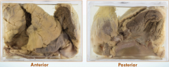
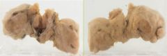
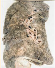
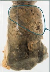
Identify |
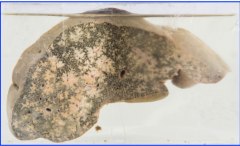
Nutmeg liver |
|
|
What is another name given to it? |
Chronic venous congestion |
|
|
Occurs due to? |
Right heart failure |
|
|
Describe the jar? |
It is filled with dark brown and yellow coloration. Dark brown due to necrosis and congestion Yellow due to fatty liver |
|
|
Where would the yellow and brown coloration be found? |
Yellow color on the periphre and dark brown in the center. |
|
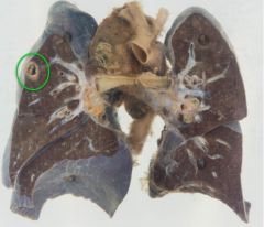
Identify the following: |
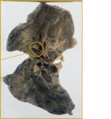
Pulmonary embolisim |
|
|
What causes this ? |
DVT of the L.L |
|
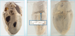
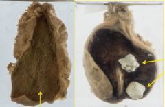
Identify the object |
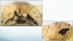
Cerebral intraventricular hemorrhage
|
|
|
Explain the jar? |
It is a dilated ventrical with brown blood clots inbetween. |
|
|
What causes this? |
In adults = Cerebrovascular accident In young = Trauma |
|
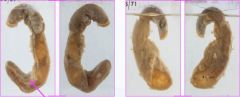
Identify the jar: |
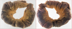
Gangrenous intestine |
|
|
Explain the jar? |
It is a swollen and dark (Gangrenous) intestine |
|
|
What causes the gangrene? |
Ischemia and infection |
|
|
Ischemia in which vien? |
Mesentric vien |
|
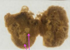
Identify the object: |
M.I in a heart with coronary artery thrombosis |
|
|
Describe the jar? |
It is a Heart with coronary artery thrombosis and a black discoloration due to necrosis. |
|
|
What type of necrosis is it? |
Coagulative necrosis |
|
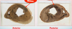
Identify the jar |
Myocardial infarction |
|
|
Explain the jar? |
A transverse cut section of the heart showing dark brown coloration affecting the left ventricle wall |
|
|
What type of necrosis is it? |
Coagulative necrosis |
|
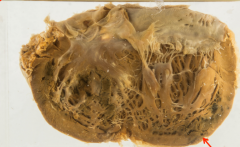
Identify the jar |
Myocardial infarction |
|
|
Explain the jar? |
Open heart showing dark brown coloration on the ventricle and endocardium. |
|
|
Type of necrosis? |
Coagulative necrosis |
|
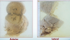
Identify the jar |
Left ventricular hypertrophy |
|
|
Explain the jar? |
Enlarged heart showing increased thicknesn in the left ventricle wall leading to narrowing of lumen (up to 5 cm) |
|
|
Most common cause of this? |
Hypertension Aortic valve stenosis |
|
|
Type of adaptation? |
Hypertrophy |
|
Identify the jar: |
Brown atrophy of the heart |
|
|
Explain the jar? |
Brown heart showing decrease in size of ventricles and normal size of blood vessels. |
|
|
What causes the brown color? |
Lupofuscin pigmentation |
|
|
What causes the atrophy? |
Aging |
|
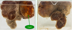
Identify the jar: |
Multinodular goiter |
|
|
Explain the jar? |
The jar shows an enlraged thyroind gland with multiple nodules. The nodules are covered by a fibrois covering. |
|
|
Cause of this? |
Iodine defiency |
|
|
Type of adaptation? |
Hyperplasia |
|
|
|
|
|
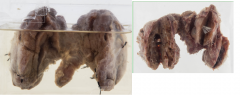
Identify the jars? |
Multinodular goiters |
|
|
Explain the jars? |
Enlarged thyroid loops, showing multiple nodules covered with fibrous tissue with cyst formation. |
|
|
Cause of this? |
Iodine defiency |
|
|
Type of adaptation? |
Hyperplasia |
|
Identify the following jars: |
|
|
|
- |
|
|
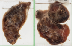
Identify the jars? |
Benign prostatic hyperplasia |
|
|
Explain the jars |
Grey coloration with multiple nodules covered with fibrous tissues |
|
|
The patient will complain of? |
Dysuria |
|
|
What type of adaptation? |
Hyperplasia |
|
Identify the jar |
Primary TB |
|
|
Explain the jar? |
Primary TB infection showing Ghons focus with granulomatous caseous necrosis. |
|
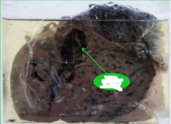
Identify the jar |
Milliary TB |
|
|
Explain the jar? |
Showing multiple small tubercles all over the lung |
|
|
Miliary TB is an example of which type of inflammation? |
Chronic specific granulomatous inflammation |
|
Identify the jar |
Tubercolous lymphadenitis |
|
|
Exlain the jar? |
Lymph node, showing multiple cheesy like material |
|
|
What type of necrosis? |
Caseous necrosis |
|
|
Type of diagnosis? |
Tubercolous lymphadenitis |
|
|
Type of inflammation? |
Chronic specific inflammation |
|
|
Diagnostic morphology? |
Caseous, granulomatous epitheloid macrophages, with langerhans giant cells. (Because its chronic) |
|
Identify the following: |
Acute suppurative appendicitis |
|
|
Explain the jar? |
Acute suppurative appendicitis showing yellow pus. |
|
|
What type of inflammation? |
Acute suppurative inflammation |
|
|
The swelling is due to? |
Exudate |
|
|
The redness is due to? |
Vasodilation |
|
|
The pain is due to? |
Prostaglandin and bradykinin |
|
Identify the jar? |
Lung abscess |
|
|
Explain the jar? |
A part of the lung showing pus material |
|
|
Which type of necrosis? |
Liquefactive necrosis |
|
|
The puss contains what? |
Neutrophils and dead cells. |
|
Identify the jar? |
Lobar pneumonia |
|
|
Explain the jar? |
A piece of the lung showing white coloration and loss of spongy feature |
|
|
What type of inflammation is it? |
Fibrinous inflammation |
|
Identify the jar |
Chronic cholecystitis with and without stones |
|
|
Explain the jar? |
Enlarged gall bladder with increased thickness of the wall. |
|
|
What is the cause of the increased wall thickness? |
Fibrosis |
|
|
What type of inflammation is it? |
Chronic non specific inflammation |