Reading...
![]()
Play button
![]()
Play button
![]()
Use LEFT and RIGHT arrow keys to navigate between flashcards;
Use UP and DOWN arrow keys to flip the card;
H to show hint;
A reads text to speech;
42 Cards in this Set
- Front
- Back
|
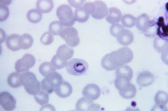
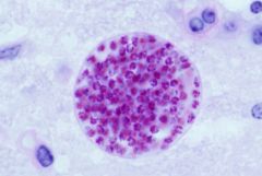
Plasmodium vivax trophozoite/ring form
- One chromatin dot with ring on it - Schuffner's dots in cytoplasm - Yellow-brown material pigment (malarial pigment) |
|
|
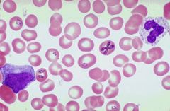
Plasmodium vivax schizont
- 12-24 merozoites |
|
|
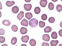
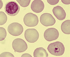
Plasmodium vivax gametocyte
- Round with "granules" |
|
|
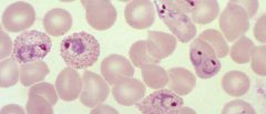
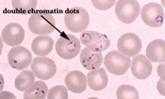
Plasmodium falciparum trophozoite
- Headphone ring with to chromatin dots - Mauer's clefts (dark blue staining of headphones) - Applique form: ring on membrane of RBC |
|
|
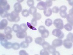
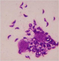
Plasmodium falciparum gametocyte
- Cigar shaped (sausage or crescent) |
|
|
Plasmodium ovale comet form
- Chromatin dot with comet shaped ring |
|
|
Plasmodium ovale trophozoite
- Shuffner's dots - Oval shaped - Ragged edges |
|
|
Plasmodium ovale schizont
- Oval shaped - 4-12 merozoites clustered around malarial pigment |
|

|
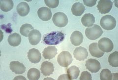
Plasmodium malariae trophozoite
- elongated "band" form in RBC - Ziemann's stippling (no malarial pigment) |
|
|
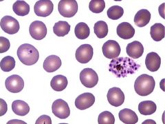
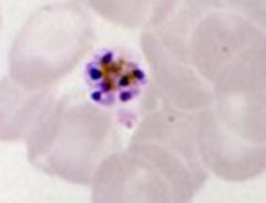
Plasmodium malariae schizont
- 6-12 merozoites - Daisy appearance - Large nuclei with malarial pigment |
|
|
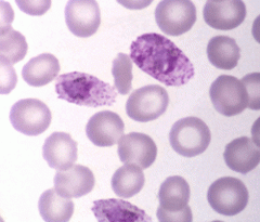
Plasmodium malariae gametocyte (left) and ring (right)
|
|
|
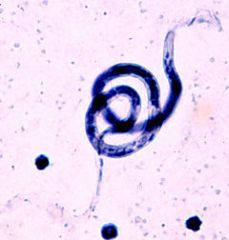
Babesia sp. ring form
- Extracellular rings distinguish from malaria - Maltese cross with 4 rings (looks like a chromosome) |
|
|
Toxoplasma gondii tachyzoite
|
|
|
Toxoplasma gondii bradyzoite
|
|
|
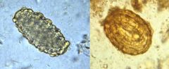
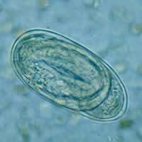
Enterobius vermicularis (pinworm) ova
- Oval, flattened on one side - Thich, colorless shell - 50-60 micrometers by 20-30 micrometers |
|
|
Eneterobius vermicularis (pinworm) adult
- Long, pointy tail - 7-13 mm female (males smaller) |
|
|
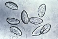
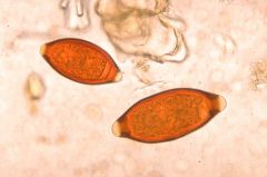
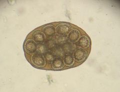
Fertilized (left) and Decorticated (right) Ascaris lumbricoides (Large intestinal roundworm) ova
- Fertilized ovum * Rounded * Yellow-brown mammillated shell - Decorticated ovum * Same but without mammillated shell |
|
|
Ascaris lumbricoides (Large intestinal roundworm) unfertilized (left) and fertilized (right) ova
- Unfertilized ovum * Elongated * Yellow-brown mammillated shell - Fertilized ovum * Rounded * Yellow-brown mammillated shell |
|
|
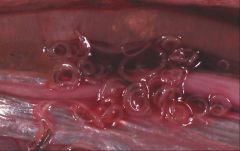
Anisakis sp. endoscopy
- Pink and tightly coiled larvae |
|
|
Trichuris trichiura (whipworm) ova
- Barrel shaped - Thock smooth shell - Clear "plugs" at each end - 45-55 micrometers by 20-23 micrometers |
|
|
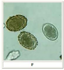
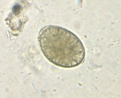
Hookworm primitive embryo ova
- 55-75 micrometers by 35-40 micrometers - Thin, clear, smooth colorless shell - Clear space between eggshell and embryo |
|
|
Hookworm embryonated ovum
- Embryo growing inside - Same as primitive embryo ovum |
|
|
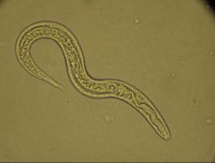
Hookworm rhabditiform larvae
- Long buccal cavity - Small genital primordium |
|
|
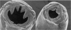
Ancylostoma duodenale (Old world hookworm) (left)
- 4 teeth for mouth Necator americanus (New world hookworm) (right) - 4 cutting plates for mouth |
|
|
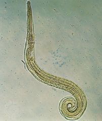
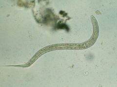
Strongyloides tercoralis (threadworm) rhabditiform larvae
- Short buccal cavity - Large genital primordium |
|
|
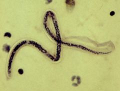
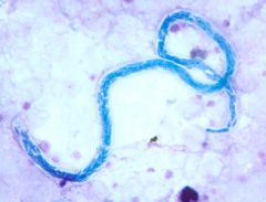
Wuchereria boncrofti microfilariae
- Sheathed - 245-300 micrometers - Nuclei DO NOT extend to tip of tail |
|
|
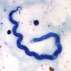
Brugia malayi microfilariae
- Sheathed - 200-275 micrometers length - Nuclei extend to tip of tail - Two terminal nuclei may be seen separate from the other nuclei at the end of one tail |
|
|
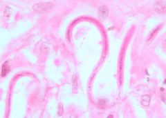
Loa loa microfilariae
- Sheathed - 250-300 micrometers - Body nuclei continuous to tip of tail |
|
|
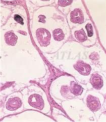
Onchocerca volvulus microfilariae in skin nodule
- Only seen from a skin biopsy where infection is |
|
|
Mansonella spp.
- Unsheathed - M. perstans * Seen in blood * No shepherd's hook - M. ozzardi * Seen in blood * Shepherd's hook at end of tail - M. streptocerca * Seen in skin biopsy * Shepherd's hook seen in tail |
|
|
Diphyllobothrium latum ovum
- Operculated egg - Boss (knob) at opposite end of operculum - Develop embryo when shed |
|
|
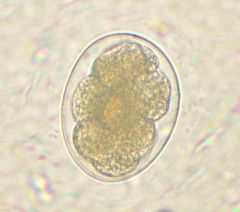
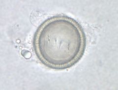
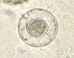
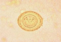
Taenia sp. ovum
- Radially striated shell - Onchosphere with 6 hooklets in 3 pairs (hexacanth embryo) - INFECTIVE (cysticercosis) |
|
|
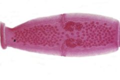
Taenia scolium (pork tapeworm) proglottid
- 7-14 branches on each side |
|
|
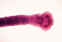
Taenia scolium (pork tapeworm) scolex
- 4 suckers - 2 rows of hooks |
|
|
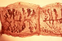
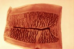
Taenia saginata (Beef tapeworm) proglottid
- 15-25 uterine branches on each side |
|
|
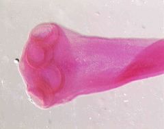
Taenia saginata scolex
- 4 suckers - NO TEETH |
|
|
Hymenolepsis nana (dwarf tapeworm) ovum
- Inner envelope - 6 hooked oncospheres contained inside - INFECTIVE |
|
|
Hymenolepsis diminuta (rat tapeworm) ovum
- Appear like nana but are LARGER (60-80 micrometers) |
|
|
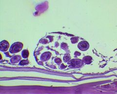
Dipylidium caninum ovum packet
- Membrane-bound sacks with 5-15 eggs with oncosphere embryos |
|
|
Dipylidium caninum proglottid
- 2 genital pores - Resemble maggots when passed |
|
|
Echinoccocus granulosus (hydatid tapeworm) hydatid cyst
- Laminated outer layer - Inner germinal center (daughter cysts and brood capsules) - Fluid with hydatid sand (loose germinal tissue with inverted scolices) |
|
|
Echinococcus multilocularis (fox/coyote tapeworm) cyst
|