![]()
![]()
![]()
Use LEFT and RIGHT arrow keys to navigate between flashcards;
Use UP and DOWN arrow keys to flip the card;
H to show hint;
A reads text to speech;
271 Cards in this Set
- Front
- Back
- 3rd side (hint)
|
Neurocystocercosis |
Eggs of T. solium |
|
|
|
Baermann culture technique for?? |
S. stercoralis larvae |
|
|
|
Achromatic granules |
I. butchilii |
|
|
|
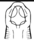
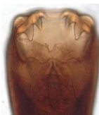
Pair of median teeth |
A. braziliense |
|
|
|
CATT ,(card agglutination) for |
Trypanosoma |
|
|
|
Infectious stage of malaria to its Definitive host |
Gametocyte |
|
|
|
Diamond medium for |
Trypanosoma |
|
|
|
Threadlike |
T. trichiura |
|
|
|
Comma shaped trophozoite |
P. falciparum |
|
|
|
Diagnostic stage for borborygmi agent |
Unembrayonated egg |
C. philippinensis |
|
|
Flat poles |
C. philippinensis |
|
|
|
Most dangerous fluke |
H. heterophyes |
|
|
|
Bile fluid spx |
C. sinensis |
|
|
|
Ectoparasites |
Mosquito |
|
|
|
Differential test for E. histolyica and dispar |
PCR |
|
|
|
Progressive motility |
E. histolytica |
|
|
|
Leningrad's curse |
G. intestinalis |
|
|
|
Delhi boil |
L. tropica |
Old world cutaneous leishmaniasis |
|
|
Iron deficiency anemia |
Trichuris |
|
|
|
Longest nematode |
D. medinensis |
|
|
|
Largest nematode |
Ascaris |
|
|
|
Congenital/vertical transmission |
A. duodenale |
|
|
|
Small wingless insect in dark |
Bedbugs |
|
|
|
Stool biopsy not stool spx for? |
T. spiralis |
|
|
|
All trematodes have 2 host except ___ |
Schistosomes |
|
|
|
2nd intermediate host of H. heterophyes, C. sinensis, O. felineus |
Fish |
|
|
|
2nd intermediate host of P. westermani |
Crab / crayfish |
|
|
|
2nd Intermediate of F. gigantica, hepatica and buski |
Plant |
|
|
|
E. ilocanum 2nd IH |
Snail |
Garrison |
|
|
Large lateral terminal |
S. mansoni |
Dako ang mansion |
|
|
Small lateral terminal |
S. japonicum |
Japanese gamay oten |
|
|
PTB like illness in acoholics males of sorsogon |
P. westarmani |
|
|
|
Bipolar thickening, bipolar filaments |
H. nana |
Bipolar si nana |
|
|
Bipolar thickening WITHOUT bipolar filaments |
H. diminuta |
|
|
|
15 -20 uterine branches, tree like appearance |
T. saginata |
Tree sa saging |
|
|
7-12 uterine branches, finger/dendritic appearance |
T. solium |
pag Finger og SOLo |
|
|
Severe anemia in the philippines |
Spirometral |
|
|
|
Destroyes trophozoite |
Iodine (quansels iodine) |
|
|
|
Gay bowel syndrome |
G. lamblia |
|
|
|
Malaria rapid diagnostic test |
Malaquick (HRP - histones rich protein) |
|
|
|
Malaquick (HRP - histones rich protein) for?? |
P. falciparum |
|
|
|
Kato thick size |
50-60g |
|
|
|
Periplaneta americana |
Cockroach |
CATPES |
|
|
Common intestinal flagellate shape |
Pear shaped |
|
|
|
Cercaria without the tail |
Schistosomule |
|
|
|
Germ layer eggs |
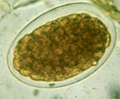
Hookworm |
Morula ball appearance |
|
|
Gardia special test |
String test |
|
|
|
4 classes of protozoans are differentiated by |
Motility |
|
|
|
The concentration procedure using ether or ethyl acetate as lipid removing agent and formalin as fixative |
Ridley Allen technique |
|
|
|
Definitive diagnosis for cryptosporidiosis |
Identification of spherical oocyts |
|
|
|
Male or female Big, blunt tail |
Female |
|
|
|
Male or female Small, curved tail |
Male |
|
|
|
Organism isolated from AIDS with keratoconjunctivitis |
Nosemca corneum |
|
|
|
Aside from flagella, this aids in the identification of flagellates |
Cystone, undulating membrane |
|
|
|
Asexual reproduction in T. vaginalis |
Longitudinal binary fission |
|
|
|
External parasite of fish with direct life cycles |
Monogenea |
|
|
|
Endoparasite with ventral surface of adhesive organ |
Aspidogastrea |
|
|
|
Location of F. buski |
Intestine |
|
|
|
Location of F. hepatica and gigantica |
Liver/bile duct |
|
|
|
Location of Echinostoma spp. H. heterophyes M. yokogawai |
Intestine |
|
|
|
Location of O. viverrini |
Liver/bile duct |
|
|
|
First seen in a Taiwan boy with meningitis |
A. cantonensis |
|
|
|
Location of A. acantonensis |
Pulmonary artery of rats |
|
|
|
Harbors the sexual stage of a parasite |
Definitive host |
|
|
|
Harbors the asexual stage of a parasite |
Intermediate host |
|
|
|
Trophozoite to cyst, happens in large intestine |
Encystation |
|
|
|
Cyst to trophozoite, happens in small intestine |
Excystation |
|
|
|
All amoeba inhabit the large intestine except ____ |
E. gingivalis |
|
|
|
All amoeba develop into cystic stage except____ |
E. gingivalis |
|
|
|
E. histolytica moves by means of ______ |
Pseudopods |
|
|
|
E. histolytica motility is ______ |
Progressive, unidirectional |
|
|
|
Virulence factor of e. histolytica that degrades the ECM of intestinal walls |
Cysteine proteinase |
|
|
|
Virulence factor of e. histolytica that mediates adhesion |
GalNac lectin |
|
|
|
Non dysenteric colitis |
E. histolytica |
|
|
|
_____ shaped ulcer in E. histolytica |
Flask |
|
|
|
Best iodine for trophozoite |
Quensels iodine |
|
|
|
Treatment for E. histolytica that has metallic/bitter taste and makes urine black |
Metronidazole |
|
|
|
Witch broom stick is seen on cyst of |
E. coli |
|
|
|
E. coli motility is ______ |
Sluggish |
|
|
|
Small race of E. histolytica |
E. hartmanni |
|
|
|
Amoeba that may ingest bacteria but not RBC |
E. hartmanni |
|
|
|
Kissing amoeba |
E. gingivalis |
|
|
|
Amoeba that has no cyst stage |
E. gingivalis |
|
|
|
Amoeba that has characteristic of glycogen vacuoles, large endosome described as bouquet of flowers |
Iodamoeba butchili |
|
|
|
Smallest amoeba, CROSS-EYED CYST |
Endolimax nana |
|
|
|
Primary amoebic meningoencephalitis (PAM) causative agent |
Naegleria fowleri |
|
|
|
Granulomatous amoebic enciphalitis (GAM) causative agent |
Acanthamoeba |
|
|
|
Cyst has doubled WRINKLED wall |
Acanthamoeba |
|
|
|
Free living amoeba associated with contact lens |
Acanthamoeba |
|
|
|
Neurologic manifestation such as ataxia and hemiparesis (partial paralysis) |
Acanthamoeba |
|
|
|
Treatment for N. fowleri and Acanthamoeba |
SXT |
|
|
|
Largest intestinal protozoa |
Balantidium coli |
|
|
|
Virulence factor of B. colo |
Hyaluronidase |
|
|
|
Trophozoite has 2 nuclei (Kidney bean shaped Macro and spherical micro) |
B. coli |
|
|
|
Thrown ball/ Rolling ball motility |
B. coli |
|
|
|
Round base and wide neck ulceration |
B. coli |
|
|
|
Only protozoan intestinal flagellate |
Gardia lamblia |
|
|
|
Only protozoan with sucking disk attachment |
Gardia lamblia |
|
|
|
Pear shaped/tear drops, 4 pairs of flagella (old man w/ eye glasses) smiling face |
Gardia lamblia |
|
|
|
Clinical manifestation Steatorrhoea Malabsorption Leningrad's curse (travellers disease) Rotten egg due to increase in H2S |
Gardia lamblia |
|
|
|
Sheppard's crook on trophozoite Cyst has lemon/nipple shaped |
Chilomastix mesnili |
|
|
|
Rosette shaped nuclei Binucleated trophozoite |
Dientamoeba fragilis |
|
|
|
Clinical manifestations
Colicky abdominal pain Irretable bowel syndrome |
D. fragilis |
|
|
|
Protozoa co-infectio with enterovirus and Ascaris (co-infection) |
D. fragilis |
|
|
|
No cystic stage, has undulating membrane |
Trichomonas |
|
|
|
Motility of T. vaginalis |
Jerky and tumbling |
|
|
|
Pathology Strawberry cervix Ping pong disease |
T. vaginalis |
|
|
|
Smear for T. vaginalis |
Giemsa Papanicolaou Acridine orange |
|
|
|
Falling leaf motility |
G. lamblia |
|
|
|
Square chromatoidal bars with splintered ends (witch broom stick) |
E. coli |
|
|
|
Vector of T. brucei gambiense |
Tsetse fly |
|
|
|
special feature of this trypanosoma is "winterbottom's sign"
|
T. brucei gambiense |
|
|
|
special feature of this trypanosome is death before CNS involvement |
T. brucei rhodesiense |
|
|
|
Causative agent of Chaga's disease |
T. cruzi |
|
|
|
Vector of chaga's disease |
Reduvid bug kissing bug assasin bug killer bug |
|
|
|
culture for T. cruzi |
weiman's medium |
|
|
|
Infective stage of T. cruzi |
metacylic trypomastigote |
|
|
|
Vector of leishmania tropica |
sand flies |
|
|
|
causative agent old world cutaneous leishmaniasis |
leishmania tropica |
|
|
|
causative agent of new world cutaneous leishmaniasis |
Leishmania mexicana |
|
|
|
causative agent of mucocutaneous leishmaniasis |
Leishmania braziliensis |
|
|
|
causative agent of viceral leishmaniasis, kala azan, dum-dum, black fever |
Leishmania donovani |
|
|
|
Diagnosis of leishmania |
smear of lesion stained with Wright or Giemsa of AMASTIGOTE |
|
|
|
Screening test for leishmaniasis |
Formol gel test Complement fixation test Fluorescent Antibody test |
|
|
|
Asexual coccidian form |
Sporogony |
|
|
|
sexual coccidian form |
Schizonogy |
|
|
|
infective stage of leishmania |
Promastigote |
|
|
|
diagnostic stage of leishmania |
Amastigote |
|
|
|
Encephalitis is the most common manifestation of this coccidian |
Toxoplasma gondii |
|
|
|
Toxoplasma initial stage, fast multiplying |
Tachyzoite |
|
|
|
Toxoplasma stage that's slow multiplying that and forms cyst |
Bradyzoite |
|
|
|
WBC transfusion is one of the mode of tans mission of this coccidian |
T. gondii |
|
|
|
Diagnosis of this coccidian is through methylene blue sabin feldman and direct microscopy |
T. gondii |
|
|
|
Diagnosis is through ZNSO4 and sheaters sugar |
Isospora belli |
|
|
|
Coccidian that causes diarrhea in immunocompromised host |
Cryptosporidium hominis Cyclospora |
|
|
|
Coccidian MOT:
|
Cryptosporidium hominis |
|
|
|
Kinyoun Acid Fast Method is Red-pink donut shape in blue background |
Cryptosporidium hominis |
|
|
|
Opportunistic agents in AIDS patient |
C. parvum |
|
|
|
Oocyst appear as Blue or Green circles under fluorescence microscopy |
Cyclospora cayetensis |
|
|
|
Acquired by ingestion of uncooked beef or pork containing tissue cysts |
Sarcosystic hominis |
|
|
|
Coccidian infective stage |
Sporulated oocyst |
|
|
|
Coccidian diagnostic stage |
unsporulated oocyst |
|
|
|
Clinical features: Cold paraxysm Temperature fall Jaundice leading to splenomegaly |
Plasmodium |
|
|
|
Plasmodium infective stage to its definitive host: |
Gametocyte |
|
|
|
Plasmodium infective stage to its intermediate host: |
Sporozoite |
|
|
|
Principal vector of Plasmodium |
Anopheles minimus var. flavirostis (LOW NIGHT FLYERS) |
|
|
|
Secondary vector of plasmodium |
Anopheles litoralis balacensis mangyunas maculatus |
|
|
|
Gold standard of plasmodium |
Thick and thin smear |
|
|
|
Quantitative buffy coat (+) on malaria appears as |
bright green and yellow |
|
|
|
Most common plasmodia in the world |
P. vivax |
|
|
|
Most common plasmodia in the Philippines |
P. falciparum |
|
|
|
Plasmodia in malay peninsula boreno |
P. knowlesi |
|
|
|
Plasmodia that affects young rbc |
P. vivax |
|
|
|
Schuffner's dots is seen on peripheral blood smear what plasmodium is this |
P. vivax |
|
|
|
Maurers dots is seen on peripheral blood smear what plasmodium is this |
P. falciparum |
|
|
|
Ziemant's dots is seen on peripheral blood smear what plasmodium is this |
P. malariae |
|
|
|
James dots is seen on peripheral blood smear what plasmodium is this |
P. ovale |
|
|
|
Refers to the absence of free flagellum but with kinetoplast |
Amastigote |
|
|
|
Largest nematode |
Ascaris lumbricoides |
|
|
|
Loeflers syndrome is seen in |
A. lumbricoides |
|
|
|
Treatment for A. lumbricoides |
Albendazole |
|
|
|
Whipworm = ? |
Trichiuris trichiura |
|
|
|
Ova resembles lemon, football with two prominent plugs at both ends |
Trichiuris trichiura |
|
|
|
Pinworm = ??? |
Enterobius vermicularis |
|
|
|
What nematode does autoinfection externally
|
E. vermicularis |
|
|
|
What nematode does autoinfection internally |
Strongyloides |
|
|
|
Patient is experiencing pruritius ani, what parasite? |
E. vermicularis |
|
|
|
Diagnosis for E. vermicularis |
Graham's cellophane/Scotch tape |
|
|
|
Diagnostic feature of E. vermicularis |
cephalic alae and distinct esophageal bulb |
|
|
|
D shaped asymmetrical ova |
E. vermicularis |
|
|
|
Pudoc/Bagsik/Mystery = |
Capillaria philippinensis |
|
|
|
Infective stage of C. philippinensis |
L3 larvae |
|
|
|
Primary host of C. philippinesis |
Herons |
|
|
|
Ova with flattened bipolar plug, peanut shaped |
C. philippinensis |
|
|
|
Clinical manifestation: |
C. philippinensis |
|
|
|
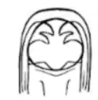
New world hookworm |
Necator americanus |
|
|
|
Old worm hookworm |
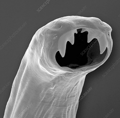
A. duodenale |
|
|
|
Infective stage of hookworms |
L3 larvae |
|
|
|
Diagnostic is egg/larvae in old stool |
hookworm |
|
|
|
Diagnostic is egg/larvae in new stool |
strongyloides |
|
|
|
N. americanus |
semiluran cutting plates |
|
|
A. duodenale |
2 pairs of ventral teeth |
|
|
1st and feeding stage of hookworm |
Rhabtidiform larva |
|
|
|
Hookworm |
Ova is transparent and ovoid, 2-8 germ layers, thin hyaline stage (morula ball) |
|
|
Clinical manifestation wakamas disease |
Hookworm larvae |
|
|
|
Clinical manifestation Iron deficiency anemia hypoalbuminemia GIT bleeding diarrhea |
hookworm adult |
|
|
|
threadworm = ?? |
Strongyloides stercolaris |
|
|
|
Nematode capable of parthenogenesis |
S. stercoralis |
|
|
|
Diagnosis DFS |
S. stercoralis |
|
|
|
Treatment for S. stercoralis |
Ivermictin |
|
|
|
Common parasite of pigs accidental host is man Final host is rat |
Trichinella spiralis |
|
|
|
Diagnosis of T. spiralis |
Bachman intradermal test Muscle biopsy |
|
|
|
Clinical manifestation inflammation jejunum and duedeum highest level of eosinophil encapsulated larvae on muscle |
T. spiralis |
|
|
|
Rat/rodent,lung worm |
Angiostrongylus cantonensis |
|
|
|
Habitat of A. cantonensis |
pulmonary artery of rat |
|
|
|
Causes eosinophilic meningoencephalitis |
A. cantonensis |
|
|
|
Intermediate host of A. cantonensis |
Giant african snail |
|
|
|
Male of A. cantonensishas well developed ________ |
KIDNEY shaped caudal bursa |
|
|
|
Famale morphology of A. cantonensis has ________ due to the looping of milky white uterine tubules around the blood filled intestine |
Barbers pole |
|
|
|
Guinea/ Israeli/Dragon / Fiery serpent worm = |
Dracunculus medinensis |
|
|
|
Intermediate host D. medinensis |
cyclops, copepods |
|
|
|
Longest nematode (up to 1m) |
D. medinensis |
|
|
|
A. brasiliense |
Cat hookworm pair of large teeth and pair of inconspicuous median teeth |
|
|
A. caninum |
3 pairs of ventral teeth |
|
|
African eye worm = ?? |
Loa loa |
|
|
|
Vector of Loa loa |
Horseflies deerflies mangoflies |
|
|
|
Blinding worm = |
onchocerca volvulus |
|
|
|
Vector of O. volculus |
Black fly |
|
|
|
Causes blinding filariasis or "river blindness" |
O. volvulus |
|
|
|
Snake-like microfilaria with graceful curves |
Wuchereria bancrofti |
|
|
|
Clinical manifestation tropical pulmonary eosinophilia elephantiasis hydrocele dermatolymphangioadenitis |
filarial nematodes |
|
|
|
Diagnostic of filarial nematodes |
microfilariae |
|
|
|
Treatment for filarial nematodes |
Diethylcarbamazine |
|
|
|
Filarial nematode Sheathed with NO nuclei |
Wuchereria bancrofti |
|
|
|
Filarial nematode Sheated with 2 caudal nuclei |
Brugia malayi |
|
|
|
Filarial nematode Unsheated, tail tapers to a thin filament containing column of 4-6 ovoid nuclei |
Mansonella ozzardi |
|
|
|
Filarial nematode
Unsheated, no nuclei |
Onchocerca volvulus |
|
|
|
Causative agent of elephantiasis |
Wuchereria bancrofti |
|
|
|
Infective stage of trematodes |
Metacercaria |
|
|
|
These parasites are hermaphroditic |
Trematodes |
2 sex |
|
|
Temperate/sheep liver fluke |
Fasciola hepatica |
|
|
|
Tropical/Giant liver fluke |
F. gigantica |
|
|
|
Chinese/oriental liver fluke |
Clonorchis sinensis |
|
|
|
Cat liver fluke |
Opisthorchis felineus |
|
|
|
Oriental lung fluke |
Paragonimus westermani |
|
|
|
Largest trematode, Giant intestinal fluke |
Fasciolopsis buski |
|
|
|
Garrison fluke |
Echinostoma ilocanum |
|
|
|
Von Seibolds fluke, smallest fluke and most dangerous |
H. heterophyes |
|
|
|
Grasshopper fluke |
Eurthema pancreaticum |
|
|
|
Oriental blood flukes |
S. japonicum |
|
|
|
Vesicle blood fluke |
S. haematobium |
|
|
|
Microscopic findings Intestine: branched/dendritic Ova: ovoid, well rounded posterior end (hen egg shaped) |
F. hepatica |
|
|
|
Halzoun-pharyngeal fasciolosis |
F. hepatica |
|
|
|
Trematode associated with carcinoma of gallbladder |
C. sinensis |
|
|
|
Findings: Obstruction of small intestine Blood loss |
F. buski |
|
|
|
Microscopic: Ventral suckers is smaller than oral Prominent opercular shoulder Comma shaped aboperculum Elongated adult |
C. sinensis |
|
|
|
Microscopy: Lancet shaped Oral sucker is smaller than ventral |
Opisthorchis felineus |
|
|
|
Findings: Cirrhosis/liver cancer |
O. felineus |
|
|
|
1st and 2nd intermediate host is snail |
E. ilocanum |
|
|
|
Collar of spines (49-51 horse shoe shape) around oral suckers |
E. ilocanum |
|
|
|
May be present in Peptic ulcer disease |
H. heterophyes |
|
|
|
Most romantic parasites |
Schistosomes |
|
|
|
Schistosome in superior mesenteric vein |
S. japonicum |
|
|
|
Schistosome in mesenteric vein of large intestine |
S. mansoni |
|
|
|
Schistosome in portal vein urinary bladder |
S. haematobium |
|
|
|
Clinical manifestation Swimmer itch Katayama fever Cercarial itch |
S. japonicun |
|
|
|
Clinical manifestation Cercardial dermatitis |
S. mansoni |
|
|
|
Mistaken as pulmonary tubercolosis |
P. westermani |
|
|
|
Treatment for flukes |
Praziquantel |
|
|
|
Eggs are mistaken as D. latum |
P. westermani |
|
|
|
No circulatory system and No digestive system |
Cestodes |
|
|
|
Larval stage where In 1st IH, globular with scolex invaginated into the body |
Procercoid |
|
|
|
Larval stage in 2nd IH, elongated with head free |
Plerocercoid |
|
|
|
Broad fish tapeworm |
D. latum |
|
|
|
Scolex is SPATULATE with 2 bothria; dark rosette like coiled uterus |
D. latum |
|
|
|
Clinical manifestation Vitamin b12 defiency Thrombocytopenia Leukopenia |
D. latum |
|
|
|
Autoinfection parasite |
Strongyloides Hymenolepis Enterobius T. solium |
|
|
|
Morphology Elongated, ivory white, ribbon like larva by piling of uterus |
Sparganum mansoni |
|
|
|
Larva: Cysticercus cellulase |
Taenia solium |
|
|
|
Ova has 3 pair of hooklets (hexacanth) |
T. solium |
|
|
|
Larva: Cysticercus bovis |
T. saginata |
|
|
|
Only tapeworm with 2 genital pores, has 2 sets of reproductive organs |
D. caninum |
|
|
|
Gravid proglottids resemble "PUMPKIN" or Vase shaped |
D. caninum |
|
|
|
Segments appear like grain of salt or rice grain shape |
Raillietina garrisoni |
|
|
|
Smallest tapeworm, dwarf tapeworm |
H. nana |
|
|
|
Only cestode with complete life cycle without IH |
H. nana |
|
|
|
Morphology Unarmed rostellum, bipolar thickening but no bipolar filaments Fried egg appearance |
H. diminuta |
|
|
|
Morphology Scolex shape: rhomboidal 2-4 cm |
H. nana |
|
|
|
Hydatid disease is associated with? |
Echinococcus granulosus |
|
|
|
Diagnosis is through Bentonite flocculation, casoni intradermal test Biopsy |
E. granulosus |
|
|
|
Fox tapeworm |
Echinococcus multilocularis |
|