Reading...
![]()
Play button
![]()
Play button
![]()
Use LEFT and RIGHT arrow keys to navigate between flashcards;
Use UP and DOWN arrow keys to flip the card;
H to show hint;
A reads text to speech;
96 Cards in this Set
- Front
- Back
|
Which 3 bones form the front of the orbit?
How far back in the orbit do they go? |
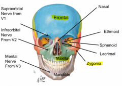
frontal, zygoma, and maxilla
a decent ways back (like half) |
|
|
What are the two holes above and below the orbit? What does through them?
|
Supra and Infra- orbital foramen
they give out the opthalmis and maxillary divisions of the trigeminal nerve respectively |
|
|
What other hole is lined up in the same saggital place as the infra and supra orbital foramen?
|
the mental foramen (for the mandibular division of CN )
|
|
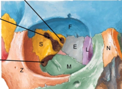
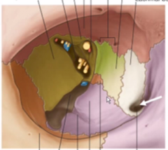
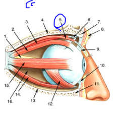
Label these (include lines)
|
Lacrimal
Ethmoid Sphenoid |
|
|
Which of those previous bones are prone to blowout fractures? Why?
|
the lacrimal and ethmoid bones because they are paper thing and can break easily during traume
|
|
|
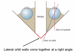
What do the medial and lateral walls of the orbit look like? (what angles are they at relative to one another?
|
|
|
|
What implication does the axis of the orbit not lining up with the axxis of the eyeball have?
|
the muscles and nerves that come out of there will have to attach obliquely
|
|
|
What are the 3 layers of the eyeball?
|
1. Sclera
2. Choroid 3. Retina |
|
|
What type of tissue is the sclera made of? Why is this functionally important?
|
collagen so it can seamlessly integrate with tendons of all the occular muscles
(this is also why the eyeball is white) |
|
|
What is contained in the choroid?
What does choroid generally refer to? |
all the vessels and nerves that aren't the optic nerve
choroid generally containing many blood vessels |
|
|
What is contained in the retina?
|
all the neural stuff. light and mechanical receptors and their connections.
|
|
|
What provides the sensory innervation of the eye?
|
CN V1
|
|
|
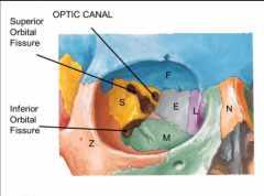
What travels in the optic canal with the optic nerve?
|
the opthalmic artery
|
|
|
What major artery does the opthalmic artery come off of and does it only supply the eye?
|
the internal carotid and no it supplies a lot of the face
|
|
|
What is in the superior orbital fissure vs inferior orbital fissure? Which is more important?
|
superior- (more superior haha) has all of the other neurons and the superior opthalmic vein
inferior- only the inferior opthalmic vein |
|
|
What is the path of tears in the eye?
|

lacrimal gland in upper lateral orbit (not shown) secretes them and this washes accross to the nasolacrimal canal to be drained
|
|
|
What is the common tendonous ring? Where is it very functionally placed.
|
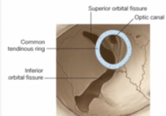
a tendonous ring attachment for all eye muscles. it is placed over the superior orbital fissure where all the motor nerves come out!
|
|
|
What is the only muscle not attached to the tendonous ring?
|
the inferior oblque
|
|
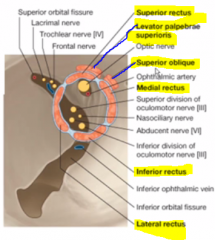
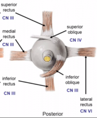
Name these muscles
|
|
|
|
Which muscles does CN 3 innervate? How many divisions in the superior fissure does it have?
|
superior, inferior, and me dial rectus
and Levator palpebrae superioris this is the levatator palpebrae superioris |
|
|
What nerves that aren't involved in the orbit come out of the superior orbital fissure?
|
the sensory branches of CN5 for the top of the face
|
|
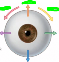
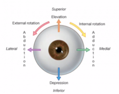
Name these actions
|
|
|
|
What is the main point of rotating your eyes? (besides being exasperated)
|
to maintain eye position while turning your head
|
|
|
ither terms for internal and internal rotation?
|
intorsion and extorsion
|
|
|
What part of the eyeball so the rectus vs oblique muscle attach?
|
rectus- anterior
oblique- posterior |
|
|
Show how the superior oblique runs.
Also compare this to the inferior oblique |
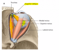
1. parralell to the medial rectus
2. threaded through troclea 3. attached to back of eye past the superior rectus Inferior oblique- the same medial course except it attaches underneath the eye |
|
|
Show a posterior view of the muscles
|
|
|
|
What is functionally significant about the obliques attching to the back of the eye?
|
they will move the eyeball to the opposite side of their pull
(imagine if back of eyeball was the front) |
|
|
Does the action of the obliques stay constant?
|
no they change with position of the ye
|
|
|
what is their role when the eye in straight?
|
superior- intorsion
inferior- extorsion |
|
|
What is their role when the eye is adducted?
|
superior- depression
inferior- elevation |
|
|
So why would a lesion of CN 3 cause you to be down and out? (lateral strabismus)
|
because then the actions of the SO and LR are unopposed.
laterall will pull to the side SO will depress and internally rotate |
|
|
What part of the eye are we referring to when we talk about internal and externalroation?
|
the top of the eyes
|
|
|
The occular muscles all have secondary actions moving the eye as long as they are not along the same action of rotation.
|
let's talk about this
|
|
|
Do either of the obliques have medial adduction power?
|
no
|
|
|
Do they have lateral abducion power?
|
yes, weakly
|
|
|
What phenomenon does this explain?
|
why lazy eyes are usually to the side
|
|
|
Which actions are controlled completely by CN IIi with nothing else being able to compensate?
|
adduction, elevation, and extorsion
|
|
|
Which two muscles control the eyelid opening?
Show |
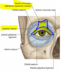
superior tarsal and Levatato papebrae superioris
|
|
|
What kind of muscle is each and what nerve innervates them?
|
superior tarsal- SM controlled by sympathetics
levatator palepebrae superioris- skeletal muscle controlled by CN 3 |
|
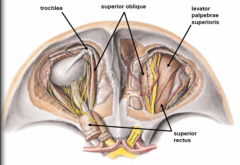
This is what you will see when you remove the frontal bone.
What surrounds this that is not shown? |
a bunch of fat to pad the eye
|
|
|
there are nerves that go into the eyeball? What do they do?
|
smooth muscle control in the eyeball or sensory info for CN 5
|
|
|
How does CN 3 split up? Which muscles does each division innervate?
|
splits into inferior and superior at the tendonous ring
superior- levator papebrae superioris and superior rectus (look up and rise lids) inferior- everything else |
|

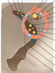
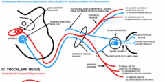
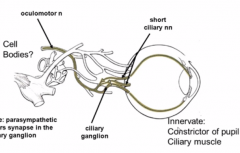
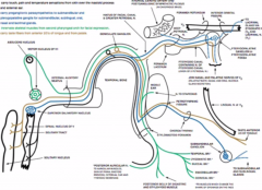
Describe the blue lines here.
|
the are the S and PS innervations.
PS- have to stop at ciliary ganglion first S- are post ganglionic and split into 2 branches. One for the pupil dilator via the ring and the other for the superior tarsal. |
|
|
How do the CN 4 and 6 travel?
|
pretty straighforward except that 4 crosses behind the midbrain.
They both come out through the superior orbital fissure and innervate their one muscle. |
|
|
How do the pupil smooth muscles get their innervation? (what layer of the eyeball do the nerves travel in?)
|
the choroid layer
|
|
|
describe how the pre and post sympathetic fibers travel to the eye.
|
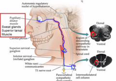
1. come out of T1
2. travel through the sympathetic trunk to synapse of the superior cervical ganglia 3. travel up the carotid plexus (around external carotid artery) to the eyeball 4. they have to travel through the ciliary ganglion, but they don't synapse there |
|
|
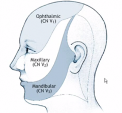
Imagine the territories of the 3 division of the trigeminal
|

they all come down from a common point in the lateral head bone (lolw/e)
they end before the ear mouth is boundary between maxillary and mandibular. opthalmic and maxillary meet at the tip of the nose |
|
|
Where does the trigeminal ganglion sit in the skull and which foramen do it's 3 divisions go through?
|
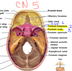
in the middle cranial fossa
V1- superior orbital fissure V2- Foramen Rotundum V3- Foramen Ovale |
|
|
What is the cowboy ross mnemonic?
|
cowboy ROS sits on the sella tucica saddle
His eyes- look through the optic canal His legs- he laces up his boots and puts them through the foramen lacerum (L for legs) Between this, there are 3 holes that spell his name from rostral to caudal Rotundum Ovale Spinosum Horse- he rides on his horse CLIVUS who hold two JUGs of water Crib- He is going to to his CRIB(iform plates) where he is going to get some crystal (CRISTA GALLI) |
|
|
Show and tell where he puts his hand and leans back on.
|
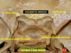
holds onto two anterior clinoid processes
leans back on posterior clinoid processes |
|
|
Where is the superior orbital fissure in all this?
|
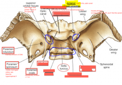
underneath the optic canal and the anterior clinoid process
|
|
|
What is the mnemonics for what the 3 different CN5 divisions supply? (don;t give what they stand for yet)
|
1. Opthalmic- NFL (watch NFL)
2. Maxillary- ZIP (maxillary zips the other two together) 3. Mandibular- BAIL (use your mouth to bail you out of jail) |
|

What does V1 NFL stand for?
|
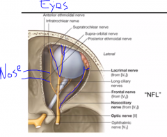
Nasalciliary
Frontal Lacrimal |
|
What does V2 ZIP stand for?
|

Zygoma
Infraorbital Pterygopalatine |
|

What does V3 BAIL stand for?
|

Bucchal
Auricotemporal Inferior Alveolar Lingulal |
|
|
Which is the only nerve that innervates the eyeball?
|
Nasociliary
|
|
|
What is pretty awesome about the location of the parasympathetic ganglia (4) in the face?
|
they are right along the rooute of the sensory nerve of it's reflex arc
|
|
|
Show where the opthalmis artery travels.
|
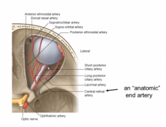
in the optic canal
|
|
|
What would happen if you had an occlusion of the opthalmic artery? WhY?
|
complete blindness in that eye since this is the only artery that supplie the retina
|
|
|
Show how the dura line the eye (and the orbital fat)
what is the orbital fat analogous to in the spine? |
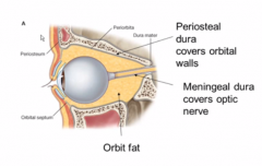
periosteal dura lines the pyrimid that is the orbit of the eye while the meningeal dural travels along with it on the sclera.
Fat fills in space between the dura the same as epidural fat in the spinal column |
|
|
Where do the ophthalmic veins come from and drain toward?
What is a risky feature they share with the spinal veins? |
they come from the superior and inferior orbital fissures
they drain toward the nose outside the eye. they don't have valves so infection can spread |
|
|
What does the superior opthalmic vein also drain?
Why do we care about this small sinus? |
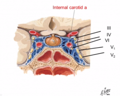
the cavernous sinus
a lot of sensitive stuff is there. (All the eye cranial nerves, V1 and 2, pituitary gland, and internal carotid artery) |
|
|
Which nerve would be affected first in a thrombolytic cavernous sinus lesion? WHy?
|

CN 6 becasue it is right in the middle next to the internal carotid whereas the other are on the side
|
|
|
NOW WE TALK ABOUT THE EAR
|
YAY
|
|
|
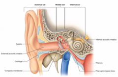
Imagine what the external, middle, and internal ear look like
|
|
|
|
What are the meatuses made of and filled with?
|
they are are air-filled cartilaginous tubes.
|
|
|
Where does the internal acoustic meatus exit into the skull?
|
the posterior cranial fossa
|
|
|
What goes through the internal auditory meatus?
|
CN 8
|
|
|
What are the 3 ear bones connected by?
|
synovial joints
|
|
|
Mnemonic for order of the 3 bones?
|
MISO soup!
From TM: Malleolus Incus Stapes Oval Window |
|
|
How does the middle ear concentrate sound?
|
the attachment of the mallelus to the TM is larger than the stapes to the oval window, making the pressure bigger
PHYSICS! |
|
|
What is conductive hearing loss?
|
a problem with the conduction system in the external or middle ear
|
|
|
What do the 3 bones look like? Menmonic.
|
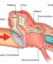
a shoulder- malleus
bent arm- incus shovel hand- stapes |
|
|
What is there is a problem in the cochlea? WHat is it called?
|
sensory hearing loss.
|
|
|
What are the two smooth muscles of the ear? What do they attach to?
|
tensor tympani attaches to maaleus
stapedius attached to (surprise) stapes |
|
|
function of the tensor tympani?
|
limit movement of the malleus to dampen the sound of our own voice. (it is attached to vibrations in our skull)
|
|
|
function of the stapedius?
|
contracts reflexively to loud sounds to disarticulate the stapes from the incus to dull transmission.
|
|
|
What CN control which SM?
|
CN 5- tensor tympani
CN 7- Stapedius |
|
|
What auditory sx would we get with a CN 5 lesion?
|
our own voice may sound louder, but nothing significant
|
|
|
What auditory sx would we get with a CN 7 lesion?
|
we wouldn't be able to damped loud sounds and we would get HYPERACUSIS (startled by loud sounds)
|
|
|
What is the chorda tympani?
|
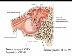
mixed branch of CN 7 along the TM. Just passing through!
|
|
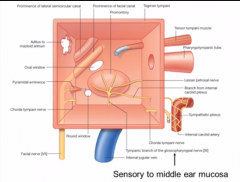
View from TM
What give sensation to the middle ear mucosa? |
CN 9
|
|
|
Where does CN 7 come through the skull?
|
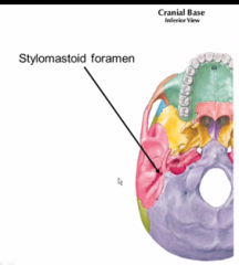
stylomastoid foramen
(between spikey styloid process and mastoid bone) |
|
|
Who do I know who had this nerve nicked?
Show pic of location. |
that girl, Ana Maria. She had a surgert that nicked here.
|
|
|
Which parts of CN 7 come through this foramen?
|
all the skeletal muscles it innervates
but not the stapedius |
|
|
Mnemonic for the 5 MUSCULAR branches of the facial nerve?
|
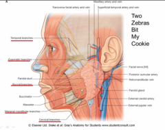
TWO- temporal
ZEBRAS- zygomatic BIT- buccal MY- marginal mandibular COOKIE- cervical |
|
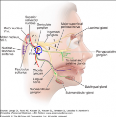
Narrate this.
|
CN 7 has 2 major routes of mixed fibers.
Muscles, salivary glands, and taste go through the lower route. They drop off the stapezius branch as they pass the middle ear. Nasal and oral mucosal secretion and taste go throught the upper branch They lower branch goes through the stylomastoid foramen and joins with the superior division at the genticulate ganglion. They both happily go through the internal auditory meatus into the brainstem |
|
|
Why was that necessary to know?
|
you can determine where the lesion on CN 7 is basen on sx
|
|
|
What if you only had the facial muscles paralyzed and nothing else? Where is the lesion?
|
at the lower branch of the facial nerve
|
|
|
What if you lost all taste?
|
the it would have to be somewhere before the genticulate ganglion.
|
|
|
What is a secondary function of the nerves of CN 5?
|
other nerves send fibers there to hitchhike to their destination
|
|
|
Where is the internal auditory meatus inside the skull? Menmonic?
|
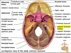
in the posterior cranial fossa anterior to the jugular foramen.
The horse (clivus) needs to listen for danger. |
|
|
Mnemonic for what the middle/outer ear resembles
|
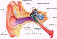
a snail getting suckerpunched
|
|
|
In the brainstem, what CN does CN 7 follow out closely? Why?
|
CN 8 because it wants to go to the middle ear to drop off it's stapedius connection before going on to do everything else
|
|
|
Where would the lesion/infection be if you had left facial paralysis AND hyperacusis?
|

before the genticulate genu (bend of CN 7) and the part where CN 7 goes through the middle ear.
|