Reading...
![]()
Play button
![]()
Play button
![]()
Use LEFT and RIGHT arrow keys to navigate between flashcards;
Use UP and DOWN arrow keys to flip the card;
H to show hint;
A reads text to speech;
75 Cards in this Set
- Front
- Back
|
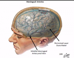
What provides the majority of the blood supply of the meninges in the head?
|
Middle meningeal artery and vein
|
|
|
What pathology is this vessel famous for? What layer is it in?
|
epidural hemorrhage in a blow to the temple
|
|
|
Show a pic of this vessel
|
|
|
|
can you dissect the pia off?
|
no, it's too close
|
|
|
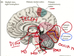
Show the secondary vesecles of development. Where do the optic cups come out of? How do you know?
|
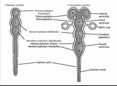
optic cups come out of the diencephalon, which is where the hypothalamus is
|
|
|
Which of these vesicles will divide into two lateral halves and which will be very thin? How did you deduce this?
|
halves- telencephalon to make lateral ventricles
thin- mesencephalon to make cerebral aqueduct |
|
|
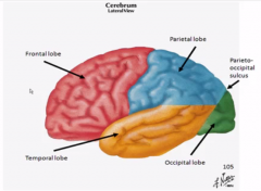
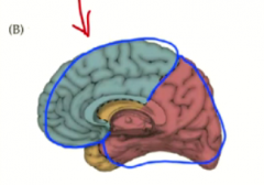
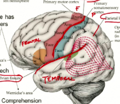
Show the lobes? Which one is really small?
|
|
|
|
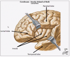
What do you see if you pry open the sylvian fissure? How do you remember from najeeb?
|
the insula (like a secret lobe)
Najeeb showed this as a place that slow pain fibers go. It was in the groove that was the sylvian fissure. |
|
|
Show a pic.
|
|
|
|
What sensation goes here and what purpose does it serve according to dr. white?
|
visceral sensations like taste come here and get processed into the limbic system for memory
(autonomic=visceral to link with najeeb) |
|
|
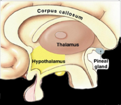
Show a view of what the diencephalon looks like
|
|
|
|
Where is the 3rd ventricle?
|
in between the thalami, so not visible when the thalamus is.
|
|
|
What was Dr. Najeeb wrong about in terms of location?
|
The hypothalamus which is below (hypo), not anterior to the thalamus
|
|
|
What is that little dot in the thalamus?
|
the interthalamic adhesion
|
|
|
Can you see the subthalamus?
|
no, it is too tiny
|
|
|
Where does the corpus callosum sit on top? Where does the cingulate gyrus sit on top?
|
corpus callosum on top of the lateral ventricle
cingulate gyrus on top of the corpus callosum |
|
|
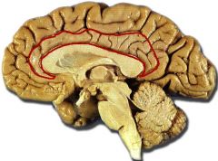
Show where the cingulate gyrus is on a real brain.
|
|
|
|
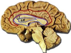
Show where the corpus callosum and lateral ventricle is.
|
|
|
|
Sorry, I messed up the last pic. What is the bottom part of the "CC" that I mislabeled?
|
the fornix
|
|
|
What can you say about the orientation of the LV vs the CC?
|
the frontal horn of the LV extends into the anterior part of the CC
|
|
|
What is the uncus? Where is it?
|
the tip of the temporal lobe on the medial view. (very tiny)
|
|
|
What does the uncus help you find?
|
the amygdala
|
|
|
Is the primary visual cortex on the lateral and medial surface?
|
no, just the medial
|
|
|
What are the top and bottom parts of the visual cortex called?
|
top- lingulate gyrus
bottom- cuneate gyrus |
|
|
According to white, is CN 11 really a CN? Why is it called one?
|
No, it is a misnamed spinal nerve that exits with CN 9 and 10 through the jugular foramen.
|
|
|
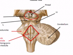
Which are the pur motor nerves and where do they all exit and originate?
|
3, 6, and 12
Originate dorsomedially and exit ventromedially |
|
|
Which are the mixed nerves and where do they all exit and originate?
|
5, 7, 9, and 10
exit ventrolaterally |
|
|
What sits on top of the superior colliculi?
|
|
|
|
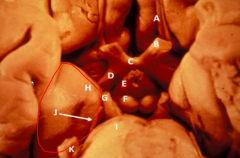
Show what structures the uncus is lateral to.
|
|
|
|
Describe the last pic.
|
The uncus is so medial and low that it is right next to the midbrain and where CN 3 comes out.
The optic chiasm, mamillary bodies, and pituitary gland are very close by too. |
|
|
Why is this pathologically significant in a tumor situation?
|
If you have a lateral tumor pushing the uncus to the middle, you will compress CN3.
|
|
|
What sign will you see outwardly from this compression?
|
inability for ipsilateral eye to look medially
|
|
|
Which arteries do the internal carotid vs the vertebral arteries come off of?
|
IC- aorta on left, bracheocephalic on right
vertebral- subclavian on both sides |
|
|
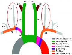
Show a pic of the vertebral arteries (red circle) and the IC's (brown)
|
|
|
|
What are the striate arteries? What do they supply?
|
a bunch of parralell arteries that supply the diencephalon and BG
|
|
|
What do the striate arteries branch off of and at what portion?
|
The three cerebral arteries befor they reach the cortex
|
|
|
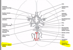
Name which striate arteries come off of each of the 3 CA's
|
ACA- medial striate arteries
MCA- lenticulostriate arteries PCA- thalamo-perforating arteries |
|
|
Show the arteries? Which are the only ones outside the circle of willis? Deduce?
|
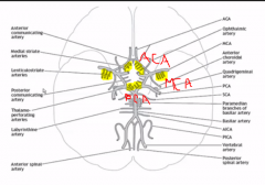
the lenticulostriate arteries which supply the internal capsule and lentiform which are more lateral structures
|
|
|
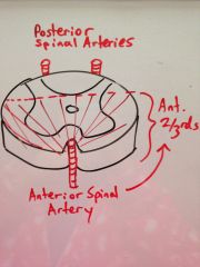
Remember the spinal arteries? Show them in the SC crossection.
|
|
|
|
What artery do the spinal arteries arise from? How do you deduce this?
|
the vertebral arteries at the top of the medulla. This is pretty much the top of the spinal cord.
|
|
|
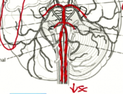
Show how the spinal arteries branch off the vertebral arteries.
|
|
|
|
What extra structures does the posterior and anterior spinal arteries supply respectively?
|
anterior- ventral medulla
posterior- lateral medulla |
|
|
Most lobes have ___ blood supplies. Which is the exception?
|
most lobes are supplied by 2 cerebral arteries.
the exception is occipital which has one (but macula is MCA) |
|
|
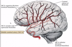
Show the course of the MCA (lateral surface)
|
|
|
|
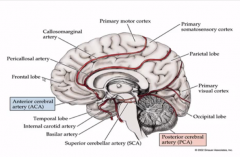
Show the course of the PCA and ACA (medial surface)
|
|
|
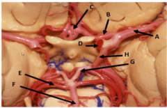
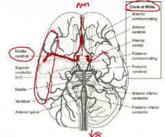
Name these structures
|
A- MCA
B- ACA C- Anterior Communicating Artery D- IC E- Superior cerebellar artery F- Basilar Artery G- PCA H- Posterior Communicating Artery |
|

What is pathological in this pic and why does this show why the circle of willis is advantageous?
|
The PCA is pale and infarcted. So the posterior Communicating Artery expanded and took over the blood supply to the bottom and back cortex areas. (not how big it is)
|
|
|
Why is there watershed zones in the brain? What is necessary for a watershed?
|
because you are supplying the brain with two different blood supplies that fuse together.
|
|
|
What are the two pairs of blood vessels?
|
internal carotid and vertebral
|
|
|
What parts of the brain does each artery supply?
|
carotid- anterior
vertebral- posterior |
|
|
What openings does the vertebral artery pass through?
|
C6-C1 transverse foramina
foramen magnum |
|
|
What is the first anastamosis formed by the arteries?
|
the circle of willis
|
|
|
WHat shape is the circle of willis?
|
a hexagon
|
|
|
What cerebral arteries does each artery branch into?
|
internal carotid- anterior cerebral artery, MCA
vertebral- PCA |
|
|
Which one of the carotid branches looks like a continuation of it and supplies a lot of stuff and is larger?
|
MCA
|
|
|
Imagine and show the direction of the MCA and ACA braching from the carotid.
|
|
|
|
Imagine and show the position of the vertebral, basilar, and PCA's.
|
vertebral- go up the ventral medulla
basilar- midline of pons |
|
|
PCA- split off posterolaterally at top of pons
|
|
|
|
How many posterior communicating arteries are there? WHat do they connect?
|
2 and they connect the PCA's with the internal carotids
THEY ARE THE ANASTOMOSIS |
|
|
How many anterior communicating arteries are there? WHat do they connect?
|
only a small one
it connects the two ACA |
|
|
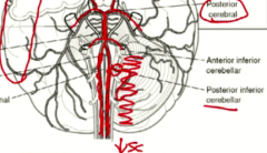
What is a very important and prominent artery coming off the vertebral?
|
the posterior inferior cerebellar artery (PICA)
|
|
|
Show the PICA
|
|
|
|
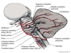
What arteries come off the bottom and top of the basilar artery?
|
bottom- anterior inferior cerebellar artery
top- superior cerebellar artery |
|
|
Where does the superior cerebellar vs the PCA branch off?
|
they both branch off the top of the basilar artery in about the same direction
|
|
|
Show these cerebellar arteries
|
|
|
|
Which artery covers most of the bottom of the brain? Show it!
|
|
|
|
Where do all these cerebral arteries lie in the meninges?
|
underneath the arachnoid.
|
|
|
Which CA supplies the primary visual cortex?
|
PCA (it's main claim to fame)
|
|
|
What does homunculus mean?
|
little person
|
|
|
Which CA supplies the trunk in sensory and motor homunculi?
|
ACA
|
|
|
Where is the trunk homunculi in the cortex?
|
the most superior and medial part
|
|
|
Show the trunk homunculi.
|
|
|
|
Is the sensory homunculus at the same levels as the motor?
|
yes
|
|
|
What is the calcarine fissure and why is it geographically significant?
|
a fissure in the occipital lobe significant b/c it has the primary visual cortex on either side of it.
|
|
|
Show the PVC and the calcarine fissure
|
|