![]()
![]()
![]()
Use LEFT and RIGHT arrow keys to navigate between flashcards;
Use UP and DOWN arrow keys to flip the card;
H to show hint;
A reads text to speech;
299 Cards in this Set
- Front
- Back
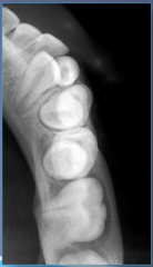
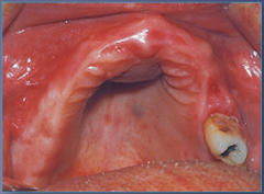
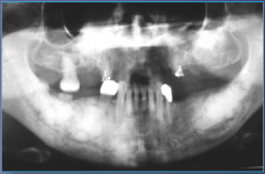
|
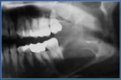
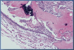
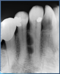
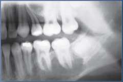
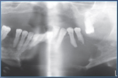
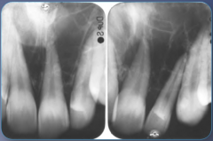
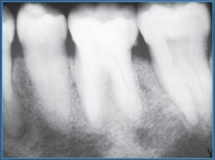
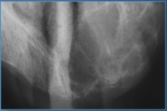
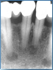
Bone Scar left by tooth extracted with Condensing Osteitis |
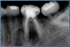
|
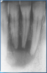
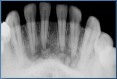
Asymptomatic |
Chronic Apical Periodontitis |
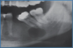
|
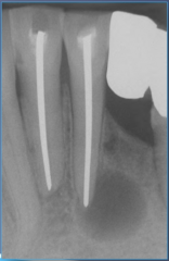
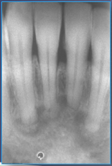
Asymptomatic |
Chronic Apical Periodontitis |
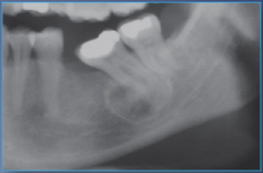
|
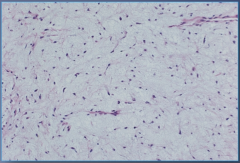
|
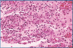
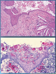
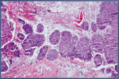
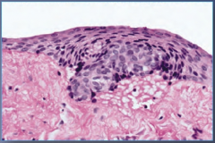
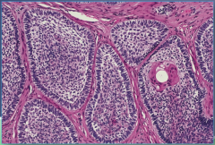
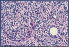
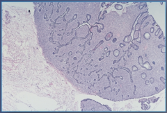
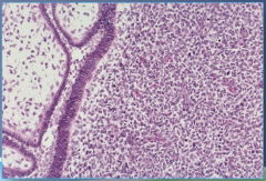
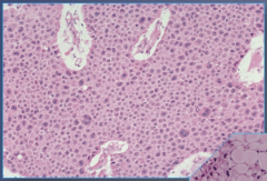
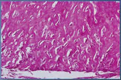
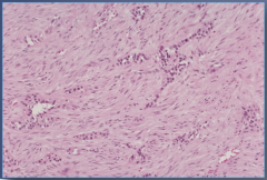
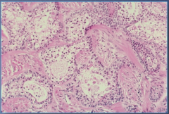
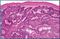
Chronic Apical Periodontitis
Look for fibroblasts, lymphocytes, and plasma cells. |
|
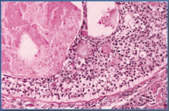
|
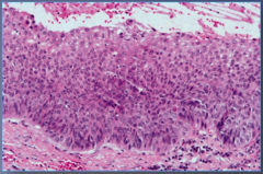
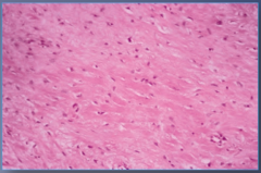
Chronic Hyperplastic Pulpitis |
|
|
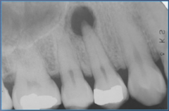
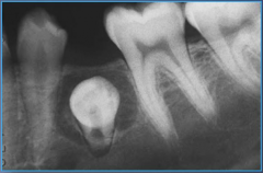
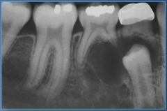
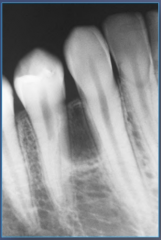
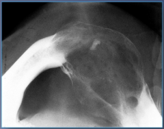
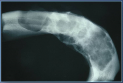
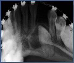
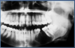
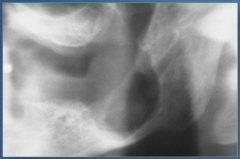
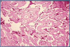
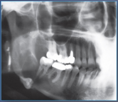
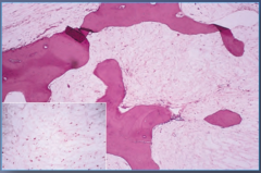
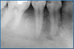
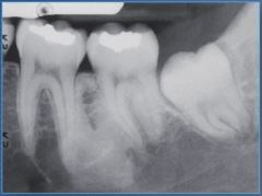
Condensing Osteitis
Fused with Lamina Dura. Entire root outline is visible. |
|
|
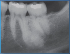
Condensing Osteitis
Fused with Lamina Dura. Entire root outline is visible. |
|
|
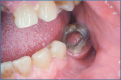
Dental Abscess |
|
|
Dental Abscess |
|
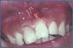
|
Dental Abscess |
|
|
Dental Abscess |
|
|
Dental Abscess |
|
|
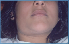
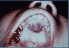
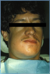
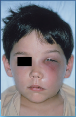
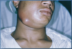
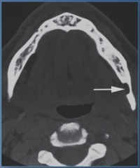
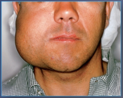
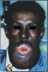
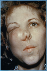
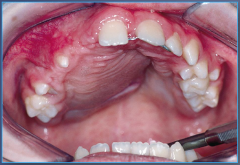
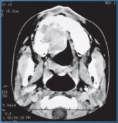
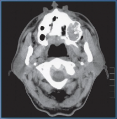
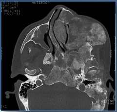
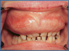
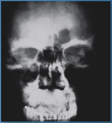
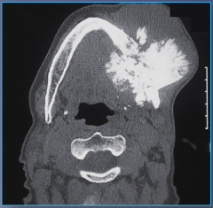
Ludwig's Angina (Cellulitis) |
|
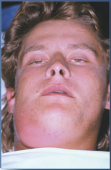
|
Ludwig's Angina (Cellulitis) |
|
|
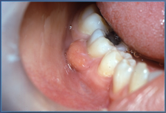
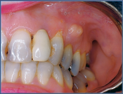
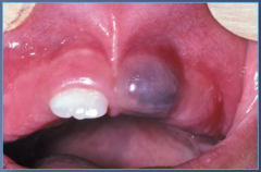
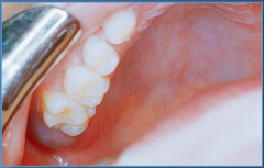
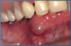
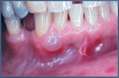
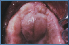
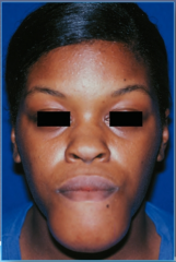
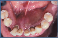
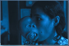
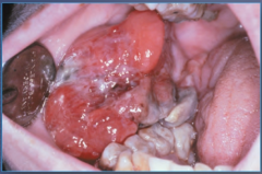
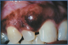
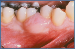
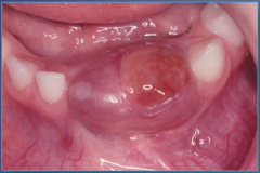
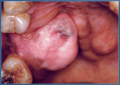
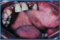
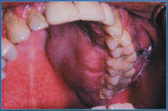
Parulis |
|
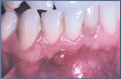
|
Parulis |
|
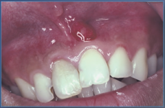
|
Parulis |
|
|
Parulis |
|
|
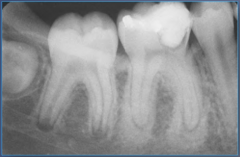
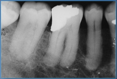
Periapical Abscess |
|
|
Periapical Abscess |
|
|
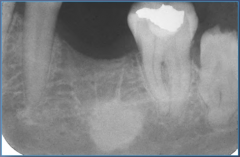
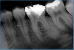
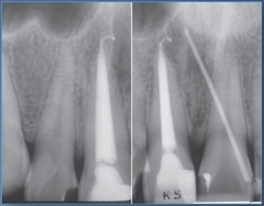
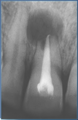
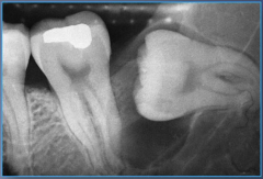
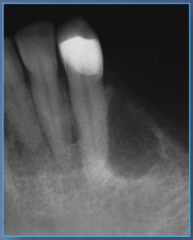
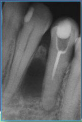
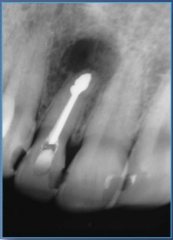
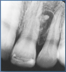
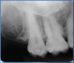
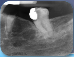
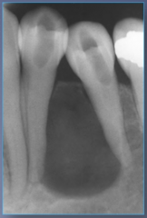
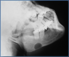
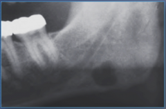
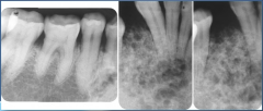
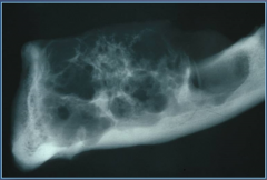
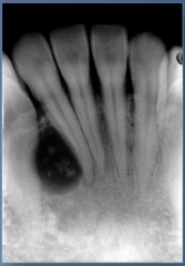
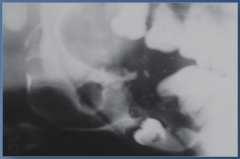
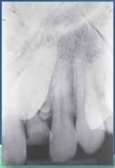
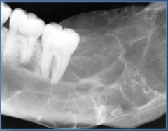
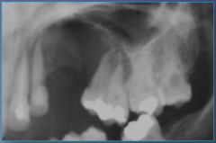
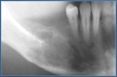
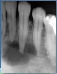
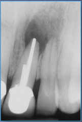
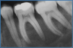
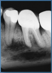
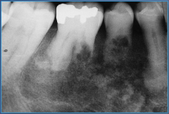
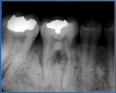
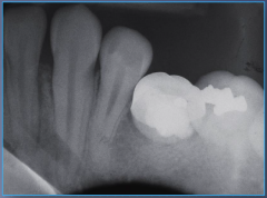
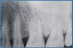
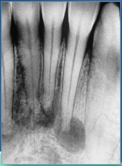
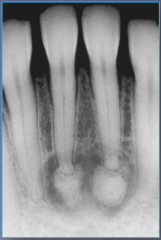
Periapical Abscess
Notice the widened PDL to large alveolar radiolucency. |
|
|
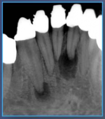
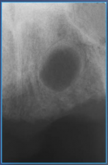
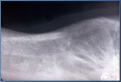
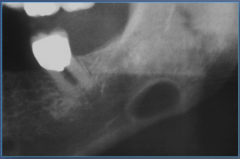
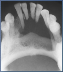
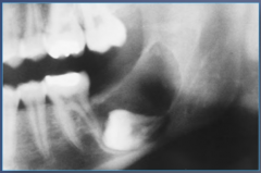
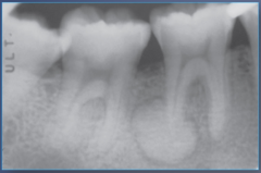
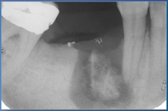
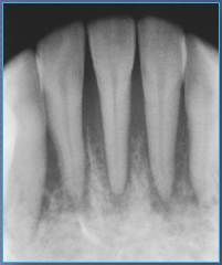
Radicular Cyst
Loss of Lamina Dura. Usually at apex, but can be lateral to root. |
|
|
Radicular Cyst
Loss of Lamina Dura. Usually at apex, but can be lateral to root. |
|
|
Radicular Cyst
Loss of Lamina Dura. Usually at apex, but can be lateral to root. |
|
|
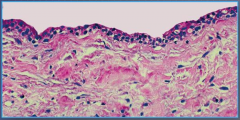
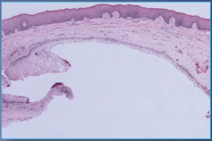
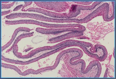
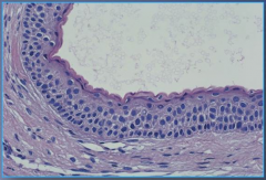
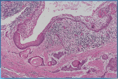
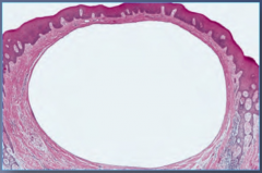
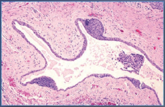
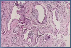
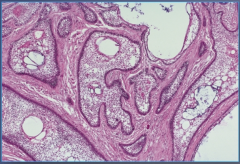
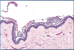
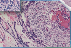
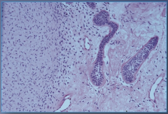
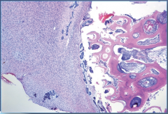
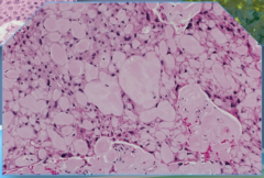
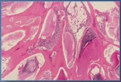
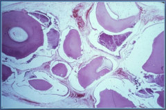
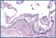
Radicular Cyst
Lined by stratified squamous epithelium. May have cholesterin clefts. |
|
|
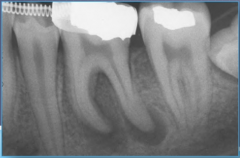
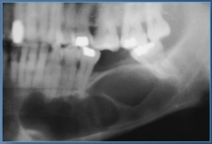
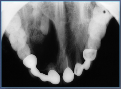
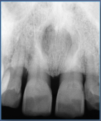
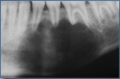
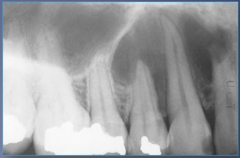
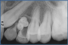
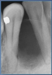
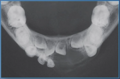
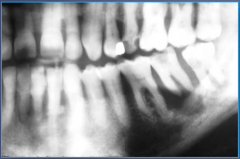
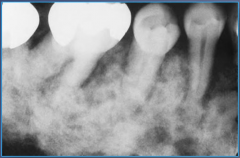
Chronic Apical Periodontitis |
|
10-30 years old |
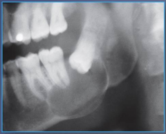
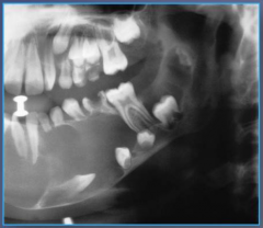
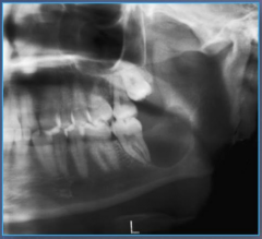
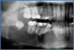
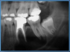
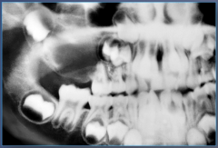
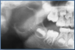
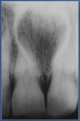
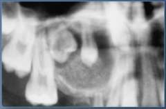
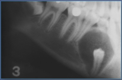
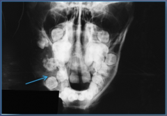
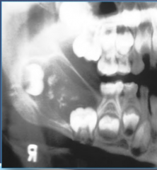
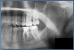
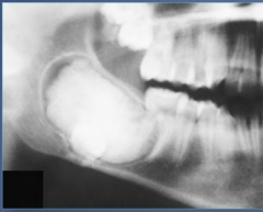
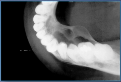
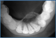
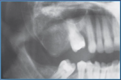
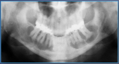
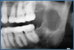
Dentigerous Cyst
Notice mandibular 3rd molar site predilection, well defined lucency, unilocular, on crown, corticated, expands |
|
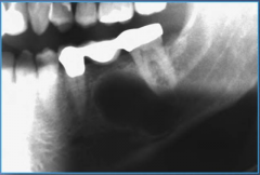
10-30 years old |
Dentigerous Cyst
Notice mandibular 3rd molar site predilection, well defined lucency, unilocular, on crown, corticated, expands |
|
10-30 years old |
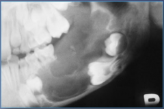
Dentigerous Cyst
Notice mandibular 3rd molar site predilection, well defined lucency, unilocular, on crown, corticated, expands |
|
10-30 years old |
Dentigerous Cyst
Well defined lucency, unilocular, on crown, corticated, expands |
|
10-30 years old |
Dentigerous Cyst
Notice mandibular 3rd molar site predilection, well defined lucency, unilocular, on crown, corticated, expands |
|
10-30 years old |
Dentigerous Cyst
Notice mandibular 3rd molar site predilection, well defined lucency, unilocular, on crown, corticated, expands |
|
10-30 years old |
Dentigerous Cyst
Well defined lucency, unilocular, on crown, corticated, expands |
|
10-30 years old |
Dentigerous Cyst
Notice mandibular 3rd molar site predilection, well defined lucency, unilocular, on crown, corticated, expands |
|
|
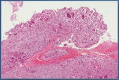
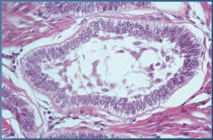
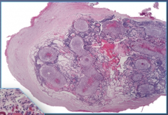
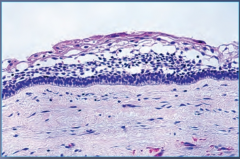
Dentigerous Cyst
Non-keratinized epithelium
|
|
|
Eruption Cyst |
|
|
Eruption Cyst |
|
|
Eruption Cyst |
|
|
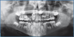
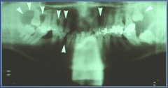
Nevoid Basal Cell Carcinoma Syndrome
Multiple Odontogenic Keratocysts |
|
|
Nevoid Basal Cell Carcinoma Syndrome
Rib anomalies |
|
Adolescent and young adults. No pain or pain, swelling |
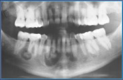
Odontogenic Keratocyst
Well defined, partially corticated border, unilocular when small->multilocular when bigger. Also notice posterior jaw predilection. |
|
Adolescent and young adults. No pain or pain, swelling |
Odontogenic Keratocyst
Well defined, partially corticated border, unilocular when small->multilocular when bigger |
|
Adolescent and young adults. No pain or pain, swelling |
Odontogenic Keratocyst
Well defined, partially corticated border, unilocular when small->multilocular when bigger. Also notice posterior jaw predilection. |
|
Adolescent and young adults. No pain or pain, swelling |
Odontogenic Keratocyst
Well defined, partially corticated border, unilocular when small->multilocular when bigger. Also notice posterior jaw predilection. |
|
Adolescent and young adults. No pain or pain, swelling |
Odontogenic Keratocyst
Well defined, partially corticated border, unilocular when small->multilocular when bigger. Also notice posterior jaw predilection. |
|
Adolescent and young adults. No pain or pain, swelling |
Odontogenic Keratocyst
Well defined, partially corticated border, unilocular when small->multilocular when bigger. Also notice posterior jaw predilection. |
|
Adolescent and young adults. No pain or pain, swelling |
Odontogenic Keratocyst
Well defined, partially corticated border, unilocular when small->multilocular when bigger |
|
|
Odontogenic Keratocyst |
|
|
Odontogenic Keratocyst |
|
|
Odontogenic Keratocyst |
|
|
Radicular Cyst
Loss of Lamina Dura. Usually at apex, but can be lateral to root. |
|
|
Radicular Cyst
Loss of Lamina Dura. Usually at apex, but can be lateral to root. |
|
|
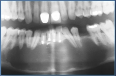
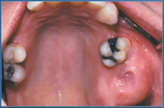
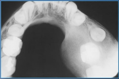
Residual Cyst |
|
|
Residual Cyst |
|
|
Residual Cyst |
|
Children + Young adults under 30, swelling |
Aneurysmal bone cyst
Expansile, multilocular, destruction, and high predilection in the mandible |
|
Children + Young adults under 30, swelling |
Aneurysmal bone cyst
Expansile, multilocular, destruction, and high predilection in the mandible |
|
Children + Young adults under 30, swelling |
Aneurysmal bone cyst
Expansile, multilocular, destruction, and high predilection in the mandible |
|
|
Aneurysmal bone cyst
Scattered multinucleated giant cells |
|
|
Aneurysmal bone cyst
Scattered multinucleated giant cells |
|
5-13 years old |
Buccal bifurcation cyst
Corticated, unilocular |
|
5-13 years old |
Buccal bifurcation cyst
Corticated, unilocular |
|
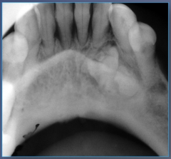
~33 (10-30 years old), asymptomatic, swelling |
Calcifying odontogenic cyst
Well defined lesion, corticated, often pure lucency but sometimes has opaque areas |
|
~33 (10-30 years old), asymptomatic, swelling |
Calcifying odontogenic cyst
Well defined lesion, corticated, often pure lucency but sometimes has opaque areas |
|
~33 (10-30 years old), asymptomatic, swelling |
Calcifying odontogenic cyst
Well defined lesion, corticated, often pure lucency but sometimes has opaque areas |
|
~33 (10-30 years old), asymptomatic, swelling |
Calcifying odontogenic cyst
Well defined lesion, corticated, often pure lucency but sometimes has opaque areas |
|
~33 (10-30 years old), asymptomatic, swelling |
Calcifying odontogenic cyst
Well defined lesion, corticated, often pure lucency but sometimes has opaque areas |
|
|
Calcifying odontogenic cyst |
|
|
Calcifying odontogenic cyst |
|
|
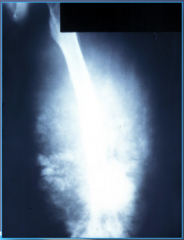
Carcinoma arising in a cyst
Islands of invasive epithelial cells with epithelial dysplasia |
|
|
Carcinoma arising in a cyst
Islands of invasive epithelial cells with epithelial dysplasia |
|
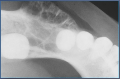
Adult, no expansion |
Focal osteoporotic bone marrow defect |
|
Adult, no expansion |
Focal osteoporotic bone marrow defect |
|
|
Gingival cyst of the adult |
|
|
Gingival cyst of the adult |
|
|
Gingival cyst of the adult |
|
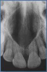
40-60 years old, Male, asymptomatic |
Lateral Periodontal Cyst
Notice the mandibular bicuspid/cuspid area. Corticated, unilocular, contiguous with the PDL space |
|
40-60 years old, Male, asymptomatic |
Lateral Periodontal Cyst
Notice the mandibular bicuspid/cuspid area. Corticated, unilocular, contiguous with the PDL space |
|
40-60 years old, Male, asymptomatic |
Lateral Periodontal Cyst
Notice the mandibular bicuspid/cuspid area. Corticated, unilocular, contiguous with the PDL space |
|
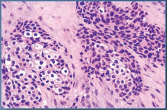
|
Lateral Periodontal Cyst
Focal nodular thickenings with swirling cells |
|
|
Lateral Periodontal Cyst
Focal nodular thickenings with swirling cells |
|
|
Lateral Periodontal Cyst
Focal nodular thickenings with swirling cells |
|
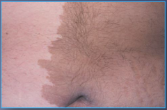
Male |
Lingual Mandibular Salivary Gland Depression |
|
Male |
Lingual Mandibular Salivary Gland Depression |
|
Male |
Lingual Mandibular Salivary Gland Depression |
|
Male |
Lingual Mandibular Salivary Gland Depression |
|
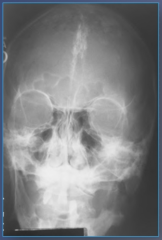
30-50 years old, Male, asymptomatic |
Nasopalatine Duct Cyst |
|
30-50 years old, Male, asymptomatic |
Nasopalatine Duct Cyst |
|
30-50 years old, Male, asymptomatic |
Nasopalatine Duct Cyst |
|
30-50 years old, Male, asymptomatic |
Nasopalatine Duct Cyst |
|
30-50 years old, Male, asymptomatic |
Nasopalatine Duct Cyst |
|
|
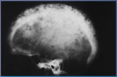
Nevoid Basal Cell Carcinoma Syndrome
Calcified Falx Cerebri |
|
|
Nevoid Basal Cell Carcinoma Syndrome
Multiple Odontogenic Keratocysts |
|
|
Nevoid Basal Cell Carcinoma Syndrome
Multiple Odontogenic Keratocysts |
|
|
Nevoid Basal Cell Carcinoma Syndrome
Odontogenic epithelial rests in cyst wall |
|
|
Radicular Cyst
Loss of Lamina Dura. Usually at apex, but can be lateral to root. |
|
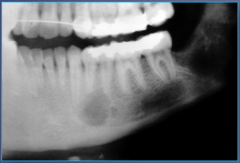
10-25 years old, asymptomatic, vital teeth, no expansion |
Simple bone cyst
Notice predilection of body of mandible to posterior mandible, unilocular, scalloping between roots, no border reaction |
|
10-25 years old, asymptomatic, vital teeth, no expansion |
Simple bone cyst
Notice predilection of body of mandible to posterior mandible, unilocular, scalloping between roots, no border reaction |
|
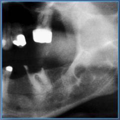
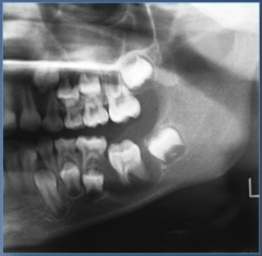
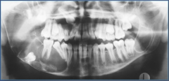
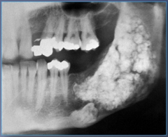
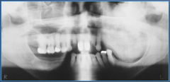
>30 |
Infiltrating Ameloblastoma
Notice how it is in the posterior mandible |
|
>30 |
Infiltrating Ameloblastoma |
|
>30 |
Infiltrating Ameloblastoma |
|
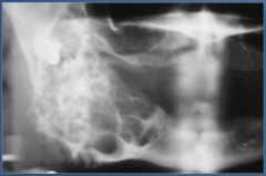
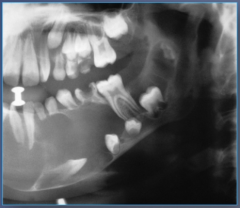
>30 |
Infiltrating Ameloblastoma
Notice how it is in the posterior mandible, well defined, multilocular |
|
>30 |
Infiltrating Ameloblastoma
Notice how it is in the posterior mandible, well defined, multilocular |
|
>30 |
Infiltrating Ameloblastoma
Well defined, multilocular |
|
>30 |
Infiltrating Ameloblastoma
Well defined, multilocular |
|
>30 |
Infiltrating Ameloblastoma |
|
>30 |
Infiltrating Ameloblastoma
Well defined, multilocular |
|
>30 |
Infiltrating Ameloblastoma
Well defined, multilocular. Notice the honeycomb appearance |
|
|
Infiltrating Ameloblastoma |
|
|
Infiltrating Ameloblastoma |
|
|
Infiltrating Ameloblastoma |
|
>30 |
Infiltrating Ameloblastoma
Notice how it is in the posterior mandible, well defined, multilocular |
|
>30 |
Infiltrating Ameloblastoma
Notice how it is in the posterior mandible, well defined, multilocular |
|
>30 |
Infiltrating Ameloblastoma
Notice how it is in the posterior mandible |
|
>30 |
Infiltrating Ameloblastoma
Notice how it is in the posterior mandible |
|
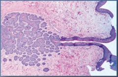
|
Unicystic Ameloblastoma
Palisading of basal cell layer with reverse polarization |
|
|
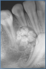
Adenomatoid Odontogenic Tumor
Pseudoducts and rosettes, calcified materials |
|
|
Adenomatoid Odontogenic Tumor
Pseudoducts and rosettes, calcified materials |
|
|
Adenomatoid Odontogenic Tumor
Pseudoducts and rosettes, calcified materials |
|
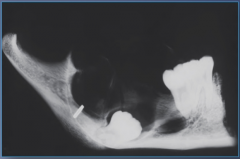
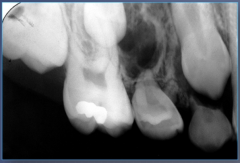
2nd decade, female |
Adenomatoid Odontogenic Tumor
Unilocular, notice extension to apex of tooth |
|
2nd decade, female |
Adenomatoid Odontogenic Tumor
Unilocular, notice extension to apex of tooth, anterior maxilla |
|
2nd decade, female |
Adenomatoid Odontogenic Tumor
Unilocular, notice extension to apex of tooth, anterior maxilla |
|
2nd decade, female |
Adenomatoid Odontogenic Tumor
Unilocular, notice extension to apex of tooth |
|
50s |
Ameloblastic carcinoma
Looks like infiltrating ameloblastoma, cortical destruction |
|
50s |
Ameloblastic carcinoma
Looks like infiltrating ameloblastoma, cortical destruction |
|
|
Ameloblastic fibroma
scattered epithelium in stroma |
|
6-14 |
Ameloblastic Fibroma
posterior mandible, well-defined, uni or multilocular |
|
|
Ameloblastic fibroma
scattered epithelium in stroma |
|
6-14 |
Ameloblastic Fibroma
posterior mandible, well-defined, uni or multilocular |
|
|
Ameloblastic Fibro-odontoma
Can be mixed with odontoma |
|
~10, asymptomatic, swelling |
Ameloblastic Fibro-odontoma |
|
~10, asymptomatic, swelling |
Ameloblastic Fibro-odontoma |
|
~10, asymptomatic, swelling |
Ameloblastic Fibro-odontoma |
|
|
Ameloblastic fibrosarcoma
numerous mitotic figures |
|
27.5, males |
Ameloblastic fibrosarcoma
mandible, poorly defined, destructive borders |
|
27.5, males |
Ameloblastic fibrosarcoma
mandible, poorly defined, destructive borders |
|
27.5, males |
Ameloblastic fibrosarcoma
mandible, poorly defined, destructive borders |
|
30-50 |
Calcifying epithelial odontogenic tumor
Posterior mandible, uni or multiocular, honeycombed |
|
30-50 |
Calcifying epithelial odontogenic tumor
Posterior mandible, uni or multiocular, honeycombed |
|
30-50 |
Calcifying epithelial odontogenic tumor
Posterior mandible, uni or multiocular, honeycombed |
|
|
Calcifying epithelial odontogenic tumor
amyloid deposits |
|
|
Calcifying epithelial odontogenic tumor
amyloid deposits |
|
|
Cementoblastoma |
|
|
Cementoblastoma |
|
|
Cementoblastoma |
|
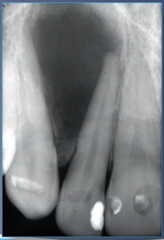
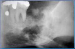
<25, painless, expansile, vital |
Cementoblastoma
mand molar/bicuspid region (mand 1st molar highest), well defined, lucent rim, periapical |
|
<25, painless, expansile, vital |
Cementoblastoma
mand molar/bicuspid region (mand 1st molar highest), well defined, lucent rim, periapical |
|
~40 (4-80), female, asymptomatic, expansion, loose teeth |
Central Odontogenic Fibroma |
|
~40 (4-80), female, asymptomatic, expansion, loose teeth |
Central Odontogenic Fibroma |
|
|
Central Odontogenic Fibroma
WHO: Has odontogenic epithelial rests |
|
|
Central Odontogenic Fibroma
Simple: No odontogenic epithelial rests |
|
>50 |
Clear cell odontogenic carcinoma
uni or multiocular, ill defined margines |
|
|
Clear cell odontogenic carcinoma
glycogen rich, palisading |
|
|
Clear cell odontogenic carcinoma
glycogen rich, palisading |
|
|
Clear cell odontogenic carcinoma
glycogen rich, palisading |
|
|
Complex Odontoma |
|
|
Complex Odontoma |
|
<20 |
Complex Odontoma
Radiolucent rim |
|
<20 |
Complex Odontoma
Radiolucent rim, posterior mandible |
|
<20 |
Complex Odontoma
Radiolucent rim, posterior mandible |
|
|
Compound Odontoma |
|
<20 |
Compound Odontoma
anterior maxilla |
|
<20 |
Compound Odontoma
anterior maxilla |
|
<20 |
Compound Odontoma
anterior maxilla |
|
25-30, asymptomatic, expansion |
Odontogenic Myxoma
uni or multilocular, honeycomb, wispy (right angles), scalloped margins |
|
25-30, asymptomatic, expansion |
Odontogenic Myxoma
uni or multilocular, honeycomb, wispy (right angles), scalloped margins |
|
25-30, asymptomatic, expansion |
Odontogenic Myxoma
uni or multilocular, honeycomb, wispy (right angles), scalloped margins |
|
25-30, asymptomatic, expansion |
Odontogenic Myxoma
uni or multilocular, honeycomb, wispy (right angles), scalloped margins |
|
25-30, asymptomatic, expansion |
Odontogenic Myxoma
uni or multilocular, honeycomb, wispy (right angles), scalloped margins |
|
|
Odontogenic myxoma
myxoid appearance |
|
|
Odontogenic myxoma
myxoid appearance |
|
Middle aged adult |
Peripheral Ameloblastoma
Usually in mandibular gingiva in posterior areas |
|
|
Peripheral Ameloblastoma
Interconnecting cords of epithelium |
|
|
Peripheral odontogenic Fibroma |
|
~37 (11-67) |
Squamous odontogenic tumor
Lateral to a root surface, notice how it destroys up to and past the alveolar crest, (can be on multiple sites) |
|
~37 (11-67) |
Squamous odontogenic tumor
Lateral to a root surface, notice how it destroys up to and past the alveolar crest |
|
~23 (<30) |
Unicystic ameloblastoma
Posterior mandible, always unilocular |
|
~23 (<30) |
Unicystic ameloblastoma
Posterior mandible, always unilocular |
|
~23 (<30) |
Unicystic ameloblastoma
Posterior mandible, always unilocular |
|
~23 (<30) |
Unicystic ameloblastoma
Posterior mandible, always unilocular |
|
|
Unicystic ameloblastoma
Palisading of basal cell layer and reverse polarization |
|
|
Unicystic ameloblastoma
Palisading of basal cell layer and reverse polarization |
|
Pain, swelling of soft tissue |
Acute Osteomyelitis
irregular areas of lucency, ill defined margins, moth-eaten appearance |
|
Pain, swelling of soft tissue |
Acute Osteomyelitis
irregular areas of lucency, ill defined margins, moth-eaten appearance |
|
Adolescents and young adults <30, jaw swelling, teeth displacement, may or may not have pain |
Central Giant Cell Granuloma |
|
Adolescents and young adults <30, jaw swelling, teeth displacement, may or may not have pain |
Central Giant Cell Granuloma
well defined, multilocular, soap bubble appearance, unilocular in smaller stages, common in mandible |
|
Adolescents and young adults <30, jaw swelling, teeth displacement, may or may not have pain |
Central Giant Cell Granuloma
well defined, multilocular, soap bubble appearance, unilocular in smaller stages, common in mandible |
|
Adolescents and young adults <30, jaw swelling, teeth displacement, may or may not have pain |
Central Giant Cell Granuloma
well defined, multilocular, soap bubble appearance, unilocular in smaller stages, common in mandible |
|
Adolescents and young adults <30, jaw swelling, teeth displacement, may or may not have pain |
Central Giant Cell Granuloma |
|
Adolescents and young adults <30, jaw swelling, teeth displacement, may or may not have pain |
Central Giant Cell Granuloma
well defined, multilocular, soap bubble appearance, unilocular in smaller stages, common in mandible |
|

Adolescents and young adults <30, jaw swelling, teeth displacement, may or may not have pain |
Central Giant Cell Granuloma
well defined, multilocular, soap bubble appearance, unilocular in smaller stages, common in mandible |
|
Adolescents and young adults <30, jaw swelling, teeth displacement, may or may not have pain |
Central Giant Cell Granuloma
well defined, multilocular, soap bubble appearance, unilocular in smaller stages, common in mandible |
|
Adolescents to middle-aged adults, expansible jaw lesion, displacement of teeth, facial deformities |
Central Ossifying Fibroma
Common in mandible, well circumscribed, smooth borders, early stages purely lucent, opacities with maturation |
|
Adolescents to middle-aged adults, expansible jaw lesion, displacement of teeth, facial deformities |
Central Ossifying Fibroma
Common in mandible, well circumscribed, smooth borders, early stages purely lucent, opacities with maturation |
|
Adolescents to middle-aged adults, expansible jaw lesion, displacement of teeth, facial deformities |
Central Ossifying Fibroma |
|
Adolescents to middle-aged adults, expansible jaw lesion, displacement of teeth, facial deformities |
Central Ossifying Fibroma
Common in mandible, well circumscribed, smooth borders, early stages purely lucent, opacities with maturation |
|
Adolescents to middle-aged adults, expansible jaw lesion, displacement of teeth, facial deformities |
Central Ossifying Fibroma
Common in mandible, well circumscribed, smooth borders, early stages purely lucent, opacities with maturation |
|
Adolescents to middle-aged adults, expansible jaw lesion, displacement of teeth, facial deformities |
Central Ossifying Fibroma
Common in mandible, well circumscribed, smooth borders, early stages purely lucent, opacities with maturation |
|
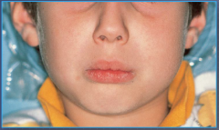
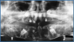
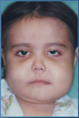
Early childhood, males, pigmented skin lesions, missing/displaced/delayed eruption of teeth |
Cherubism
Bilateral posterior mandible, symmetrical moderate to large multilocular, may be diffuse |
|
Early childhood, males, pigmented skin lesions, missing/displaced/delayed eruption of teeth |
Cherubism
Bilateral posterior mandible, symmetrical moderate to large multilocular, may be diffuse |
|
Early childhood, males, pigmented skin lesions, missing/displaced/delayed eruption of teeth |
Cherubism
Bilateral posterior mandible, symmetrical moderate to large multilocular, may be diffuse |
|
Early childhood, males, pigmented skin lesions, missing/displaced/delayed eruption of teeth |
Cherubism
Bilateral posterior mandible, symmetrical moderate to large multilocular, may be diffuse |
|
Early childhood, males, pigmented skin lesions, missing/displaced/delayed eruption of teeth |
Cherubism
Bilateral posterior mandible, symmetrical moderate to large multilocular, may be diffuse |
|
Adults, slow growing expansile mass, may or may not have pain or loose teeth |
Chondrosarcoma
Ill-defined mixed, resembles snowstorm |
|
Adults, slow growing expansile mass, may or may not have pain or loose teeth |
Chondrosarcoma
Ill-defined mixed, resembles snowstorm |
|
|
Chronic Osteomyelitis
irregular areas of lucency, ill-defined margins, moth-eaten appearance, may be extensive |
|
|
Chronic Osteomyelitis
irregular areas of lucency, ill-defined margins, moth-eaten appearance, may be extensive |
|
|
Chronic Osteomyelitis
irregular areas of lucency, ill-defined margins, moth-eaten appearance, may be extensive |
|
|
Chronic Osteomyelitis
irregular areas of lucency, ill-defined margins, moth-eaten appearance, may be extensive |
|
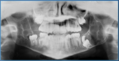
Childhood to young adulthood, asymptomatic jaw enlargement |
Fibrous dysplasia
Will not cross midline, ground glass appearance |
|
Childhood to young adulthood, asymptomatic jaw enlargement |
Fibrous dysplasia
Will not cross midline, ground glass appearance |
|
Childhood to young adulthood, asymptomatic jaw enlargement |
Fibrous dysplasia
Will not cross midline, ground glass appearance |
|
Childhood to young adulthood, asymptomatic jaw enlargement |
Fibrous dysplasia
Will not cross midline, ground glass appearance |
|
Childhood to young adulthood, asymptomatic jaw enlargement |
Fibrous dysplasia
Will not cross midline, ground glass appearance |
|
Childhood to young adulthood, asymptomatic jaw enlargement |
Fibrous dysplasia
Will not cross midline, ground glass appearance |
|
Childhood to young adulthood, asymptomatic jaw enlargement |
Fibrous dysplasia
Will not cross midline, ground glass appearance |
|
Childhood to young adulthood, asymptomatic jaw enlargement |
Fibrous dysplasia
Will not cross midline, ground glass appearance |
|
Childhood to young adulthood, asymptomatic jaw enlargement |
Fibrous dysplasia
Will not cross midline, ground glass appearance |
|
Childhood to young adulthood, asymptomatic jaw enlargement |
Fibrous dysplasia
Will not cross midline, ground glass appearance |
|
Middle-aged and older black females, asymptomatic, pain when secondarily infected |
Florid Cemento-osseous Dysplasia
Lesions in all four quadrants, multiple round lesions of various sizes |
|

Middle-aged and older black females, asymptomatic, pain when secondarily infected |
Florid Cemento-osseous Dysplasia
Lesions in all four quadrants, multiple round lesions of various sizes |
|
Middle-aged and older black females, asymptomatic, pain when secondarily infected |
Florid Cemento-osseous Dysplasia
Lesions in all four quadrants, multiple round lesions of various sizes |
|
Middle-aged and older black females, asymptomatic, pain when secondarily infected |
Florid Cemento-osseous Dysplasia
Lesions in all four quadrants, multiple round lesions of various sizes |
|
Middle-aged and older females, asymptomatic, usually vital teeth |
Focal Cemento-osseous Dysplasia
Common in mandibular teeth, solitary well-defined periapical lesion, retain a thin radiolucent rim |
|
Middle-aged and older females, asymptomatic, usually vital teeth |
Focal Cemento-osseous Dysplasia
Common in mandibular teeth, solitary well-defined periapical lesion, retain a thin radiolucent rim |
|
Middle-aged and older females, asymptomatic, usually vital teeth |
Focal Cemento-osseous Dysplasia
Common in mandibular teeth, solitary well-defined periapical lesion, retain a thin radiolucent rim |
|
Primary type in middle aged females, secondary in older adult male, asymptomatic |
Hyperparathyroidism of Bone
Diffusely involves all four quadrants, ground glass bone, loss of lamina dura |
|
Primary type in middle aged females, secondary in older adult male, asymptomatic |
Hyperparathyroidism of Bone
Diffusely involves all four quadrants, ground glass bone, loss of lamina dura |
|
Primary type in middle aged females, secondary in older adult male, asymptomatic |
Hyperparathyroidism of Bone
Diffusely involves all four quadrants, ground glass bone, loss of lamina dura |
|
Primary type in middle aged females, secondary in older adult male, asymptomatic |
Hyperparathyroidism of Bone
Diffusely involves all four quadrants, ground glass bone, loss of lamina dura |
|
Primary type in middle aged females, secondary in older adult male, asymptomatic |
Hyperparathyroidism of Bone
Diffusely involves all four quadrants, ground glass bone, loss of lamina dura |
|
Primary type in middle aged females, secondary in older adult male, asymptomatic |
Hyperparathyroidism of Bone
Diffusely involves all four quadrants, ground glass bone, loss of lamina dura |
|
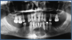
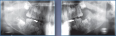
Adolescents and young adults, asymptomatic, vital |
Idiopathic Osteosclerosis
Mandibular first molar periapical region, confluent with lamina dura, does not obscure PDL |
|
Adolescents and young adults, asymptomatic, vital |
Idiopathic Osteosclerosis
Mandibular first molar periapical region, confluent with lamina dura, does not obscure PDL |
|
Usually adults, often older adults, pain, swelling, or pathologic fracture, may be asymptomatic |
Metastatic bone tumor
Common in mandible, diffuse radiolucencies, moth-eaten, well circumscribed |
|
Usually adults, often older adults, pain, swelling, or pathologic fracture, may be asymptomatic |
Metastatic bone tumor
Common in mandible, diffuse radiolucencies, moth-eaten, well circumscribed |
|
Usually adults, often older adults, pain, swelling, or pathologic fracture, may be asymptomatic |
Metastatic bone tumor
Common in mandible, diffuse radiolucencies, moth-eaten, well circumscribed |
|
Usually adults, often older adults, pain, swelling, or pathologic fracture, may be asymptomatic |
Metastatic bone tumor
Common in mandible, diffuse radiolucencies, moth-eaten, well circumscribed |
|
Middle aged and older adults, rare before 40, bony enlargement, pathologic fracture |
Osteitis deformans
Cotton wool opacities in bones, in jaws you have more mixed lesions, loss of lamina dura, hypercementosis |
|
Middle aged and older adults, rare before 40, bony enlargement, pathologic fracture |
Osteitis deformans
Cotton wool opacities in bones, in jaws you have more mixed lesions, loss of lamina dura, hypercementosis |
|
Middle aged and older adults, rare before 40, bony enlargement, pathologic fracture |
Osteitis deformans
Cotton wool opacities in bones, in jaws you have more mixed lesions, loss of lamina dura, hypercementosis |
|
Middle aged and older adults, rare before 40, bony enlargement, pathologic fracture |
Osteitis deformans
Cotton wool opacities in bones, in jaws you have more mixed lesions, loss of lamina dura, hypercementosis |
|
Middle aged and older adults, rare before 40, bony enlargement, pathologic fracture |
Osteitis deformans
Cotton wool opacities in bones, in jaws you have more mixed lesions, loss of lamina dura, hypercementosis |
|
Middle aged and older adults, rare before 40, bony enlargement, pathologic fracture |
Osteitis deformans
Cotton wool opacities in bones, in jaws you have more mixed lesions, loss of lamina dura, hypercementosis |
|
Young adult, pain and swelling, may have loose teeth, may have tooth ache, may have paresthesia |
Osteosarcoma
More common in mandible, diffuse borders, 25% show sunburst, widened periodontal ligament, spiky resorption, moth eaten appearance |
|
Young adult, pain and swelling, may have loose teeth, may have tooth ache, may have paresthesia |
Osteosarcoma
More common in mandible, diffuse borders, 25% show sunburst, widened periodontal ligament, spiky resorption, moth eaten appearance |
|
Young adult, pain and swelling, may have loose teeth, may have tooth ache, may have paresthesia |
Osteosarcoma
More common in mandible, diffuse borders, 25% show sunburst, widened periodontal ligament, spiky resorption, moth eaten appearance |
|
Young adult, pain and swelling, may have loose teeth, may have tooth ache, may have paresthesia |
Osteosarcoma
More common in mandible, diffuse borders, 25% show sunburst, widened periodontal ligament, spiky resorption, moth eaten appearance |
|
Young adult, pain and swelling, may have loose teeth, may have tooth ache, may have paresthesia |
Osteosarcoma
More common in mandible, diffuse borders, 25% show sunburst, widened periodontal ligament, spiky resorption, moth eaten appearance |
|
Young adult, pain and swelling, may have loose teeth, may have tooth ache, may have paresthesia |
Osteosarcoma
More common in mandible, diffuse borders, 25% show sunburst, widened periodontal ligament, spiky resorption, moth eaten appearance |
|
Young adult, pain and swelling, may have loose teeth, may have tooth ache, may have paresthesia |
Osteosarcoma
More common in mandible, diffuse borders, 25% show sunburst, widened periodontal ligament, spiky resorption, moth eaten appearance |
|
Young adult, pain and swelling, may have loose teeth, may have tooth ache, may have paresthesia |
Osteosarcoma
More common in mandible, diffuse borders, 25% show sunburst, widened periodontal ligament, spiky resorption, moth eaten appearance |
|
Young adult, pain and swelling, may have loose teeth, may have tooth ache, may have paresthesia |
Osteosarcoma
More common in mandible, diffuse borders, 25% show sunburst, widened periodontal ligament, spiky resorption, moth eaten appearance |
|
Young adult, pain and swelling, may have loose teeth, may have tooth ache, may have paresthesia |
Osteosarcoma
More common in mandible, diffuse borders, 25% show sunburst, widened periodontal ligament, spiky resorption, moth eaten appearance |
|
Young adult, pain and swelling, may have loose teeth, may have tooth ache, may have paresthesia |
Osteosarcoma
More common in mandible, diffuse borders, 25% show sunburst, widened periodontal ligament, spiky resorption, moth eaten appearance |
|
Young adult, pain and swelling, may have loose teeth, may have tooth ache, may have paresthesia |
Osteosarcoma
More common in mandible, diffuse borders, 25% show sunburst, widened periodontal ligament, spiky resorption, moth eaten appearance |
|
Young adult, pain and swelling, may have loose teeth, may have tooth ache, may have paresthesia |
Osteosarcoma
More common in mandible, diffuse borders, 25% show sunburst, widened periodontal ligament, spiky resorption, moth eaten appearance |
|
Young adult, pain and swelling, may have loose teeth, may have tooth ache, may have paresthesia |
Osteosarcoma
More common in mandible, diffuse borders, 25% show sunburst, widened periodontal ligament, spiky resorption, moth eaten appearance |
|
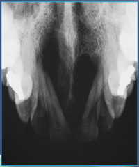
Middle aged black females, also Hispanics and Asians. Asymptomatic, vital teeth |
Periapical Cemento-osseous Dysplasia
Periapical areas of mandibular anterior teeth, corticated border in early stages, surrounding radiolucent zone in intermediate and mature |
|
Middle aged black females, also Hispanics and Asians. Asymptomatic, vital teeth |
Periapical Cemento-osseous Dysplasia
Periapical areas of mandibular anterior teeth, corticated border in early stages, surrounding radiolucent zone in intermediate and mature |
|
Middle aged black females, also Hispanics and Asians. Asymptomatic, vital teeth |
Periapical Cemento-osseous Dysplasia
Periapical areas of mandibular anterior teeth, corticated border in early stages, surrounding radiolucent zone in intermediate and mature |
|
Middle aged black females, also Hispanics and Asians. Asymptomatic, vital teeth |
Periapical Cemento-osseous Dysplasia
Periapical areas of mandibular anterior teeth, corticated border in early stages, surrounding radiolucent zone in intermediate and mature |
|
Middle aged black females, also Hispanics and Asians. Asymptomatic, vital teeth |
Periapical Cemento-osseous Dysplasia
Periapical areas of mandibular anterior teeth, corticated border in early stages, surrounding radiolucent zone in intermediate and mature |
|
|
Child or young adult below 25 who presents with bony hard swellings in the mandible. There are onion-skin layerings of bone over cortex there is a source of infection. |
Garre's osteomyelitis
Pure radiolucency (Diffuse and/or Mottled) |
|
|
Child or young adult below 25 who presents with bony hard swellings in the mandible. There are onion-skin layerings of bone over cortex there is a source of infection. |
Garre's osteomyelitis
Pure radiolucency (Diffuse and/or Mottled) |
|
|
Child or young adult below 25 who presents with bony hard swellings in the mandible. There are onion-skin layerings of bone over cortex there is a source of infection. |
Garre's osteomyelitis
Pure radiolucency (Diffuse and/or Mottled) |
|
|
Child or young adult below 25 who presents with bony hard swellings in the mandible. There are onion-skin layerings of bone over cortex there is a source of infection. |
Garre's osteomyelitis
Pure radiolucency (Diffuse and/or Mottled) |
|
|
Older adult with pain, fistulation, and pathologic fracture in the mandible. There are ill-defined radiolucencies with foci of opacities. It looks moth-eaten. |
Osteoradionecrosis
Pure radiolucency (Diffuse and/or Mottled) |
|
|
Older adult with pain, fistulation, and pathologic fracture in the mandible. There are ill-defined radiolucencies with foci of opacities. It looks moth-eaten. |
Osteoradionecrosis
Pure radiolucency (Diffuse and/or Mottled) |
|
|
Older adult with pain, fistulation, and pathologic fracture in the mandible. There are ill-defined radiolucencies with foci of opacities. It looks moth-eaten. |
Osteoradionecrosis
Pure radiolucency (Diffuse and/or Mottled) |
|
|
Older adult with pain, fistulation, and pathologic fracture in the mandible. There are ill-defined radiolucencies with foci of opacities. It looks moth-eaten. |
Osteoradionecrosis
Pure radiolucency (Diffuse and/or Mottled) |
|
|
Older adult with sequestration of dead bone through mucosa in the mandible. Ill-defined or moth-eaten radiolucency. |
BRONJ
Pure radiolucency (Diffuse and/or Mottled) |
|
|
Older adult with sequestration of dead bone through mucosa in the mandible. Ill-defined or moth-eaten radiolucency. |
BRONJ
Pure radiolucency (Diffuse and/or Mottled) |
|
|
Older adult with sequestration of dead bone through mucosa in the mandible. Ill-defined or moth-eaten radiolucency. |
BRONJ
Pure radiolucency (Diffuse and/or Mottled) |
|
|
Older adult with sequestration of dead bone through mucosa in the mandible. Ill-defined or moth-eaten radiolucency. |
BRONJ
Pure radiolucency (Diffuse and/or Mottled) |
|
|
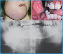
Patient comes in with cutaneous and facial epidermal cysts. The radiograph shows many osteomas, multiple odontomas, supernumerary teeth, and impactions. |
Gardner syndrome
(She may also tell you polyps, but I didn't because that's a giveaway) |
|
|
Patient comes in with cutaneous and facial epidermal cysts. The radiograph shows many osteomas, multiple odontomas, supernumerary teeth, and impactions. |
Gardner syndrome
(She may also tell you polyps, but I didn't because that's a giveaway) |
|
|
Patient comes in with cutaneous and facial epidermal cysts. The radiograph shows many osteomas, multiple odontomas, supernumerary teeth, and impactions. |
Gardner syndrome
(She may also tell you polyps, but I didn't because that's a giveaway) |
|
|
Patient comes in with cutaneous and facial epidermal cysts. The radiograph shows many osteomas, multiple odontomas, supernumerary teeth, and impactions. |
Gardner syndrome
(She may also tell you polyps, but I didn't because that's a giveaway) |
|
|
An adolescent/young adult less than 30 comes in with jaw pain and swelling. The radiograph shows mixed and well defined. Also there could be some obscure root outlines. |
Osteoblastoma if larger than 2 cm Osteoid Osteoma if less than 2 cm |
|
|
An adolescent/young adult less than 30 comes in with jaw pain and swelling. The radiograph shows mixed and well defined. Also there could be some obscure root outlines. |
Osteoblastoma if larger than 2 cm Osteoid Osteoma if less than 2 cm |
|
|
An adolescent/young adult less than 30 comes in with jaw pain and swelling. The radiograph shows mixed and well defined. Also there could be some obscure root outlines. |
Osteoblastoma if larger than 2 cm Osteoid Osteoma if less than 2 cm |
|
|
An adolescent/young adult less than 30 comes in with jaw pain and swelling. The radiograph shows mixed and well defined. Also there could be some obscure root outlines. |
Osteoblastoma if larger than 2 cm Osteoid Osteoma if less than 2 cm |
|
|
Female adolescent comes in with discolored gingiva. Radiograph presents with honey-comb or soap bubble appearance and patient is asymptomatic. |
Central Hemangioma
Pure Radiolucency, multilocular |
|
|
Female adolescent comes in with discolored gingiva. Radiograph presents with honey-comb or soap bubble appearance and patient is asymptomatic. |
Central Hemangioma
Pure Radiolucency, multilocular |
|
|
Female adolescent comes in with discolored gingiva. Radiograph presents with honey-comb or soap bubble appearance and patient is asymptomatic. |
Central Hemangioma
Pure Radiolucency, multilocular |
|
|
Female adolescent comes in with discolored gingiva. Radiograph presents with honey-comb or soap bubble appearance and patient is asymptomatic. |
Central Hemangioma
Pure Radiolucency, multilocular |
|
|
Your patient who is between 10-19 comes in with pain and swelling. They also have some loose teeth in the mandible. The radiograph shows expansile destructive radiolucent lesions with ill-defined margins. |
Ewing Sarcoma
Onion skin layering is uncommon in jaws.
Pure radiolucencies (Diffuse and/or mottled) |
|
|
Middle-aged/older adult comes in with pain and pathologic bone fracture. Oral symptoms include numbness, loose teeth, and gingival enlargement in the mandible. Radiograph shows multiple small round sharply-demarcated lucencies (punched out) |
Multiple Myeloma
Pure radiolucency (Unilocular, well-defined) |
|
|
A boy less than 10 years old comes into your office with periapical pathosis and radiograph shows teeth just floating. |
Langerhans Cell Disease |
|
|
A boy less than 10 years old comes into your office with periapical pathosis and radiograph shows teeth just floating. |
Langerhans Cell Disease |
|
|
A boy less than 10 years old comes into your office with periapical pathosis and radiograph shows teeth just floating. |
Langerhans Cell Disease |
|
|
A boy less than 10 years old comes into your office with periapical pathosis and radiograph shows teeth just floating. |
Langerhans Cell Disease |