Reading...
![]()
Play button
![]()
Play button
![]()
Use LEFT and RIGHT arrow keys to navigate between flashcards;
Use UP and DOWN arrow keys to flip the card;
H to show hint;
A reads text to speech;
56 Cards in this Set
- Front
- Back
|
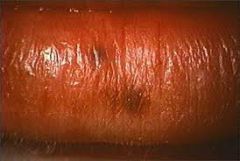
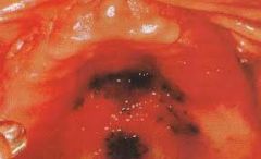
What is a clinical term to describe a red patch?
|
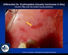
Erythroplakia
|
|
|
What type of Pt might you see erythroplakia in?
|
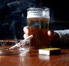
A chronic tobacco/alcohal user
**This is a possible pre-maligancy 85% of the time.** |
|
|
Is erythroplakia inflammatory in nature?
|
No!
|
|
|
What type of TX might be considered for erythroplakia?
|
Removal of the lesion,
Chemo/radiation, Close follow up |
|
|
What is a clinical term to describe a collection of blood within tissue space?
|
Hematoma
|
|
|
Will a hematoma blanch?
Why or why not? |
No, because its not a vascular lesion.
|
|
|
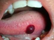
What is a hemangioma?
|
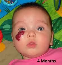
A benign proliferation of blood vessels.
*Remember, is seen in young children/infants* |
|
|
What might a red or brown lesion be an indicator of?
|
Possible maligancy.
|
|
|
What is Telangiectasia?
What age group is more likely to have it? |
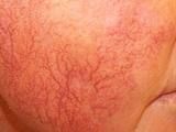
Spider veins in the face.
Typically more in the elderly but = in males/females. |
|
|
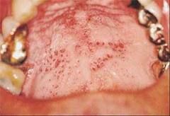
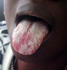
What is erythematous candidiasis?
|
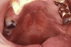
A common, red looking, candidiasis.
(May look like herpes if on commisures) |
|
|
Candidiasis can appear as ________ & ________ lesions.
|
Red & White.
|
|
|
Patients who have ____________are often more likely to experience Erythematous Candidiasis.
|
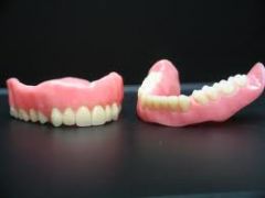
Dentures
*Be sure the PT is cleaning them regularly, may need to disinfect.* |
|
|
Which populations would Oral Submucous Fibrosis be found in?
|
Usually in developing countries, but with immigration it is being seen more in the US.
|
|
|
What does Oral Submucosis Fibrosis look like?
What is a DD of it? |
Has a white marble appearance in the buccal mucosa.
DD: lichen planus |
|
|
What are 2 pigmented lesions that are variants of normal?
|
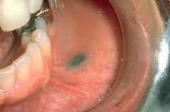
Amalgam Tattoo Oral Pigmentation
|
|
|
What might the tissue around a titanium implant look like?
|
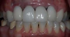
Grey-blue tissue around the gingiva.
(The tissue is so thin the metal shows through.) |
|
|
What type of pigmentation would have Grey, Blue or Black around the gingival margin?
|
Heavy metal pigmentation.
|
|
|
What are 2 examples of medications where pigmentation is involved?
|
Minocycline tetracycline
|
|
|
What is another name for Minocycline tissue staining?
|
Black bone staining
|
|
|
What is another name for a benign mole?
Where in the oral cavity is likely to be found? |
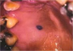
Nevus
The hard palate is the most common intraoral site. |
|
|
What is the DD for a Nevus?
What is the TX? |
DD: Amalgam tattoo, melanoma, hemangioma
Tx: Monitor |
|
|
Wht is the term for a freckle on the lip?
|
Ephelis (macule)
|
|
|
How would you describe an oral melanoma?
|
Its usually located on the hard palate and maxillary gingiva. Usually has irregular/asemmetical boarders. Usually dark in color
|
|
|
Who is more likely to have an oral malanoma?
|
Men 40+, Can be deadly so keep an eye on it.
*Always investigate a dark lesion.* |
|
|
How might someone get Frictional Keratosis?
|
Cheek Biting, Ortho Tx, Broken or Missing teeth, Bruxing
|
|
|
Is Frictional Keratosis rare or common?
|
Common
|
|
|
Why can't you just wipe it off?
|
Keratin is produced to protect the site and won't simply wipe away.
The condition must be alleviated. |
|
|
What is the DD for Frictional Keratosis?
|
Leukoplakia, Lichen planus, hyperplastic candidiasis.
|
|
|
What is the most common white lesion in pipe or heavy smokers?
|
Nicotine Stomatitis
|
|
|
What is nicotine stomatitis?
|

Hyperkeratosis on the palate with possible inflamed minor salivary gland ducts.
(Looks like red or brown spots in the keratin) |
|
|
Although its relatively rare, which population would you see chemical burns?
|
The elderly.
Often from sucking on an asprin to alleviate a toothache. |
|
|
Thermal burns are a result of?
|
Hot Food!
|
|
|
What might a chemical or thermal burn look like?
|
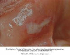
Localized white plaques.
DO NOT RUB THEM OFF--can cause raw or bleeing "under tissue". |
|
|
What is the DD for Chemical/Thermal burns?
|
Frictional keratosis, hyperplastic candidiasis, lichen planus
|
|
|
What is Candidiasis?
|
A fungal infection caused gy the Candida C. albicans
|
|
|
Where can candidiasis be found?
|
Mouth, GI tract & vagina.
(Same bacteria can be in all locations) |
|
|
What type of infection is candidiasis?
|
Opportunistic!
*You might see this in Pt's with asthma in the back of the throat due to the inhaler* |
|
|
What is the usually appearance of candidiasis?
|
White apperance but has an erythematous form as well.
|
|
|
If the candidiasis is wiped off what will the appearance be?
|
Red, ulcertative look to the underlying tissues.
|
|
|
What is the Tx for this fungus?
|
Antifungal
|
|
|
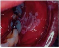
What is another name for Acute pseudomembranous candidiasis?
|

Thrush
Its common in newborns. |
|
|
Where would Thrush be located?
Can it be wiped off? |
It has both extra and intra oral characteristics.
(Can often be found in skin/fat folds) Yes it can be wiped off, a bleeding base is rare. |
|
|
DD?
Tx? |
Most lesions that wipe off are candidiasis.
Tx: antifungal |
|
|
How would you treat dentures or partials for a Pt with acute pseudomembranous candidiasis?
|
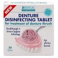
They need to be disinfected so as to avoid re-infecting the area.
|
|
|
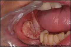
What is the rarest form of candidiasis?
|
Chronic Hyperplastic Candidiasis
** It is the only form to show possibility of pre-malignancy in a low amount of cases** |
|
|
What characteristic of Chronic Hyperplastic Candidiasis is unlike other forms of candidiasis?
|
It won't rub off!!
(Has a thick, raised, white plaque.) |
|
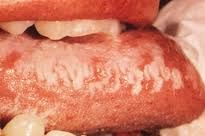
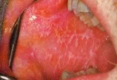
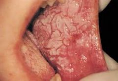
What disease states is Hairy Leukoplakia associated with?
|
EBV (Epstein Barr Virus) and HIV.
|
|
|
What is the most common site for Hairy Leukoplakia?
|
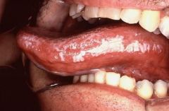
Lateral boarder of tongue, often is Bilateral!
Won't wipe off! |
|
|
What would be the DD of Hairy Tongue?
|
Frictional keratosis, lichen planus, candidiasis, possible burns.
|
|
|
What is the origin of Lichen Planus?
|
This inflammatory disorder has an unknown cause.
|
|
|
Which sex is more likely to have lichen planus and what surface is it usually located?
|
Female 90%
Buccal mucosa |
|
|
What conditions can exaserbate lichen planus?
|

Stress & Anxiety
|
|
|
What are the 6 forms of Lichen Planus? Which is most common?
|
1) Atrophic (red patches with white)
2) Bullous 3) Erosive (angry red ulcerated look) 4) Papular 5) Plaque like 6) Reticular- Wickam striae **Most common form** |
|
|
What is White Sponge Nevus?
|
A mutation in the keratin gene. It's genetic
|
|
|
Is White Sponge Nevus usually seen laterally or bilatterally?
Will it rub off? |
Most often bilaterally.
No it won't rub off. Usually is located on the buccal mucosa. |
|
|
DD For White sponge nevus? Tx?
|
DD: Cheek Chewing, Lichen planus, leukodema Tx: None
|