![]()
![]()
![]()
Use LEFT and RIGHT arrow keys to navigate between flashcards;
Use UP and DOWN arrow keys to flip the card;
H to show hint;
A reads text to speech;
36 Cards in this Set
- Front
- Back
|
Urothelial cancers of the upper urinary tract are more common in men than in women. Is this trend also true for bladder cancer? What about racial biases for bladder cancer?
|
Yes, it is about three times more common in men than in women.
More common among whites, about twice, than African Americans and Hispanic Americans. |
|
|
How does the incidence data differ from mortality data for bladder cancer?
|
Even though whitey males get it more commonly than all others, the mortality from the disease is much higher in Caucasian women, and even higher still in African American women. Women have a higher than 30% increased risk of dying from bladder cancer than men do.
The prognosis for bladder cancer in Hispanics is better than Caucasians. |
|
|
What are risk factors for bladder cancer?
|
Age, as well as Caucasian race and male gender
Occupations with increased risk – barbers, beauticians, physicians, drill press operators, autoworker, painter, truck driver, leather worker, metal worker, machiner, dry cleaner, etc. Cigarette smoking & exposure – fourfold higher incidence Infections & long term irritation such as chronic foley catheterization or bladder calculi Analgesic abuse, particularly with phenacetin Schistosoma cystitis Pelvic irradiation – twofold to fourfold increased risk Cyclophosphamide treatment – up to 9x increased risk Misc stuff such as Blackfoot disease, being a renal transplant patient, some chinese herb Interestingly, genetics do not appear to be a risk factor |
|
|
Explain the differences between von brunn’s nests, cystitis cystica, and cystitis glandularis
|
Von brunn’s nests are benign islands of urothelium situated in the lamina propria. Cystitis cystica occurs when the urothelium of von brunn’s nests undergo eosinophilic liquefaction in the center of the islands and appear cystic. Cystitis glandularis occurs when the islands have undergone glandular metaplasia, and this is believed to be a precursor to adenocarcinoma.
|
|
|
What is an inverted papilloma and what conditions is it associated with?
|
This is a benign lesion that is the same as a papilloma however instead of growing into the lumen it is inverted into the stroma of the bladder. It is associated with conditions such as chronic inflammation or obstruction, as such it may be covered with squamous metaplasia or cystitis cystica as well as normal urothelium.
|
|

A 71-year-old white woman presented for evaluation of recurrent irritative voiding symptoms. She reported an approximately 20-year history of this clinical problem and thought that its onset could be temporally associated with a hysterectomy she had undergone for a benign condition. At presentation, the patient reported that she had chronic intermittent vaginal burning, and urinary hesitancy, intermittency, and urgency. A culture-proven urinary tract infection (UTI) caused by Pseudomonas aeruginosa was noted during one of her marked symptomatic episodes. Multiple times, however, cultures had failed to reveal a specific infecting organism.
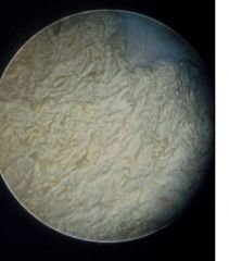
The patient was otherwise healthy and had no history of diabetes mellitus, tuberculosis, urolithiasis, cancer, pediatric UTIs, or known genitourinary tract anatomic anomalies. Previous cystoscopic examinations had shown erythematous lesions in the bladder. Biopsies of these areas had demonstrated chronic inflammation without evidence of malignancy. What lesion is shown? |

It is squamous metaplasia with marked keratinization. It is believed to be a response to a noxious stimulus in the bladder and is believed to be a pre‐malignant lesion with transformation to SCC in 20% of cases.
|
|
|
What cytogenetic associations do CIS and muscle-invasive bladder cancers have in common that supports their direct relationship?
|
Loss of chromosome 17p, deletions and/or mutations of the TP53 gene and its products
|
|
|
What is the difference between the following lesions histologically?
|
Papilloma – rare, but if it truly is a benign papilloma it is usually found by itself. It has a fibrovascular stalk with no histological abnormalities and no more than 7 cell layers. Benign, almost never recurs.
Well differentiated tumors (papillary urothelial neoplasm of low malignant potential, or PUNLMP) – contains more than 7 cell layers, mild anaplasia, rare mitotic figures, base to surface maturation, slightly irregular; often recur. Moderately differentiated tumors (Low grade urothelial carcinomas) – greater disturbance of base to surface cellular maturation, loss of cellular polarity, nuclear to cytoplasm ratio is greater, prominent nucleoli Poorly differentiated tumors (High grade urothelial carcinomas) – no differentiation from base to surface layer of cells, frequent mitotic figures, high nuclear to cytoplasm ratio |
|
|
What are risk factors for nonbilharzial SCC of the bladder?
|
Chronic infections, bladder stones, chronic indwelling catheters, bladder diverticulae, cigarette smoking
|
|
|
You are seeing a patient in Dr. Sutherland’s clinic with bladder cancer who was born with exstrophy. What type of cancer are they most likely to have? In patients with adenocarcinoma of the bladder that are not primary, what are potential origins?
|
Adenocarcinoma.
Adenocarcinoma found in the bladder is either primarily vesical, which is rare, from the urachus, or is a metastatic lesion. Potential origins include the rectum, prostate, stomach, ovary, endometrium, and breast. |
|
|
What are the most common sites of mets in bladder cancer? What are the common sites of distant vascular spread for bladder cancer?
|
Pelvic lymph nodes. Specifically, paravesical nodes in 16%, obturator nodes in 74%, external iliac nodes in 65%, and presacral nodes in 25%
Liver, lung, bone, adrenals, intestines |
|
|
True or False. Most bladder cancers are superficial papillary TCCs upon initial diagnosis.
|
True. 55-60% are superficial papillary TCCs initially (keep in mind Chapter 76 says 70%), although the majority will recur, and 10-20% will develop invasive or metastatic disease. 40‐45% of bladder cancers are high grade, more than half of which are muscle invasive or worse at diagnosis.
|
|
|
True or False. Over 25% of newly diagnosed high-grade lesions are muscle invasive or metastatic.
|
False. Of the 40-45% of newly diagnosed bladder cancers that are high-grade, more than half are muscle-invasive or metastatic at the time of diagnosis.
This question is tricky because what we usually say is that 25% of all-comers with newly diagnosed bladder cancer have muscle invasive disease. |
|
|
What blood group is associated with bladder cancer?
|
Lewis blood group
|
|
|
What are some chromosomal abnormalities that are associated with bladder cancer?
|
Deletion of chromosome 9 – This is more commonly associated with superficial bladder cancers.
Deletion of chromosome 17p – This is more commonly associated with invasive bladder cancers, this is the location of the TP53 tumor suppressor gene. Deletion or mutation of chromosome 13q – RB gene is located here, this is also associated with aggressive cancer. |
|
|
We know that cytopathology isn’t 100% sensitive for bladder cancer and that there are about 10‐20% false negative results. What about specificity, are there false positive results?
|
False positives occur in 1-12%, secondary to cellular atypia, inflammation, or changes associated with radiation or chemo.
|
|
|
A patient is referred with a CT urogram finding a filling defect in the bladder consistent with a malignancy, and he initially presented with gross hematuria. What is the next step?
|
You should set the patient up for cystoscopy under anesthesia with biopsy, flexible cysto is a waste of resources in this case.
|
|
|
You are doing a cystoscopy under general anesthesia on a patient with a lesion in a bladder diverticulum that was diagnosed on office cysto previously. Should you attempt to resect the lesion?
|
It should be sampled rather than resected based on the risk of perforation with attempt at endoscopic treatment. This patient should have either a partial cystectomy or total cystectomy.
|
|
|
You do a flexible cystoscopy on a patient with microscopic hematuria and their bladder appears to be normal. You do bladder washing at the time of cystoscopy. When you follow these washings up you determine that there is the indication of high grade lesions. You schedule the patient for cystoscopy under general anesthesia. Once you enter the bladder with the rigid cystoscope you look around to find a normal appearing bladder, what should you do?
|
Random sample biopsies. It is likely that the patient has CIS that is not visible under regular white light cystoscopy.
There isn’t a standard, but you can take a biopsy of each lateral wall, the anterior dome, the posterior wall, the trigone, and the prostatic urethra. Most people omit the prostatic urethra. |
|
|
With radical cystectomy for muscle invasive bladder cancer, should you do a lymph node dissection? If so what are your boundaries?
|
Always do complete bilateral pelvic lymphadenectomies from slightly above the iliac bifurcations to the femoral canals and from the genitofemoral nerves to the bladder pedicles.
|
|
|
From what are small cell carcinomas of the bladder derived, and what is the typical prognosis?
|
Neuroendocrine stem cells or dendritic cells, but they may be mixed with elements of TCCs
They are aggressive, and usually respond to, but are not often cured by, cisplatin-based chemo |
|
|
What is the function of the wild type or normal TP53 gene?
|
The normal protein, wild-type TP53, has a variety of functions, including acting as a transcription factor that suppresses cell proliferation (Vogelstein, 1990; Cote and Chatterjee, 1999), directing DNA damaged cells toward apoptosis before DNA replication (S phase of cell cycle) occurs (reviewed in Harris and Hollstein, 1993), contributing to the repair of damaged DNA by inducing the production of deoxyribonucleotide triphosphates in the nucleus (reviewed by Lozano and Elledge, 2000).
Because of TP53's functions of repairing damaged DNA and directing cells with other genetic abnormalities toward apoptosis, TP53 mutations have been associated with genomic instability—and hence progressive development of further mutations. For tumors to exceed 1 or 2 mm in diameter, new blood vessels must feed them. Wild-type TP53 induces the expression of a potent inhibitor of angiogenesis, thrombospondin-1 (TSP-1), a normal constituent of the extracellular matrix, whereas mutant (or absent) TP53 does not. This gene is located on the short arm of chromosome 17 or 17p. |
|
|
Which chromosomal defects are associated with superficial cancers and which are associated with high grade invasive cancers of the bladder?
|
hrough such correlations, several groups have now identified chromosome 9 (Tsai et al, 1990; Spruck et al, 1994) and particularly 9q losses (Knowles et al, 1994; Habuchi et al, 1998; Simoneau et al, 1999; Czerniak et al, 1999) as early events in the development of low-grade superficial tumors. Alternatively, high-grade cancers are much more commonly associated with TP53 abnormalities and chromosome 17p deletions.
|
|
|
Why when doing a radical cystectomy for muscle invasive bladder cancer do you take a frozen section of the prostatic urethral margin? If a patient has a frozen section of the prostatic urethra come back positive what do you do? What does this mean for their prognosis?
|
More than 40% of men with muscle invasive bladder cancer have invasion into the prostate and the majority are at the prostatic urethra.
The operation to do is a cystoprostourethrectomy, but even then about 40% of patients with prostatic urethral positivity have stromal invasion and will develop metastasis if they do about 80% of the time. |
|
|
With radical cystectomy for muscle invasive bladder cancer, should you do a lymph node dissection? If so what are your boundaries?
|
A standard lymph node dissection - includes bilateral pelvic lymphadenectomies from slightly above the iliac bifurcations to the femoral canals and from the genitofemoral nerves to the bladder pedicles.
An extended pelvic lymphadenectomy - including the common iliac nodal tissue to the aortic bifurcation, has been demonstrated to offer therapeutic advantage in patients with lymph node metastases ( Herr et al, 2002 ). Some clinicans will include in this the pre-sacral nodes |
|
|
What complications should be discussed with patients before TURBT? What complication rate can you quote?
|
The main complications include uncontrolled hematuria and clinically significant bladder perforation. These occur about 5% of the time.
|
|
|
What can be done to minimize the risk of complication for TURBT?
|
Avoid bladder overdistention: Fill bladder only enough to visualize contents. This minimizes bladder wall movement and lessens thinning of the detrusor.
Use muscle-paralyzing agents: This minimizes the risk of the obturator reflex especially when resecting lateral wall tumors. Resect large or bulky tumors in a staged manner: allows for safer resection of residual tumor if indicated |
|
|
What is a theoretical risk associated with bladder perforation during TURBT?
|
Intraperitoneal tumor seeding is a possibility and has been reported anecdotally. However, the authors seem to downplay its significance.
|
|
|
What are the theoretical advantages and risks associated with random biopsies at the time of TUR? Is this practice indicated? How often would random bladder biopsies change your treatment paradigm?
|
Since CIS may be found in normal appearing urothelium, random tissue biopsies at the time of TUR may uncover otherwise hidden CIS and consequently affect treatment of some patients. In one study, the practice of random biopsies in high-risk patients changed management in 7% of cases.
*This must be weighed against the theoretical risk that biopsies provide an exposed bed allowing for tumor seeding in otherwise unaffected areas of the bladder or prostatic urethra. The authors state that is practice is not indicated in low-risk patients (low-grade papillary tumors with negative cytology) but do not definitively state when it is indicated. |
|
|
What intravesical agent applied immediately after TUR has been show to reduce the recurrence rate of non-muscle-invasive bladder cancer by approximately 50%? What is its mechanism of action? Can BCG be used in this setting?
|
Mitomycin C; works by killing off residual tumor cells dislodged by resection that might otherwise reimplant and contribute to early recurrence.
BCG should not be used in this setting due to the risk of systemic absorption, sepsis, and death. |
|
|
You have just finished an initial TURBT on a 73 year old female that presented with gross hematuria as a referral. After you have finished the TURBT you perform a bimanual examination, during this exam you palpate a hard area on the anterior vaginal wall. What conclusions might you draw from this examination?
|
This patient most likely has extravesical disease that is T3 at least. The TURBT specimen might come back as muscle invasion staging this patient at T2, but she is more than likely T3 disease.
|
|
|
We do frozen biopsy of the urethral margin at the time of radical cystectomy to determine if the patient is a candidate for orthotopic diversion. We don’t we do a frozen section of the distal ureteral margins at the time of surgery, why?
|
Many people believe that one should take frozen sections of the distal ureteral margins and cut back till the margins are negative before undergoing the diversion. However, many studies have shown that this may not provide any long term benefit in regards of better outcomes. This is Dr. Pruthi’s argument for not doing the frozen sections of the ureteral margins.
|
|
|
How is prognosis related to both number of nodes removed at lymphadenectomy and number of positive nodes? What is lymph node density and how does it relate to prognosis?
|
Prognosis improves with the number of nodes removed at pelvic lymphadenectomy
Prognosis worsens with the number of positive nodes found at the time of surgery Lymph node density is simply the ratio of positive nodes to the total number of nodes removed at surgery; prognosis worsens as lymph node density rises. |
|
|
What is the mortality rate for radical cystectomy? What is the overall complication rate?
|
Mortality = 1-2%
Complication rate = 25% |
|
|
How common is a uretero-enteric stricture encountered after an ileal conduit?
|
Strictures in a refluxing anastomosis is very uncommon, about 3%.
|
|
|
How common is a bowel obstruction seen after radical cystectomy?
|
his is also relatively rare at about 4-10% of patients, but less than 10% will require surgery to correct the problem
|

