![]()
![]()
![]()
Use LEFT and RIGHT arrow keys to navigate between flashcards;
Use UP and DOWN arrow keys to flip the card;
H to show hint;
A reads text to speech;
41 Cards in this Set
- Front
- Back
|
What does retroperitoneal mean? Which organs are retroperitoneal? |
Organs that lie behind the peritoneal cavity - eg. the pancreas, duodenum, kidneys |
|
|
What does subperitoneal mean? Give some subperitoneal organs |
Below the peritoneum - eg. bladder |
|
|
The peritoneum is composed of which two membranes? |
Visceral (organ-lining) and parietal (body wall-lining) peritoneum |
|
|
At what spinal level is the hilum of the kidney? |
L1 |
|
|
Describe the path of the ureter |
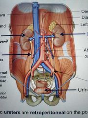
- crosses above psoas major - crosses over bifurcation of common iliac artery - enters posterior aspect of bladder Follows tips of lumbar spinous processes |
|
|
The kidneys are found between which spinal levels? Which one is slightly lower and why? |
Between T11/12 and L2/3. Right kidney is slightly lower due to the prescence of liver |
|
|
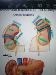
Draw a diagram showing the relationships of the kidneys to its anterior structures |
|
|
|
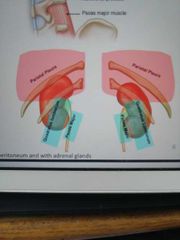
Draw a diagram showing the relationships of the kidneys to their posterior structures |
|
|
|
What is the difference between perinephric and paranephric fat? What separates the two? |
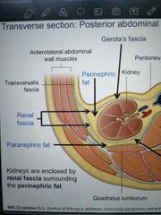
Perinephric fat immediately surrounds the kidney, followed by renal fascia, followed by paranephric fat |
|
|
Which fascia encloses the kidneys on the anterior surface? The posterior surface? |
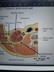
Gerota's fascia anteriorly, Zuckerkandl's fascia posteriorly |
|
|
What is nephroptosis? |
A fallen or dropped kidney |
|
|
What is a perinephric abscess? |
An infection of perinephric fat |
|
|
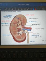
Draw a diagram of a cross section of the kidney |
|
|
|
Describe the path of urine flow through the kidney |
From the renal pyramid (medulla), minor calyx, major calyx, renal pelvis, ureter |
|
|
The IVC lies to the left or right of the descending aorta? |
Right |
|
|
What is an ARA? |
Accessory renal artery, an extra artery supplying the kidney as well as the renal artery. May or may not present a clinical problem |
|
|
The left renal vein passes underneath which structure? |
The superior mesenteric artery |
|
|
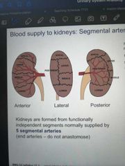
Draw a diagram of the segmental arrangement of the kidneys |
|
|
|
How many segmental arteries are there in each kidney? Do they interact? |
5 - one for each segment No, they are end arteries |
|
|
What is a pelvic kidney? What is the incidence of a pelvic kidney? |
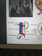
A kidney located within the pelvic cavity, having failed to ascend past the arterial fork formed by the umbilical arteries. 1 in 2000 |
|
|
What is a horseshoe kidney? What is its prevalence? |
A fusion of the two kidneys at the inferior poles, forming a horseshoe shape - 1 in 500-600 |
|
|
Which structure does the ureter develop from? |
Ureteric bud of mesonephric duct |
|
|
Name the arteries supplying the ureters |
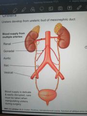
Renal Gonadal Aortic Iliac Vesical |
|
|
What is a bifid ureter? |
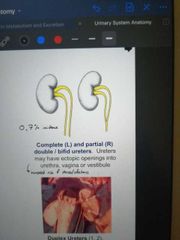
A divided ureter, either complete or partial |
|
|
What are the 3 main constriction points of the ureter? |
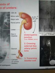
- ureteropelvic Junction - crossing of external iliac artery - traversing of bladder Wall |
|
|
What is a calculus? |
A kidney stone |
|
|
If a calculus is present in the kidney, where will the pain be referred to? What about in the ureter? |
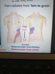
From the kidney - T10 From the ureter - T11 - L2 |
|
|
What is the trigone? |
A smooth, triangular portion at the base of the bladder that contains both entry points for the ureters and the exit point for the urethra |
|
|
What is the bladder muscle also called? |
Detrusor muscle |
|
|
What causes contraction of the detrusor muscle? |
Parasympathetic innervation from S2, 3, 4 |
|
|
What causes the reflex relaxation of the external urethral sphincter? |
Parasympathetic innervation from S2, 3, 4 |
|
|
What is micturition? |
Passing urine |
|
|
What causes relaxation of the detrusor muscle? |
Sympathetic innervation from the hypogastric nerve |
|
|
What causes contraction of the internal urethral sphincter? |
Sympathetic innervation |
|
|
What causes contraction of the external urethral sphincter? |
Somatic innervation from the S2-4 pudendal nerve |
|
|
What are the 3 parts of the male urethra? How long are each of them? |
Prostatic (3cm), membranous (2cm), penile (15cm) |
|
|
Where is the internal urethral sphincter located? |
At the base of the bladder |
|
|
Where is the external urethral sphincter located? |
Around the membranous urethra, upon exit from the peritoneal cavity |
|
|
When inserting a catheter, what regions of the male urethra do you need to be aware of and why? |
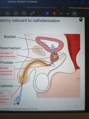
1. External urethral meatus, because it is the narrowest part of the urethra 2. Change in angle of urethra, because there is an unprotected portion of the urethral wall which may be at risk of rupture 3. Membranous urethra, because it is the 2nd narrowest part of the urethra and the least distensible |
|
|
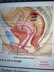
How long is the female urethra? |
3 - 4cm |
|
|
What is the vesicouterine pouch? |
The fold of the peritoneum between the superior bladder and the Inferior uterus |