![]()
![]()
![]()
Use LEFT and RIGHT arrow keys to navigate between flashcards;
Use UP and DOWN arrow keys to flip the card;
H to show hint;
A reads text to speech;
63 Cards in this Set
- Front
- Back
|
What are the two arches at the back of the throat? Why do they form? |
Palatoglossal arch anteriorly Palatopharyngeal arch posteriorly They form because the muscles for which they are named run along those arches |
|
|
What can be found in between the two arches of the throat? |
Palatine tonsils |
|
|
What are the 3 functions of the uvula? |
- To help block off the Nasopharynx upon swallowing - to aid in mucus Secretion - sensory fibres to initiate the gag reflex |
|
|
What are epiglottic valleculae? Why are they clinically important? |
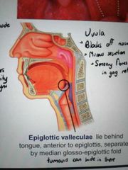
Two small pouches between the posterior surface of the tongue and the epiglottis. Tumours can hide in there and be very difficult to see |
|
|
What marks the boundary between the oral cavity and the Oropharynx? |
The Palatopharyngeal arch |
|
|
What are the superior and Inferior labial frenulums? Why are they clinically important? |
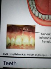
Small tags of connective tissue above and below your teeth. If they are missing it is a sign of Ehlers Danlos Syndrome |
|
|
How many teeth do adults have? How many of each type are there? |
32 - per quadrant there are 2 incisors, 1 canine, 2 premolars, 3 molars |
|
|
How many deciduous teeth do children have? How many of each are there? |
20 - per quadrant, 2 incisors, 1 canine, 2 molars |
|
|
What is a solitary maxillary median incisor? What is it a sign of? |
A single central incisor in the upper row of teeth, indicating possible mutations in the Shh gene responsible for bilateral symmetry |
|
|
What is the lingual frenulum and what is ankyloglossia? |
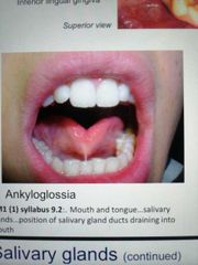
The tag of tissue that anchors your tongue to the floor of your mouth. Ankyloglossia is when this tag is too strong and restricts your tongue movement, leading to speech and swallowing problems |
|
|
What are sublingual caruncles? |
Bulges on the lingual frenulum that contain openings for the submandibular duct |
|
|
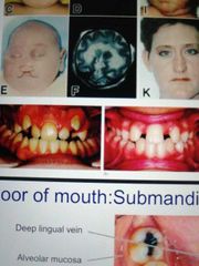
Where do sublingual ducts open in the mouth? |
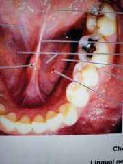
On the sublingual fold - red spot in image is a good example |
|
|
Where is the sublingual gland located? |
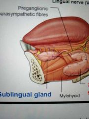
Inferior to the anterior portion of the tongue |
|
|
Where are the submandibular glands located? Why does it have two parts? |
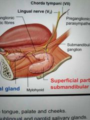
Underneath the posterior part of the tongue. It wraps around the mylohyoid muscle. The part that lies above is the deep part, that which lies below is the superficial part |
|
|
What are the 3 salivary glands? |
Sublingual, submandibular, parotid |
|
|
How are the sublingual and submandibular glands innervated? |
Facial nerve CN7 |
|
|
Describe the location of the parotid gland |
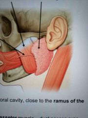
Outside the oral cavity, close to the ramus of the mandible |
|
|
How is the parotid gland innervated? |
Glossopharyngeal nerve CN9 |
|
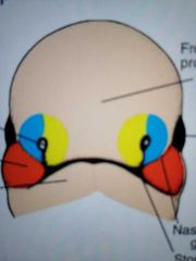
Describe the coloured regions in the developing face |
Red = maxillary prominence Blue = lateral nasal prominence Yellow = medial nasal prominence Bottom peach = mandibular prominence Top peach = frontonasal prominence Eyes are on the side |
|
|
Describe how the face develops over weeks 5 to 10 |
The nasal pits move closer together and the medial nasal prominences fuse around week 7. The maxillary prominences extend upwards to form the cheeks. The eye sockets move medially |
|
|
How does the hard palate develop? |
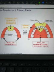
Initially, maxillary processes are linked by the medial nasal prominence. This grows posteriorly while the mandibular prominences grow medially to fuse, giving a triangular primary palate which will contain the 4 incisors, and the secondary palate that contains all the other teeth |
|
|
What is the philtrum of the lip? |
The depression in the middle of the top lip |
|
|
When does the secondary palate fuse? |
Around 10 weeks |
|
|
What can result from a failure of the palatine shelves to fuse? |
Cleft lip/palate |
|
|
What are the 4 extrinsic tongue muscles? |
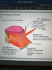
Palatoglossus (forms anterior arch) Styloglossus Hyoglossus Genioglossus |
|
|
How is genioglossus innervated and what does it do? |
Hypoglossal (CN12) Protrudes tongue |
|
|
How is Hyoglossus innervated and what does it do? |
Hypoglossal CN12 depresses and retracts tongue |
|
|
How is Styloglossus innervated and what does it do? |
Hypoglossal (CN12) retracts and elevates tongue |
|
|
How is palatoglossus innervated and what does it do? |
Vagus (odd muscle out) Elevates tongue |
|
|
What cranial nerve provides motor innervation to the entire tongue? |
Hypoglossal (CN12) |
|
|
How is the tongue innervated with sensory fibres? |
Ant 2/3rds: General sensation by Trigeminal V3 (lingual) Taste buds by facial nerve CN7
Pos 1/3: Sensation and taste by glossopharyngeal CN9
Small region at back of tongue: Sensation and taste by vagus CN10 |
|
|
What are the 4 movements of the mandible? |
Protrusion Depression Retraction Elevation |
|
|
What is special about the temporomandibular joint? |
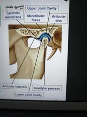
It has a double synovial joint |
|
|
Which two bones form the temporomandibular joint? |
Condylar process of mandible Mandibular fossa of temporal bone |
|
|
Which movements occur at the upper temporomandibular capsule? |
Protrusion/retraction |
|
|
Which movements occur at the lower temporomandibular capsule? |
Hinge movements of elevation and depression |
|
|
What are the four major mastication muscles? |
Temporalis Masseter Lateral pterygoid Medial pterygoid |
|
|
How are all 4 mastication muscles innervated? |
Mandibular division of Trigeminal nerve CN5 |
|
|
What is the function of temporalis? |
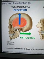
To elevate and retract the mandible |
|
|
What is the purpose of the Masseter muscle? What are its two parts? |
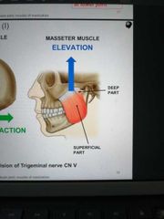
To elevate the mandible Superficial and deep |
|
|
What is the function of the lateral pterygoid muscles? How many parts does it have? |
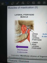
Protrusion of mandible Upper and lower head |
|
|
What is the function of medial pterygoid? How many parts does it have? |
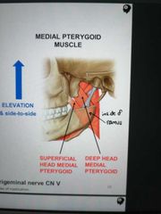
Elevation of mandible Superficial and deep parr |
|
|
What are the borders of the Nasopharynx? |
Choanae Lower border of soft palate |
|
|
How are muscles of the Nasopharynx innervated? |
CN5b and CN9 |
|
|
What are the borders of the Oropharynx? |
Lower border of soft palate Superior border of epiglottis Palatopharyngeal arch anteriorly |
|
|
What is the innervation of the Oropharynx? |
CN 9 |
|
|
What are the borders of the Laryngopharynx? |
Superior border of epiglottis Cricopharyngeus |
|
|
What is the innervation of the laryngopharynx? |
CN 9 and 10 |
|
|
What two muscles make up the soft palate? How are they innervated? |
Tensor veli palatini - CN5 Levator veli palatini - CN10 |
|
|
What are the 3 external pharyngeal muscles? How are they arranged? |
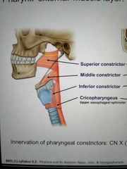
Superior, middle and inferior pharyngeal constrictors. Arranged like nested cups, one inside the other |
|
|
What is the midline raphe? |
A band of connective tissue running down the posterior pharynx that provides a point of attachment for the external pharyngeal muscles |
|
|
How are the external pharyngeal muscles innervated? |
CN10 |
|
|
Which 3 muscles make up the internal pharyngeal wall? |
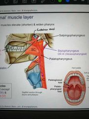
Salpingopharyngeus Stylopharyngeus Palatopharyngeus (forms an oral cavity arch) |
|
|
Where is the origin of the salpingopharyngeus muscle? How is it innervated? |
Eustachian canal CN10 |
|
|
Where is the origin of Stylopharyngeus? How is it innervated? |
Styloid process of temporal bone CN9 |
|
|
What is the origin of Palatopharyngeus? How is it innervated? |
Soft palate CN10 |
|
|
What is the purpose of the external pharyngeal muscles? |
To constrict the pharynx |
|
|
What is the purpose of the internal pharyngeal muscles? |
To shorten and widen the pharynx |
|
|
What is the technical term for swallowing? |
Deglutition |
|
|
Describe the 1st phase of deglutition |
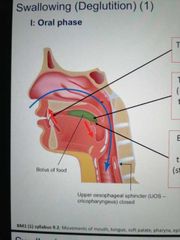
Oral phase
- tip of tongue is pressed against hard palate - palatal arches open and posterior tongue is depressed - bolus moves backwards onto post 1/3 of tongue, triggering swallowing reflex |
|
|
Describe the 2nd phase of deglutition |
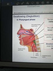
Pharyngeal phase - tongue pulled up and back forcing bolus into oropharynx. - soft palate drawn up - elevation of hyoid and larynx causes closure of the epiglottis as it pushes against food - bolus reaches superior pharyngeal constrictor which pushes it down |
|
|
Describe the 3rd phase of deglutition |
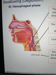
- swallowing reflex causes external pharyngeals to relax, then contract - bolus is forced through the cricopharyngeus into the oesophagus - soft palate relaxes - hyoid and larynx depress back to normal and epiglottis flips back up |
|
|
All internal pharyngeal muscles are innervated by the... nerve, except... and... which are innervated by... and... nerves |
- vagus (X) - stylopharyngeus - tensor veli palatini - glossopharyngeal (IX) - trigeminal (V) |