![]()
![]()
![]()
Use LEFT and RIGHT arrow keys to navigate between flashcards;
Use UP and DOWN arrow keys to flip the card;
H to show hint;
A reads text to speech;
135 Cards in this Set
- Front
- Back
|
This level of the nervous system is composed of the brain and spinal cord. |
Central Nervous System (CNS)
|
|
|
This level of the nervous system is composed of 12 pairs of cranial nerves and 31 pairs of spinal nerves. Divided into Somatic and Autonomic. (Autonomic is further divided into sympathetic and parasympathetic). |
Peripheral Nervous System (PNS) |
|
|
A collection of neurons within the brain or spinal cord and therefore, within the central nervous system. |
Nucleus |
|
|
Structures which are composed of thousands of neurons and are located outside of the central nervous system. |
Ganglia |
|
In a quadruped or in a biped below the diencephalic flexure, this is the term meaning towards the back (spine). |
Dorsal or Posterior |
|

In a quadruped or in a biped below the diencephalic flexure, this is the term meaning towards the front (abdomen). |
Ventral or Anterior |
|
In a quadruped or in a biped below the diencephalic flexure, this is the term meaning towards the head. |
Rostral |
|
In a quadruped or in a biped below the diencephalic flexure, this is the term meaning towards the rear (tail). |
Caudal |
|
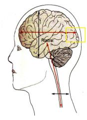
In a biped above the diencephalic flexure, this the the term meaning towards the back of the skull. |
Caudal |
|
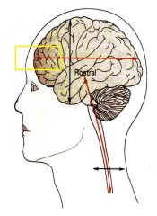
In a biped above the diencephalic flexure, this the the term meaning towards the front (eyes). |
Rostral |
|
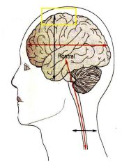
In a biped above the diencephalic flexure, this the the term meaning towards the top of the skull. |
Dorsal |
|

In a biped above the diencephalic flexure, this the the term meaning towards the feet. |
Ventral |
|
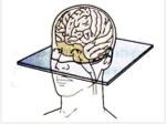
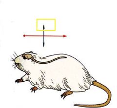
Name this plane. |
Transverse or Horizontal |
|
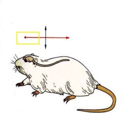
Name this plane. |
Sagittal |
|
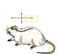
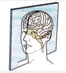
Name this plane. |
Coronal |
|
Name this major subdivision of the brain. |
Cerebellum |
|

Name this major subdivision of the brain. |
Medulla |
|
Name this major subdivision of the brain. |
Pons ("Bridge") |
|
Name this major subdivision of the brain. |
Midbrain |
|
Name this major subdivision of the brain which includes the thalamus and hypothalamus. |
Diencephalon |
|
Name this major subdivision of the brain which includes the cerebral cortex and subcortical nuclei. |
Telencephalon or Forebrain |
|
|
"Bumps" on the brain which usually indicate the location of underlying nuclei or fiber tracts. |
Gyri (or Hillocks, Tubercles, Colliculi, Peduncles, Brachium, Eminence) |
|
|
"Grooves" on the brain which divide brain into different regions or represent a midline division of the brain. |
Sulci or Fissures |
|
|
Name of the three meninges that surround the brain and spinal cord. |
Dura mater, arachnoid, and pia mater. |
|
|
Two of the specializations arising from the pia mater. |
Denticulate ligaments and filum terminale. |
|
|
The outer most meningeal layer. This layer is dense, thick, hard and largely inflexible. |
Dura mater (hard mother) |
|
|
The middle meningeal layer. It is composed of a smooth outer surface which is fused to the dura and meshwork which extends to the final layer. |
Arachnoid |
|
|
The space between the arachnoid and the pia mater. This area contains cerebrospinal fluid (CSF). It has projections of arachnoid which run throughout it and attach to the pia mater forming a meshwork. |
Subarachnoid Space |
|
|
The innermost meningeal layer which is intimately fused to the cord along the length and which forms the filum terminale. |
Pia mater (soft mother) |
|
Projections of pia mater found at all levels of the spinal cord which extend from the spinal cord to the inner surface of the dura mater. |
Denticulate ligaments. |
|
|
Name the vertebrae where the spinal cord ends. |
Around L2-L3 |
|
|
Name the vertebrae where dura and arachnoid fuse. |
Around S2 |
|
|
How many vertebrae are there in the spinal column? |
32. 7 Cervical, 12 thoracic, 5 lumbar, 5 sacral (fused), and 3 coccygeal (fused). |
|
|
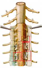
This is the site where nerves going out to the muscles or carrying sensory information into the spinal cord emerge from the spinal cord. (ie. a joint between two vertebrae). |
Intervertebral Foramen |
|
|
This constitutes the distal end of the spinal cord at the level of L2/L3. Here, the meninges continue caudually creating a resevoir of CSF until S2 where they fuse into the filum terminale. |
Conus medullaris |
|
|
This is the extension of pia mater beyond the conus medullaris. It runs through a resevoir of CSF and terminates at the coccygeal ligament. |
Filum terminale |
|
|
A collection of nerve roots that emerge from the spinal cord at levels below L2 and extend beyond the conus medullaris. They form a collection of fine filaments distal to the end of the spinal cord. |
Caudas equina |
|
|
This test, which is carried out to obtain samples of CSF for testing or for injection of anesthesia into the CSF for a spinal block, is done below L3. Why? |
It avoids the possibility of hitting the spinal cord while still gaining access to the CSF in the conus medullaris. |
|
|
This type of stain serves to identify neuronal cell bodies (grey matter). |
Nissl Stain |
|
|
This type of stain serves to identify myelinated axons (white matter). |
Fiber Stain |
|
|
This type of spinal root allows for sensory (afferent) input to enter the spinal cord. It is the only type of root that has a ganglia. |
Dorsal Root |
|
|
This type of spinal root allows for motor (efferent) output to exit the spinal cord. All neurons are within the spinal cord (CNS), therefore, this type of root doesn't have a ganglia. |
Ventral Root |
|
|
A collection of neurons without dendrites (input/afferent only) and instead have two axons, which arise in the periphery (outside of the CNS) between a spinal nerve and a dorsal root. |
Dorsal Root Ganglion (DRG) |
|
|
Dorsal and ventral roots come together in the periphery to form these. Therefore, they are composed of both sensory and motor axons. |
Spinal Nerves |
|
|
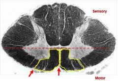
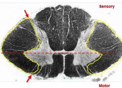
The central area of the spinal cord has a butterfly shape and is divided into the dorsal and ventral horn. It composed of which type of matter? |
Grey Matter (Neurons and Glia) |
|
|
The peripheral area of the spinal cord around the "butterfly" is divided into dorsal, lateral, and ventral columns. It is composed of which type of matter? |
White Matter (Axons and Glia) |
|
|
Neurons in this horn of the spinal cord process sensory input (afferent). |
Dorsal Horn |
|
|
Neurons in this horn of the spinal cord process motor output (efferent). |
Ventral Horn |
|
|
A bundle of axons that connects two CNS regions. Can be ascending (spinal cord to brain) or descending (brain to spinal cord). |
Tract |
|
|
A bundle of anatomically defined fibers that subserve a common function; aka tract. |
Fasciculus (Multiple) |
|
|
An area containing multiple tracts. |
Funiculus (Plural) |
|
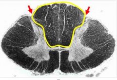
This collection of tracts contains LARGE diameter sensory axons of DRG neurons. It generally conducts afferent sensory information from the periphery. |
Dorsal White Column or Dorsal Funiculus |
|
This collection of tracts contains mainly descending axons from the brainstem to the spinal cord. It generally conducts efferent motor output to the periphery. |
Ventral White Column or Ventral Funiculus |
|
This collection of tracts contains both ascending sensory axon tracts (pain, temperature, cerebellar) and descending motor axon tracts from brain (cortex, midbrain). |
Lateral White Column or Lateral Funiculus |
|
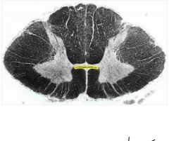
This collection of tracts contains axons crossing from one side of the spinal cord to the other. |
Anterior White Commissure |
|
|
The subdivision of the peripheral nervous system that is responsible for carrying motor and sensory information both to and from the CNS. This system is made up of nerves that connect skin, sensory organs, and all skeletal muscles. It is responsible for producing contractions of skeletal (voluntary) muscle as well as processing sensory information that arrives via external stimuli (eg, hearing, touch, and sight).
|
Somatic Nervous System (Voluntary Peripheral Control and Sensation)
|
|
|
|
Autonomic Nervous System (Involuntary Peripheral Control and Sensation)
|
|
|
One of the two functionally, chemically, and anatomically distinct subdivisions of the autonomic nervous system. Under the classic view of these systems, it serves to prepare the body for an emergency.
|
Sympathetic Nervous System |
|
|
One of the two functionally, chemically, and anatomically distinct subdivisions of the autonomic nervous system. Under the classic view of these systems, it serves to conserve and restore energy and maintain homeostasis during non-emergent situations. |
Parasympathetic Nervous System
|
|
|
The new view of these two subdivisions of the autonomic nervous system is that they are CONSTANTLY working to regulate the activity of involuntary structures. (Not one or the other depending on scenario).
|
Parasympathetic and Sympathetic Nervous Systems |
|
|
In this subdivision of the peripheral nervous system, motor neurons in the spinal cord project DIRECTLY to the skeletal muscles they innervate.
|
Somatic Nervous System |
|
|
In this subdivision of the peripheral nervous system, motor neurons in the spinal cord project to and synapse on another neuron located in a ganglion in the periphery. This neuron then innervates the target structure. (ie. Requires two neurons for signal transmission) |
Autonomic Nervous System |
|
|
This is the first of two neurons required for signal transmission in the autonomic nervous system. It is ALWAYS located in the CNS. It projects onto the second neuron.
|
Preganglionic Neuron |
|
|
This is the second of two neurons required for signal transmission in the autonomic nervous system. It is ALWAYS located in a ganglia (not a DRG). It receives information from the first neuron and projects onto the target structure. Shorter in the parasympathetic nervous system. |
Postganglionic Neuron |
|
|
Preganglionic neurons in this subdivision of the autonomic nervous system are located in the thoracic and upper lumbar part of the spinal cord (thoracolumbar). Ie. between T1 and L2. They all originate in the spinal cord and exit via ventral roots.
|
Sympathetic Preganglionic Neurons
|
|
|
Preganglionic neurons in this subdivision of the autonomic nervous system are located in the brainstem and sacral part of the spinal cord (craniosacral). Cranio portion- cranial nerves, sacral portion- "low" organs.
|
Parasympathetic Preganglionic Neurons |
|
|
This is the location of the majority of the ganglia of the sympathetic nervous system. It is located adjacent to the vertebral column and extends to the base of the coccyx. Here the ganglia are attached to adjacent spinal cords by Rami Communicante
|
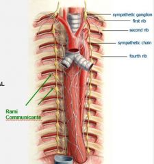
Sympathetic Chain |
|
|
This is the location of the minority of the ganglia of the sympathetic nervous system. These are called collateral (prevertebral) ganglia.
|
Ventral (in front of) the aorta
|
|
|
The connective tissue that surrounds individual axons in a spinal nerve.
|
Endoneurium
|
|
|
The connective tissue sheath that surrounds multiple bundles of endoneurium-coated axons in a spinal nerve.
|
Perineurium
|
|
|
The common thick external sheath that coats an entire spinal nerve.
|
Epineurium |
|
|
The term for axons that conduct information which is leaving the CNS (output). These cell bodies are located within the spinal cord. |
Efferent (Motor)
|
|
|
The term for axons that conduct information towards the CNS (input). These cell bodies are located in dorsal root ganglion.
|
|
|
|
True or False: a single spinal nerve can be composed of many morphologically and anatomically distinct types of axons. (ex. big/small diameter, myelinated/unmyelinated. afferent/efferent). |
TRUE |
|
|
Outgoing axons that carry motor commands to skeletal muscles.
|
General Somatic Efferent Axons (GSE) |
|
|
Outgoing axons from the autonomic nervous system that carry outgoing motor commands that innervate smooth muscle, cardiac muscles, and glandular tissue.
|
General Visceral Efferent Axons (GVE) |
|
|
Axons that transmit sensory information (touch, pressure, pain, temperature, muscle tension) from the body to the spinal cord.
|
General Somatic Afferent Axons (GSA) |
|
|
|
General Visceral Afferent Axons (GVA) |
|
|
This type of nerve can carry strictly motor or strictly sensory or both types of information. It is NOT formed from dorsal or ventral roots because they originate in the brainstem. Neurons carrying afferent information in these nerves are located in periphery ganglia analogous to DRG but not DRG. Efferent neurons are located in a nucleus somewhere in the brainstem. Components include: GSA, GVE, GVA, GSE, SSA, and SVE.
|
Cranial Nerve (12 total)
|
|
|
This type of nerve carries both motor and sensory information. It is formed from dorsal and ventral roots in the spinal cord. Neurons carrying afferent information in these nerves are located in DRG. Efferent neurons are located in the spinal cord. Components include: GSA, GVE, GVA, and GSE.
|
Spinal Nerve (31 total) |
|
|
This type of afferent axon is unique to a cranial nerve and carries sensory information unique to the head (vision, hearing, balance, taste, and etc.)
|
Special Somatic Afferent (SSA) |
|
|
This type of efferent axon is unique to a cranial nerve and carries motor information unique to the voluntary muscles which have a different embryological origin. |
Special Visceral Efferent (SVE)
|
|
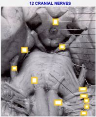
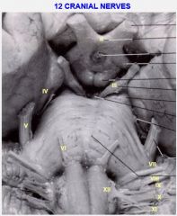
Locate all 11 cranial nerves (vagus nerve not shown).
|
|
|
|
A type of visceral efferent that innervates a subset of skeletal muscles that have a unique embryological origin. These skeletal muscles are the muscles involved in movement of the facial muscles, jaws, pharynx, and larynx. Cranial nerves V, VII, IX, and X innervate muscles derived from these arches. |
Branchial Motor [Embryological origin: Branchial (aka phayrngeal) arches] |
|
|
If a cranial nerve has a motor component (somatic or visceral) the neuronal cell bodies will be located here. |
A nucleus located inside the CNS (ie, brain) |
|
|
If a cranial nerve has a sensory component (general or special) its neuronal cell bodies will be located here. |
A ganglion outside of the CNS which is analagous to, but not, a DRG. |
|
|
This cranial nerve carries special sensory information for the sense of smell. Its sensory ganglion are located in the olfactory epithelium on the roof of the nasal cavity which project to the oflactory bulb and tract. |
I- Olfactory Nerve |
|
|
This cranial nerve carries special sensory information for vision. Its sensory ganglion are located in retinal ganglion cells in the ganglion cell layer of the retina which project through the optic chiasm into the brain. |
II- Optic Nerve |
|
|
This cranial nerve carries both somatic motor and visceral motor efferents. Its somatic motor components has cells of origin the brainstem which target muscles that move the eye (except the lateral rectus and superior oblique). Its visceral motor post ganglionic neuron is the ciliary ganglion (orbit) which targets constrictor pupillae and the ciliary muscle. |
III- Oculomotor Nerve |
|
|
This cranial nerve carries somatic motor efferents from cells of origin within the brainstem to targets in the superior obliqe muscle within the orbit. |
IV- Trochlear Nerve |
|
|
This cranial nerve carries branchial motor efferents and general sensory afferents. The branchial motor components originate in the brainstem and target to the muscles of mastication. The GSA components originate in the semilunar (trigeminal) ganglion in the middle cranial fossa and provide general sensory information for most of the head (face, nasal cavity, tongue, teeth).
|
V- Trigeminal |
|
|
This cranial nerve carries somatic motor efferents. It originates in the brainstem and the lateral rectus muscle in the orbit.
|
VI- Abducens |
|
|
This cranial nerve carries branchial motor, visceral motor, general sensory, and special sensory information. The branchial motor component targets to the muscles of facial expression. The visceral motor component comes from pterygopalatine ganglion and submandibular ganglion to target the lacrimal and salivary glands, respectively. The general sensory comes from geniculate ganglion in the temporal bone to provide information from the area just behind and below the external auditory canal. The Special sensory relays information about taste from the anterior 2/3 of the tongue and soft palate.
|
VII- Facial |
|
|
This cranial nerve carries special sensory afferents from spiral, cochlear and vestibular ganglia in the cochlea and inner ear. It relays information about hearing and balance.
|
VIII- Vesibulocochlear |
|
|
This cranial nerve carries branchial motor, visceral motor, general sensory, and special sensory information. The branchial component targets to the stylopharyngeus in the pharnx. The visceral motor component has a postsynaptic ganglion called otic ganglion which target to parotid salivary gland. The general sensory component have ganglion in the superior/inferior of jugular foramen which innervate phayrnx, tongue, oral cavity, and middle ear. The special sensory have ganglia in the same place which relay taste information from the posterior 1/3 of the tongue. |
IX- Glossopharyngeal |
|
|
This cranial nerve carries branchial motor, cisveral motor, general sensory, and special sensory information. The branchial motor targets the muscles of the pharynx and larynx. The visceral motor has postganglionic neruons in or near the target organs (viscera in thorax and abdomen). The sensory component relays general sensory from larynx and external auditory meatus and special sensory from uvula (taste) from superior/inferior ganglia in jugular foramen. |
X- Vagus |
|
|
This cranial nerve relays branchial motor efferents from the brainstem to target the sternocleidomastoid and trapezius muscles (neck).
|
XI- Accessory |
|
|
This cranial nerve relays somatic motor efferents from the brainstem to target tongue muscles.
|
XII- Hypoglossal |
|
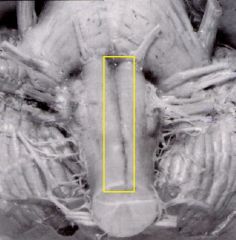
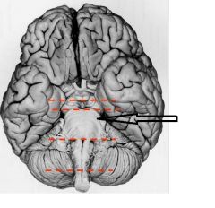
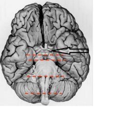
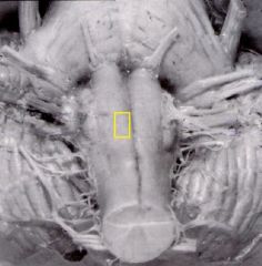
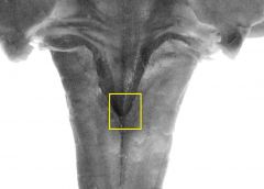
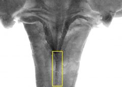
Identify this structure.
|
Anterior Median Fissure |
|
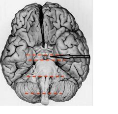
Identify this structure.
|
Pyramid
|
|

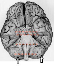
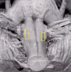
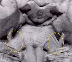
Identify these structures.
|
Olivary Eminence
|
|
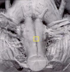
Identify these structures.
|
Preolivary Sulci
|
|
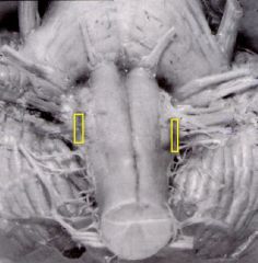
Identify these structures.
|
Postolivary sulci
|
|
Identify this structure.
|
Pyramidal decussation
|
|
Identify this structure.
|
4th Ventricle (sagittal view)
|
|
Identify this structure on the dorsal medulla (cerebellum removed).
|
Obex |
|
Identify this structure on the dorsal medulla (cerebellum removed).
|
Posterior Median Fissure
|
|
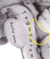
Identify this structure. |
Middle Cerebellar Peduncle (connects to ventral pons)
|
|
|
Fiber tracts that connect the cerebellum to the brainstem. They are made up of axons entering the cerebellum that originated in the spinal cord or other parts of the brinstem (eg., medulla, pons) and axons leaving the cerebellum which are destined for brainstem targets.
|
Cerebellar Peduncles |
|
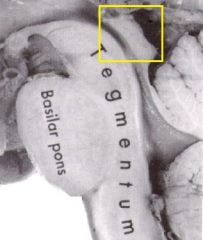
Identify this structure on the sagittal view of the brainstem. It is composed of the superior and inferior colliculi.
|
Tectum
|
|
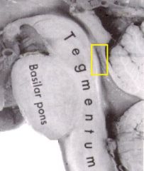
Identify this structure on the sagittal view of the brainstem.
|
Cerebral Aqueduct
|
|
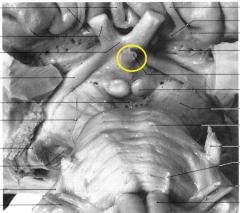
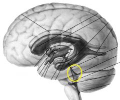
This is the stalk of the pituitary gland viewed from the ventral diencephalon. Provide its name.
|
Infindibulum |
|
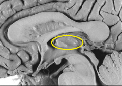
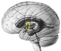
Identify this structure as seen from the sagittal view of the diencephalon.
|
Thalamus
|
|
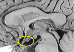
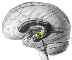
Identify this structure as seen from the sagittal view of the diencephalon.
|
Hypothalamus (below)
|
|
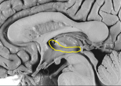
Identify this structure as seen from the sagittal view of the diencephalon.
|
|
|
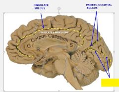
Identify this sulcus in the lateral cortex.
|
Lateral Sulcus |
|
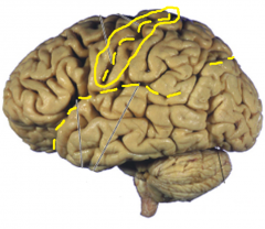
Identify this sulcus in the lateral cortex.
|
Central Sulcus
|
|
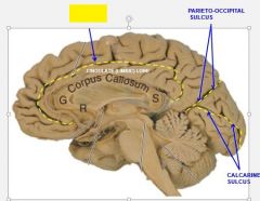
Identify this sulcus in the lateral cortex.
|
Parieto-Occipital Sulcus
|
|
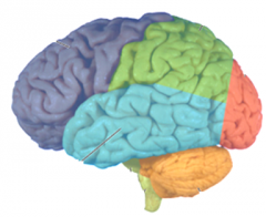
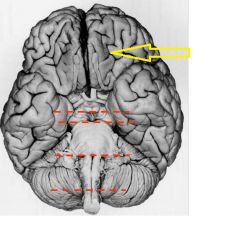
Name the dark blue lobe at the rostral end of the lateral cerebral cortex.
|
Frontal Lobe |
|

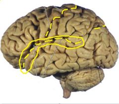
Name the green lobe at the dorsal end of the lateral cerebral cortex.
|
Parietal Lobe |
|

Name the light blue lobe at the ventral end of the lateral cerebral cortex.
|
Temporal Lobe |
|

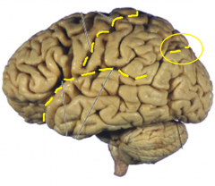
Name the red lobe at the caudal end of the lateral cerebral cortex.
|
Occipital Lobe |
|
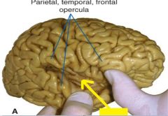
Name this cortex which is buried deep within the lateral sulcus and covered by gyri from the temporal, parietal, and frontal lobes.
|
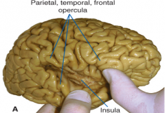
Insular Cortex (Insula) |
|
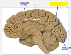
Name this sulcus seen in the medial view.
|
Parieto-occipital Sulcus |
|
Name this sulcus seen in the medial view.
|
Calcarine Sulcus
|
|
Name this sulcus seen in the medial view. |
Cingulate Sulcus |
|
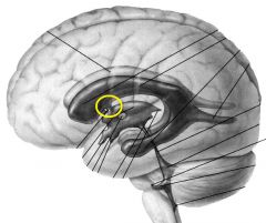
Name this component of the ventricular system.
|
Interventricular Foramen |
|
Name this ventricle. Seen from the medial view.
|
Fourth Ventricle |
|
Name this ventricle. Seen from the medial view.
|
Third Ventricle |
|
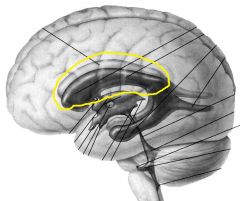
Name this ventricle. Seen from the medial view.
|
Lateral Ventricle
|
|
Name this component of the ventricular system.
|
Cerebral Aqueduct
|
|
|
This tissue arises from tufts of cells within the walls of ventricles. It serves to secrete CSF. (An active process which requires energy).
|
Choroid Plexus |
|
|
The names of the two openings in the fourth ventricle which allow CSF from outside of the brain to flow into the subarachnoid space. |
Foramen of Magendie and Foramen of Luschka
|
|
|
The name for specializations of the arachnoid layer which take up CSF and remove it from the ventricles to avoid excess buildup.
|
Arachnoid Granulations |
|
|
|
|