Reading...
![]()
Play button
![]()
Play button
![]()
Use LEFT and RIGHT arrow keys to navigate between flashcards;
Use UP and DOWN arrow keys to flip the card;
H to show hint;
A reads text to speech;
21 Cards in this Set
- Front
- Back
|
The two most important types of sensory organs within muscle, the parameter they measure and the fiber type that comprise them
|
Muscle spindle – measures the length and rate of change of muscle length, Type Ia fiber
Golgi tendon – measures the tension developed within a muscle, Type Ib fiber |
|
|
Two factors that influence conduction velocity of nerve fibers
|
Myelination and axonal diameter
|
|
|
The muscle spindle
• Three components • Their physiological roles (3) • What helps the spindle regulate muscle contraction? |
• 3 components
Intrafusal muscle fibers, a group of 2-12. Large diameter myelinated sensory axons that have receptors on the central/noncontractile regions of intrafusal fibers Small diameter myelinated motor axons that innervate the contractile regions of the intrafusal muscle fibers • Physiological roles - participate in the stretch reflex - relay proprioceptive information about the body to the CNS - regulate muscular contraction • Descending motor pathways and afferent feedback |
|
|
Three types of intrafusal fibers, how their nuclei are arranged and the parameter in which they detect
|
• Static nuclear bag fibers
- have many nuclei dispersed within a central “bag” region - respond to changes in muscle length • Dynamic nuclear bad fibers - have many nuclei dispersed within a central “bag” region - respond to changes in muscle length and rates of change in mucle length • Nuclear chain fibers - Have their nuclei arranged in a row - respond to changes in muscle length |
|
|
Discuss the afferent innervation of intrafusal fibers:
• What nerve fibers are innervating? • What types of intrafusal fibers do they innervate and where are their sensory endings found on these fibers? • How does the information they relay differ? |
Group Ia fibers iNN all three types of intrafusal fibers. Their sensory endings spiral around the central region of these fibers
Group II fibers iNN all static nuclear bag and nuclear chain fibers and some dynamic nuclear bag fibers. Their sensory endings are slightly distal to the central region of the intrafusal fibers. Unlike group Ia fibers, Group II only monitors static changes. |
|
|
Discuss the motor innervation of intrafusal fibers:
• What fiber is innervating? Include 2 characteristics of the fibers, their subtypes and what these subtypes innervate. • Where on the intrafusal fibers do the motor neurons iNN? |
The motor iNN is by Aγ-fibers. They are small and myelinated. Two subtypes are
- dynamic Aγ-motor neurons which iNN dynamic nuclear bag fibers - static Aγ-motor neurons which iNN static nuclear bag and chain fibers Aγ-motor neurons will iNN on the contractile portions of the fibers |
|
|
Motor innervation of extrafusal fibers:
• What nerve fiber is involved? Include two characteristics |
Aα-fibers which are large and myelinated.
|
|
|
Explain the differences between conduction velocities and fiber innervations of Aγ- and Aα- fibers?
|
Aα-fibers are larger than Aγ-fibers and have a higher conduction velocity. Aα-fibers iNN extrafusal muscle fibers whereas Aγ-fibers iNN intrafusal muscle fibers
|
|
|
Describe the role of the muscle spindle in the stretch reflex, starting from the activation of a tendon. Use the following terms:
• Ia afferents • homonymous, syngerist and antagonistic muscles • interneurons • reciprocal innervation |
Once the tendon has been mechanically stretched, the Ia afferent fibers propagate the APs towards their synapse where they excite Aα-fibers. Through excitation, Aα-fibers will influence the activity of two groups of muscles:
- they will cause the contract of the homonymous and synergistic muscles - In a process called reciprocal innervation, they will excite interneurons whose role is to inhibit other Aα-motor neurons from stimulating antagonistic muscles. |
|
|
Describe the activity of muscle spindles when its homonymous muscle participates in a sustained contraction.
|
The muscle spindles are continuously excited and its afferent fibers have sustained firing of APs.
|
|
|
Describe how muscle spindles can be an “unloaded” or “loaded” state depending on the neurons that are stimulated
|
The muscle spindle ends in an “unloaded” state when its corresponding homonymous muscle has its Aα-motor neurons stimulated. The stimulation causes the muscle to contract and the muscle spindle becomes slack at its ends, no longer being active.
In a “loaded” state for the muscle spindle, both the Aα-motor fibers innervating muscle and the Aγ-fibers innervating the muscle spindle are stimulated or co-activated. The muscle spindle is remains under a stimulated condition and can measure muscle length continuously. |
|
|
The Golgi Tendon Organ
• Where it’s located • What it measures • Describe the nerve that innervates it. What happens to the nerve when it enters the Golgi capsule? What kinds of structural fibers are inside the capsule? What happens to the nerve when the tendon is stretched? |
The Golgi Tendon Organ
• between the tendon and the extrafusal muscle fibers • measures muscle tension • A single Ib afferent fiber iNN the tendon organ. Once it enters the Golgi capsule, it loses its myelination and its free nerve endings interweave amongst the collagen fibers inside the capsule. When the tendon is stretched, cation channels on the nerve endings open, leading to the increase of AP firing of the Ib fiber. |
|
|
Describe the inverse myotactic reflex in which the Golgi tendon organ participates. Which nerve fiber is being inhibited? Where are the excitatory connections? What role do interneurons fulfill in the reflex?
|
In the inverse myotactic reflex, the Golgi tendon relaxes the homonymous and synergistic muscles but stimulate the antagonistic muscles. The principle nerve fiber of the Golgi tendon, Ib afferent, forms excitatory connections on interneurons. These interneuons will affect Aα-motor.
- excite Aα-motor neurons of the antagonistic muscle - inhibit Aα-motor neurons of the homonymous muscle |
|
|
Renshaw cell
• is categorized as this type of neuron • It’s source of input |
Renshaw cell
• is an inhibitory interneuron • receives input from re-current collateral axons of Aα-motor fibers |
|
|
Input to Motor Neurons (3)
|
• Muscle spindle afferents
• Local interneurons • Brainstem and cortical neurons |
|
|
The motor unit
• Its components (2) • This determines how many muscle fibers are in a motor unit • The location of the neuromuscular jxn • Explain the distribution of muscle fibers and the innervating axon |
The motor unit
• The α-motor neuron and the mextrafusal muscle fibers it innervates • The level of fine motor activity determines the number of muscle fibers – The more precise the activity, the less extrafusal muscle fibers within the unit • The NMJ is located in the middle of the muscle fiber • A motor axon distributes only one nerve ending branch to any one muscle fiber |
|
|
Two locations of motor nuclei
|
• Ventral horn of the spinal cord
• brain stem |
|
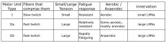
Name Three types of motor units and include
• The fibers that comprise them • The level of tension they produce (small/large) • Their response to fatigue • Whether they are aerobic/anaerobic • And describe the motor axons that innervate them |
|
|
|
The two crucial factors controlling muscle contraction
|
• The firing rate of the motor neuron – as firing rate increases, so does the force developed by the muscle
• The number of excited motor units recruited – the more recruits, the greater the force developed creases, so does the force developed by the muscle |
|
|
Hypotonia
• Def • two methods to detect • Damage to either one of these neurons can cause this • The condition can lead to this kind of paralysis |
Hypotonia
• Reduced muscle tone • Detected through palpation and resistance testing to passive stretch of the limb • Damage to either the Ia afferent pathway or the α-motor neuron * Note that either of these pathways can be spared and hypotonia can still result such as with damage to the CNS • can lead to flaccid paralysis |
|
|
LMN Syndrome
• State of the muscle fiber • These types of responses are damaged • 4 sx |
LMN Syndrome
• atrophy • Voluntary and reflexive responses • 4 sx - Hypotonia - Hyporeflexis - Fasciculations - Fibrillations |