Reading...
![]()
Play button
![]()
Play button
![]()
Use LEFT and RIGHT arrow keys to navigate between flashcards;
Use UP and DOWN arrow keys to flip the card;
H to show hint;
A reads text to speech;
90 Cards in this Set
- Front
- Back
- 3rd side (hint)
|
1, List commissural fibre tracts of the telencephalon
|
a) Corpus callosum
b) Anterior commissure c) Hippocampal commissure |
|
|
|
2, Name the associate fibre tracts of the telencephalon
|
a) cingulum fascicle
b) arcuate fascicle c) uncinate fascicle |
|
|
|
3, List the parts of the mesencephalon
|
a) Tectum - lamina quadrigemina (sup. & inf. colliculi)
b) Tegmentum - substantia nigra c) Base - the cerebral peduncles - below the substantia nigra’ |
|
|
|
4, List the parts that develop from the metencephalon
|
a) Pons
b) Cerebellum |
|
|
|
5, List the a) primary and b) secondary brain vesicles
|
a) Prosencephalon, mesencephalon, rhombencephalon
b) Telencephalon, diencephalon, mesencephalon, myencephalon, metencephalon, |
|
|
|
6, List the structures that form the anterior wall of the 3rd ventricle
|
a) Lamina terminalis
b) Lamina rostralis c) Anterior commissure d) Column of fornix |
|
|
|
7, Define the term “lamina choroidea” and Where is it located in the brain.
|
a) non differentiating parts of the wall of the brain vesicles that stay in the form of a simple columnar epithelium
b) medial wall of lateral ventricle, roof of 3rd ventricle, posterior part of the roof of the 4th ventricle |
|
|
|
8, Define the location and Brodmann’s number of a) the Broca’s speech area and b) primary auditory cortex
|
a) In the triangular & opercular parts of the inferior frontal gyrus. Brodmann Area’s 44 & 45
b) In the transverse temporal gyrus on the upper bank of the superior temporal gyrus. Brodmann Area’s 41 & 42 |
|
|
|
9, Where are the places of bony attachment of the a) falx cerebri & b) tentorium cerebelli
|
a) Crista galli, along the edges of the groove for the superior sagittal sinus, internal occipital protuberance.
b) Superior edge of the petrosal part of the occipital bone, along the groove for the transverse sinus. |
|
|
|
10, Where is the stria terminalis located
|
•Amygdala septum pellucidum
|
|
|
|
11, Where is the stria medullaris located
|
•Septum pellucium stria medullaris habenular n. fasciculus retroflexus interpeduncular nuclei spinal
cord & brainstem |
|
|
|
12, Where is the induceum griceum located
|
•Dentate gyrus fasciculus gyrus induceum griceum septum pellucidum
|
|
|
|
13, What is the origin and termination of the a) fornix, b) stria terminalis
|
a) Origin – Hippocampus, Termination – Mamillary body
b) Origin – Amygdaloid body, Termination – Medial hypothalamus, septum pellucidum |
|
|
|
14, List the histological layers of the neocortex from the outer surface to the white matter
|
•Molecular (plexiform) layer
•External granular layer •External pyramidal layer •Internal granular layer •Internal pyramidal layer •Multiform layer |
|
|
|
15, Name a) excitatory and b) inhibitory cell types in the cerebellar cortex
|
a) Pyramidal cells, horizontal cells, spiny stellate cells, spiny fusiform cells
b) Non-spiny stellate cells (basket cells, chandelier cells), non-spiny fusiform cells |
|
|
|
16, List five major cell types of the cerebellar cortex, indicate their excitatory or inhibitory character and their locations in the cortical layers
|
•Stellate cell - inhibitory - molecular layer
•Basket cell- inhibitory - molecular layer •Purkinje cell - inhibitory- Purkinje cell layer •Golgi cell - inhibitory - granular layer •Granular cells - excitatory - granular layer |
|
|
|
17, Name afferent pathways of the neostriatum, Name also the major neurotransmitters that are released by them
|
a) Fibres from the cerebral cortex - Corticospinal tract - glutamate
b) Fibres from the thalamus - Thalamostriatal tract - glutamate c) Fibres from the substantia nigra - Nigrostriatal tract - dopamine d) Fibres from the raphe nuclei - Raphe Dorsalis tract - serotonin |
|
|
|
18, Name 2 efferent pathways of the neostriatum, Name also the neurotransmitters that are released by the axon terminals of these pathways
|
a) Striatopallidal projections - GABA
b) Striatonigral projections - GABA |
|
|
|
19, Name the afferent fibre tracts of the cerebellar cortex that arise from the brainstem and terminate with a) mossy fibres
b) climbing fibres Describe also the method & location of how the mossy & climbing fibres terminate in the cerebellar cortex |
a) Pontocerebellar tract, vestibulocerebellar tract, reticulocerebellar tract
b) Olivocerebellar tract c) Mossy fibres: terminate in the stratum granulosum forming a wide arborization pattern and establishing synaptic contacts with dendrites of granule cells d) Climbing fibres: terminate in the stratum moleculare forming a narrow arborization field and establishing synaptic contacts usually with one Purkinje cell |
|
|
|
20, Name the neural elements that participate in the formation of the cerebellar glomeruli
|
Terminating mossy fibres make synaptic contact with granular cell dendrites.
|
|
|
|
21, List the cerebellar efferent pathways
|
a) Cerebellovestibular
b) Cerebelloreticular c) Cerebellothalamic (to VA, VL) d) Dentatorubral (red) |
|
|
|
22, Name the symptoms of cerebellar damage
|
a) Asynergia (no coordination)
b) Nystagmus c) Hypotonia d) Intentional tremor e) Cerebellar ataxia (walking) f) Dysarthria (speech) g) Dysmetria (inability to estimate distances) |
|
|
|
23, Name 3 non-pyramidal types of neurons in the cerebral cortex
|
a) Stellate cells
b) Fusiform cells c) Horizontal cells of Cajal |
|
|
|
24, What are the commissural pathways of the cerebrum
|
•Corpus callosum,
•Anterior commissure, •Hippocampal commissure (or commissura fornicis, or lira of David) |
|
|
|
25, Describe the origin of the neural crest, List the cell types developed from it.
|
•Develops from the neural fold (ectoderm)
•Located dorsolateral to neural tube a) Parts: during folding of the neural plate, these cells appear along the edge of the neural groove •Ectodermal origin •Forms a tempora intermediate zone between tube & surface ectoderm – laterally b) Cell types of the nervous system that develop from the neural crest a- Dorsal Root Ganglia, Sensory Ganglia b- Dorsal Sensory Root of Spinal Nerve & Dorsal Root Neurons c- Sympathetic Neuroblast d- Schwann cell e- Pigment cell f- Odontoblast g- Meninges h- Mesenchyme of Pharyngeal Arches |
|
|
|
26, Describe a) the ultrastructural composition of the Nissl-substance and b) the parts where they are present
|
a) RER in granular arrangement; This consists of endoplasmic reticulum, ribosomes & mRNA
b) found in the soma (cell body) & proximal dendritic processes •Used for basophilic protein synthesis •Motor neuron: Large Nissl body, Sensory neuron: Small Nissl body |
|
|
|
27, What is the difference between the axon hillock and initial axon segment? What are the features in which the
membrane covering the initial axon segment differs from the other parts of the cell membrane |
•Axon hillock – conically shaped region from which the axon extends
•The axon hillock contains parallel arrays of microtubules and is devoid of Nissl substance •Initial axon segment – 1st segment where the nerve impulse/AP is initiated •The initial segment has a bundle of microtubules, Ca channels & no Schwann cells |
|
|
|
28, List a) the types of all glial cells in the CNS and b) the embryonic germ layers they developed from
|
a) Astrocytes (fibrous & protoplasmic) – ectoderm
b) Oligodendrocytes – ectoderm c) Ependymal cells – ectoderm d) Microglia – mesoderm |
|
|
|
29, List the major functions of the glial cells of the CNS
|
•Structural support
•Nutritional support •Electrical insulator •Mechanical support •Uptake of neurotransmitters •Keep a constant electrical environment |
|
|
|
30, Make a drawing of a peripheral nerve
|

|
|
|
|
31, What is the histological structure and function of a perineurim
|
• Perineurium – The protective sheath surrounding a fascicle.
It is a supporting structure of peripheral nerve trunks. • 7-8 concentric layers of connective tissue and perineurial cells (epithelioid myofibroblasts) surround the fasicle. There are strong, tight intercellular junctions between the perineurial cells, that help insulate the underlying nerve fibres and form the major diffusion barrier within the nerve. |
|
|
|
32, List the layers that separate the lumen of the blood vessels from the nervous tissue in the CNS
|
Which layer forms the blood brain barrier.
a) Tight junctions between endothelial cells b) Basement membrane of endothelial cells c) Glial limiting membrane ( Glial Processes of Astrocytes) Primary element of BBB = tight junctions of endothelial cells |
|
|
|
33, What are the major differences between the fast and slow axonal transport mechanisms.
!! (3 SIDED CARD) !! |
Fast –
1. Bidirectional (anterograde & retrograde); 2. Transports macromolecules and cytoplasmic organelles Anterograde (kinesin), Retrograde (dynein) ; 3. 10-20 cm (~200 mm) per day ; 4. Uses proteins for transport; |
Slow –
1. Unidirectional (only anterograde) 2. Transports molecules that participate in the formation of the cytoskeleton ; 3. Few mm (~2 mm) per day 4. Transported molecules become part of the microtubules and are transported by them |
|
|
34, Name the macromolecules that play a substantial role in the fast axonal transport mechanism
|
a) Kinesin
b) Dynein |
|
|
|
35, Define the term “amic polarity”, List the cell compartments that can be distinguished from each other in the sense of histodynamic polarity
|
Compartments of nerve cells that can be distinguished from each other; Neurons are functionally polarized. Different parts of the
neuron are specialized for different functions; a) Part for detection of stimuli b) Transform stimuli into electrical signals c) Conduction d) Generation of AP e) Transmission of signal from cell to cell ; neuron is polarized 1) Receptive segment a) PNS – peripheral part of axon b) CNS – somatodendritic part of axon 2) Conductive segment 3) Initial segment |
|
|
|
36, List three types of neurotransmitters
|
a) Amino acids (glutamate, aspartate, GABA)
b) Biogenic amine (ACh, noradrenalin, adrenalin, dopamine, serotonin) c) Peptide (substance P, VIP, vasopressin) |
|
|
|
37, Define the term ionotropic neurotransmitter, List at least 3 ionotropic neurotransmitter receptors
|
a) Ionotropic NT – the NT receptor is directly related to an ion channel; Conformational changes of the receptor will result
in opening of the channel directly; b) nACh-R, GABAa-R, Gly-R, Glutamate-R, NMDA-R, non-NMDA-R (AMPA, kainate) |
|
|
|
38, Define the term metabotropic neurotransmitter, List at least 3 metabotropic neurotransmitter receptors
|
a) Metabotropic NT – NT receptors that are not coupled directly to ion channels; They activate G-proteins and through
this they induce various metabolic processes that result in ion exchange between the extracellular & intracellular compartments; b) GABAb-R, mGlu-R, 5-HT-R, Dopamine-R, mACh-r |
|
|
|
39, What is the role of synapsin in the process of chemical neurotransmission
|
The synapsin I protein acts as a glue between the synaptic vesicle & microfilaments; It is responsible for anchoring the
vesicle to actin filaments in the reserve zone; When Ca2+ enters, it binds to Ca2+-Calmodulin-dependent kinases (CAMK-II); The CAMK-II will phosphorylate both the synapsin I and the myosin light chain kinase causing the release of the vesicle from the microfilament and ATP-driven movement of the vesicle towards the active zone; |
|
|
|
40, Name 3 macromolecules that are integral parts of the membrane of synaptic vesicles
|
a) Synaptobrevin, synaptogyrin
b) Synaptotagmin c) Synaptophysin d) Various transporter molecules |
|
|
|
41, Make a drawing of synapse and label relevant structures
|
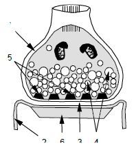
1) Axon terminal or presynaptic element
2) Postsynaptic element 3) Synaptic cleft 4) Synaptic vesicles 5) Presynaptic membrane specialization 6) Postsynaptic membrane specialization |
|
|
|
42) Electrical synapse a) width of synaptic cleft b) macromolecules interconnecting adjacent nerve cells
|
a) 4 nm
b) Laminin, collagen type IV, proteoglycans, glycoproteins, ACh, connexin 6 connexin molecules form a connexon unit. The connexon units are located at the crossing points of a hexagonal grid structure, where they establish direct contact with the other unit; Located in the membranes of adjacent cells; |
|
|
|
43, Classify synapses according to their a) ultrastructural feature, b) function and c) pre-and postsynaptic elements
|
a) symmetric and asymmetric
b) inhibitory, excitatory c) axodendritic, axoaxonic, axosomatic |
|
|
|
44, List those macromolecules that have already been identified in the synaptic cleft of the myoneural junction
|
a) Laminin
b) Agrin c) Merosin M d) Entactin (LAME) e) Sialic acid f) ACh Esterase g) Collagen type IV h) Proteoglycans (TAP1) i) Glycoproteins (SAC-PG) |
|
|
|
45, Define the term and physiological role of Ranvier nodes
|
A short interruption in the myelin sheath of a nerve fibre, occurring between two myelin coverings of different glial cells. At
this point, an axon potential can be generated and faster conductivity can be achieved |
|
|
|
46, List the major factors that inhibit the regeneration of damaged axons in the CNS
|
•Lack of BM around myelin sheaths
•Formation of glial scar tissue (Reactive gliolysis) •Presence of myelin associated glycoproteins that inhibit axonal growth •Presence of protein (Nogo A) that inhibit axonal growth |
|
|
|
47, List the major factors playing role in regeneration of myelinated neuron/cell in the nervous system
|
•Axon with filopodia
•Basement membrane of Schwann cells •Integrin protein |
|
|
|
48, List the exogenous molecules influencing axonal growth during neurohistogenesis
|
•Cell adhesion molecules (NCAM, NGCAM)
•Extracellular growth molecules (laminin & fibronectin) •Nerve Growth Factor (NGF) •Diffusible agents (glutamate, GABA) |
|
|
|
49, Define the term “Wallerian Degeneration”
|
The degenerative changes observed in the distal segment of a peripheral nerve fibre (axon and myelin) when its continuity with its cell body interrupted by a focal lesion
|
|
|
|
50, List three types of neurotransmitters
|
a) Amino acids (glutamate, aspartate, GABA)
b) Biogenic amine (ACh, noradrenalin, adrenalin, dopamine, serotonin) c) Peptide (substance P, VIP, vasopressin) |
|
|
|
51, Define the term ionotropic neurotransmitter, List at least 3 ionotropic neurotransmitter receptors
|
a) Ionotropic NT – the NT receptor is directly related to an ion channel; Conformational changes of the receptor will result in opening of the channel directly
b) nACh-R, GABAa-R, Gly-R, Glutamate-R, NMDA-R, non-NMDA-R (AMPA, kainate) |
|
|
|
52, Define the term metabotropic neurotransmitter, List at least 3 metabotropic neurotransmitter receptors
|
a) Metabotropic NT – NT receptors that are not coupled directly to ion channels; They activate G-proteins and through this they induce various metabolic processes that result in ion exchange between the extracellular & intracellular compartments
b) GABAb-R, mGlu-R, 5-HT-R, Dopamine-R, mACh-R |
|
|
|
53, List 3 neurotransmitters that play substantial roles in the endogenous pain attenuation mechanisms of the CNS
|
j) Enkephalin, GABA
k) Serotonin, Noradrenalin l) Glutamate m) Glycine |
|
|
|
54, Name at least five neurotransmitter substances, Describe the main steps of the mechanism how they are released from the presynaptic profile
|
•Acetylcholine, GABA, noradrenalin, glycine, glutamate
•Release mechanism: i) Neurotransmitter is stored in synaptic vesicles ii) The synaptic vesicles fuse with the presynaptic membrane iii) The neurotransmitter is released from the vesicle via a process of exocytosis |
|
|
|
55, Which cranial nerve does the inferior salivatory nucleus belong to? Define the location and the function of the inferior salivatory nucleus
|
•Glossopharyngeal N
•In the medulla oblongata, dorsal to the ambiguous nucleus •Parasympathetic (visceromotor) nucleus, it sends preganglionic fibres to the otic ganglion, secretomotor innervation of the parotid gland |
|
|
|
56, Name the cranial nerves that run on the anterior surface of the flocculus cerebella
|
a) Facial nerve (VII)
b) Vestibulocochlear nerve (VIII) |
|
|
|
57, List the cranial nerves a) somatomotor nuclei of which are located in the ventrolateral somatomotor cell column b) that send sensory fibres to the spinal nucleus of the trigeminal nerve
|
a) Trigeminal, facial, glossopharyngeal, vagus & accessory n (V, VII, IX, X, XI)
b) Trigeminal, facial, glossopharyngeal & vagus (V, VII, IX, X) |
|
|
|
58, List the cranial nerves that have somatomotor nuclei in the dorsomedial somatomotor cell column
|
a) Oculomotor (III)
b) Trochlear (IV) c) Abducens (VI) d) Hypoglossal (XII) |
|
|
|
59, Which cranial nerves carry sensory information from the taste buds and which nuclei receive this information
|
a) Facial, Glossopharyngeal, Vagus (VII, IX, X)
b) The taste impulses reach the nucleus of the solitary tract and then presumably reach the VPM nucleus of the thalamus; From there the impulse enters the cortical taste area in the opercular part of the inferior frontal gyrus on the ipsilateral side |
|
|
|
60, Which nuclei perform similar functions as the gracile & cuneate nuclei and superficial lamina of the dorsal horn
|
a) Principal nucleus of trigeminal nerve
b) Descending spinal nucleus of trigeminal nerve |
|
|
|
61, What is the location of those cells which ascend from the a) gracile and b) cuneate funiculus tract c) lemniscus
trigeminalis and d) where do they originate |
a) Dorsal root ganglia
b) Rexed laminae I, IV-V of the spinal gray matter c) Principal sensory and descending spinal nuclei of the trigeminal nerve d) Layer V of the premotor, primary motor and primary sensory areas of the cerebral cortex |
|
|
|
62, Define the position of the following in the CNS: a) red nucleus, b) parabrachial nucleus, c) substantia nigra &
d) dorsal nucleus of vagus |
a) In the tegmentum of the midbrain
b) In the parvocellular area of the pons c) Embedded in the cerebral hemisphere below the subthalamic nucleus d) Rostral medulla oblongata in the vagal trigone |
|
|
|
63, List the major afferent and efferent connections of the red nucleus
|
a) Afferent – corticospinal tract, dentatorubral tract
b) Efferent – rubrospinal tract |
|
|
|
64, Which nuclei are involved in sound localization
|
•Superior olivary complex; Has bipolar neurons with a lateral and medial dendrite. Lateral dendrite receives auditory input from
one ear and medial from the other ear. |
|
|
|
65, Make a drawing and label the tracts (pathways) of the lateral funiculus of the spinal cord
|
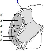
1. Dorsolateral fasciculus (of Lissauer)
2. Fasciculus proprius 3. Lateral (crossed) corticospinal tract 4. Rubrospinal tract 5. Posterior spinocerebellar tract 6. Anterior spinocerebellar tract 7. Spinothalamic tract 8. Reticulospinal tract |
|
|
|
66, Make a drawing and label the tracts (pathways) of the anterior and posterior funiculi of the spinal cord
|
1. Fasciculus gracilis
2. Fasciculus cuneatus 3. Comma tract (of Schultz) 4. Fasciculus proprius 5. Anterior (direct) corticospinal tract 6. Tectospinal tract 7. Medial longitudinal fasciculus 8. Reticulospinal tract 9. Spinothalamic tract 10. Olivospinal tract 11. Vestibulospinal tract |
|
|
|
67, List the descending pathways connecting the brainstem and the spinal cord
|
a) Medial descending pathways (axial and proximal muscles):
i) Vestibulospinal ii) Reticulospinal iii) Tectospinal b) Lateral descending pathways (distal muscles): i) Rubrospinal c) Aminergic pathways: i) Cerulospinal ii) Raphe spinal (Raphe magnus) |
|
|
|
68, List the ascending pathways of the spinal cord
|
a) Gracile fascicle
b) Cuneate fascicle c) Spinothalamic tract d) Spinocervicothalamic tract e) Spinocerebellar pathways: dorsal, ventral, rostral, cuneocerebellar tracts f) Spinoreticular tract g) Spinomesencephalic tract |
|
|
|
69, What are the functional elements of the spinal cord
|
a) C1-C3 – Head & Neck
b) C4 – Phrenic Nerve c) C5-T1 – Brachial plexus, upper limb d) T2-S2– Trunk, Thorax & Abdominal muscles + organs e) L1-S2 – Lower limb f) S2-S4 – Sacral plexus, Parasymp. |
|
|
|
70, Define the segments of the spinal cord at the level of which a) the interomediolateral nucleus and b) Clark’s column are located
|
a) T1-L2, S2-S4, rexed lamina VII
b) T1-L3, rexed lamina VII |
|
|
|
71, Define the appearance of the interomediolateral nucleus in the spinal and its neural functions
|
a) Thoracolumbar (T1-L2) & sacral (S2-S4)
b) Sympathetic & parasympathetic preganglionic motor neurons |
|
|
|
72, What makes up the pyramidal tract
|
a) Corticospinal tract
b) Corticobulbar tract |
|
|
|
73, Define the location of the pyramidal tract in the a) internal capsule b) mesencephalon c) pons d) medulla oblongata & e) spinal cord
|
a) Knee (genu) of the internal capsule
b) Middle part of the base of the mesencephalon (cerebral peduncle) c) Base of pons d) Pyramid e) Lateral & anterior corticospinal tract |
|
|
|
74, Describe the somatotopy of the pyramidal tract
|
a) Cortex: Medial—Lateral (low=medial)
b) IC: 90 rotation, anterior-posterior (low=post) c) Mesencephalon: 90 rotation again, medial-lateral (low=lat) |
|
|
|
75, Where are the pyramidal fibres located in the spinal cord
|
a) Uncrossed – descend in the medial part of the anterior funiculus ( Anterior Corticospinal tract)
b) Crossed – descend in the dorsal part of the lateral funiculus (Lateral Corticospinal tract) |
|
|
|
76, Name the motor nuclei of cranial nerves that receive exclusively contralateral innervations from the pyramidal tract
|
a) Facial motor nuclei (CN VII) - Motor neurons in the motor nucleus of the facial nerve (VII) that innervate the muscles of facial expression on the
lower half of the face b) Ambiguous nuclei - Motor neurons in the ambiguous nucleus that innervate the muscles of the soft palate c) Hypoglossal motor nuclei (CN XII) - Motor neurons in the motor nucleus of the hypoglossal nerve that innervate the genioglossus muscle |
|
|
|
77, In the pyramidal tract, where do indirect corticospinal fibres end
|
•In lamina 5, 6 & 7 on interneurons
|
|
|
|
78, Name the structures that border the internal capsule,
What is the location of the pyramidal tract in the internal capsule. |
a) Borders –
Medially: caudate nucleus, thalamus. Laterally: Lentiform nucleus (putamen & globus pallidus) b) The pyramidal tract is located at the knee of the internal capsule |
|
|
|
79, Which cortical areas does the pyramidal tract arise from? Define also the Brodmann number of these regions
|
•Primary Somatosensory cortex (Brodmann Area 1,2,3)
•Motor cortex (Brodmann Area 4) •Premotor cortex (Brodmann Area 6,8) |
|
|
|
80, List the symptoms of pyramidal tract disorder
|
a) Deviated uvula towards site of lesion
b) Tongue will deviate towards the paralyzed genioglossus (contralateral to the lesion) |
|
|
|
81, List the receptors, axons, cell groups, fibre tracts & nuclei that are parts of the sensory ascending fibre tract system called the spinothalamic tract
|
List the components from the receptors to the cerebral cortex.
a) High threshold receptors b) C and Aδ afferents c) Interneurons in the spinal dorsal horn d) Projecting neurons in the spinal gray matter e) Spinothalamic tract f) Ventral posterolateral nucleus g) Thalamocortical tract h) Postcentral gyrus (secondary and association sensory cortical territories) |
|
|
|
82, List the receptors, axons cell groups, fibre tracts & nuclei that are parts of the sensory dorsal column – medial lemniscus ascending fibre tract system
|
List the components from the receptors to the cerebral cortex.
a) Low threshold receptors b) Aα and Aβ primary afferent fibres and their cell bodies in the spinal dorsal root ganglia c) Gracile and cuneate fasciculus d) Gracile and cuneate nuclei e) Medial lemniscus f) Ventral posterolateral nucleus of the thalamus g) Thalamocortical tract h) Postcentral gyrus |
|
|
|
83, List 5 nuclei, area or fibre tracts in the CNS system that participate in the formation of the dorsal column medial lemniscus ascending sensory system
|
a) Gracile fasciculus
b) Cuneate fasciculus c) Gracile nucleus d) Cuneate nucleus e) VPL f) VPM g) Somatosensory cortex in the cerebral cortex (Brodmann area 3,1,2) |
|
|
|
84, List 3 major fibre tracts of the anterolateral ascending sensory system
|
a) SpinoMesencephalic tract
b) SpinoReticular (spinobulbar) tract c) SpinoThalamic tract "Mr. T" |
|
|
|
85, Name the ascending sensory pathways of the spinal cord that carry nerve signals generated by low threshold
receptors in the skin, muscles of the upper limb to the cerebellum |
a) Cuneocerebellar tract
b) Rostral spinocerebellar tract |
|
|
|
86, Name the ascending tracts that conduct sensory impulses from the a) lower and b) upper limb to the cerebellum
|
a) Lower limb – dorsal spinocerebellar tract and ventral spinocerebellar tract
b) Upper limb – cuneal spinocerebellar tract and rostral spinocerebellar tract |
|
|
|
87, Name the specific sensory nuclei of the thalamus that send afferent fibres to the cerebral cortex.
In which histological layer of the cerebral cortex do these afferent fibres terminate. |
a) Lateral geniculate body
b) Medial geniculate body c) Ventral posterolateral nucleus d) Ventral posteromedial nucleus e) They all terminate in the internal granular layer (Layer IV) |
|
|
|
88, List the cranial nerves that have viscerosensory nuclei.
|
a) Facial nerve (VII)
b) Glossopharyngeal nerve (IX) c) Vagus nerve (X) |
|
|
|
89, Which type of primary afferents conduct volleys to the spinal cord from a) high and b) low threshold mechanoreceptors
and c)muscle spindles |
a) C and Aδ fibres
b) Aβ and Aα fibres c) Ia |
|
|
|
90, Define the terms a) dermatome and b) head-zone
|
a) Dermatome – an area of the skin which is innervated by one spinal segment
b) Head-zone – a hypersensitive area of skin associated with inflammation of underlying viscera which projects to that area, usually due to both regions being innervated by the same cranial nerve. |
|