![]()
![]()
![]()
Use LEFT and RIGHT arrow keys to navigate between flashcards;
Use UP and DOWN arrow keys to flip the card;
H to show hint;
A reads text to speech;
95 Cards in this Set
- Front
- Back
- 3rd side (hint)
|
Central nervous system |
Brain and spinal chord |
|
|
|
Stimuli |
Things in the environment, which a person can sense and respond to |
|
|
|
CNS protection |
Skull, vertebral column and meninges |
|
|
|
Meninges |
Pia mater, arachnoid mater and dura mater |
|
|
|
Pia mater |
Rich blood supply which nourishes the cell |
|
|
|
Arachnoid |
Cerebrospinal fluid which acts as a cushion and maintains constant pressure and nourishes cell |
|
|
|
Dura mater |
Tough durable membrane for protection |
|
|
|
Grey matter |
Mostly cell bodies |
|
|
|
White matter |
Mostly axons or nerve fibers |
|
|
|
Ventricles |
Filled with cerebrospinal fluid |
|
|
|
Cerebrum |
Largest part, connected to corpus callosum and controls emotion, personality and voluntary muscles |
|
|
|
Corpus callosum |
Allows communication between right and left hemisphere of the cerebrum |
|
|
|
Cerebellum |
Controls voluntary muscular movement, posture and balance |
|
|
|
Medulla oblongata |
Maintains vital body functions such as breathing, blood pressure and heart rate |
|
|
|
Spinal chord |
Simple reflexes |
|
|
|
Sensory nerves |
Afferent, unipolar |
|
|
|
Motor neurons |
Efferent, multipolar |
|
|
|
Somatic nervous system |
Controls voluntary skeletal muscle response |
|
|
|
Autonomic nervous system |
Controls involuntary skeletal muscle response.
Divided into sympathetic and parasympathetic nervous system |
|
|
|
Sympathetic |
Controls activities that increase the amount of energy used. Works with adrenaline |
|
|
|
Parasympathetic |
Controls activities that decrease the amount of energy used. Rest and digest response |
|
|
|
Neurons |
Basic unit of the nervous system |
|
|
|
Epineurium |
Layer of connective tissues that surrounds a nerve |
|
|
|
Perineurium |
Encloses each bundle of nerve cells |
|
|
|
Endoneurium |
Delicate connective tissue inside a nerve |
|
|
|
Dendrites |
Carry nerve impulses towards cell body |
|
|
|
Cell body |
Contains nucleus, controls functions |
|
|
|
Myelin sheath |
Fatty layer for insulation which speeds up impulses |
|
|
|
Neurilemma |
Membrane which repairs a damaged neuron |
|
|
|
Schwann cell |
Nourishes the neuron |
|
|
|
Nodes of Ranvier |
Gaps between Schwann cells which speed up nerve impulses |
|
|
|
Reflex actions |
Fast involuntary responses |
|
|
|
Simple reflexes arcs |
Neuron pathways that carry impulses Receptors - sensory neuron - dorsal root - spinal chord - synapse with interneuron - motor neuron - ventral root- effectors |
|
|
|
Synaptic knob |
End of the axon which produces neurotransmitters |
|
|
|
Alzheimer’s |
Cause: old age, head injure, genetics Symptoms: loss of memory, loss of words, not doing activities, mood change Treatment: no cure, drugs slow it down |
|
|
|
Multiple sclerosis |
Causes: genetic, environmental factor Symptoms: loss of memory and coordination, visual problems and depression Treatment: drugs help with symptoms |
|
|
|
Brain injury |
Affect movement, speech, memory |
|
|
|
Spinal injury |
Loss of sensation and paralysis |
|
|
|
Stem cell |
Cells that are capable of dividing and differentiating into almost any cell type |
|
|
|
Receptors |
Involved with detecting the changes in stimuli that occurs in environment |
|
|
|
Receptors |
Involved with detecting the changes in stimuli that occurs in environment |
|
|
|
Interoceptors and exteroceptors |
Internal and external stimuli |
|
|
Front (Term) |
Back (Definition) |
|
|
|
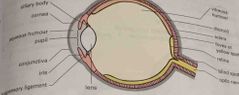
Eye muscles |
Allow eyeball to move |
|
|
|
Eye muscles |
Allow eyeball to move |
|
|
|
Eye glands |
Secrete tears to prevent it from drying out |
|
|
|
Eye muscles |
Allow eyeball to move |
|
|
|
Eye glands |
Secrete tears to prevent it from drying out |
|
|
|
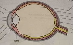
Sclera |
Outer layer, inelastic tissue, maintains shape of eye and allow muscles to attach |
|
|
|
Eye muscles |
Allow eyeball to move |
|
|
|
Eye glands |
Secrete tears to prevent it from drying out |
|
|
|
Sclera |
Outer layer, inelastic tissue, maintains shape of eye and allow muscles to attach |
|
|
|
Cornea |
Front part, transparent allowing light to pass through |
|
|
|
Choroid |
Middle layer, contains blood vessels. Front part forms iris |
|
|
|
Choroid |
Middle layer, contains blood vessels. Front part forms iris |
|
|
|
Iris |
Surround the pupil |
|
|
|
Choroid |
Middle layer, contains blood vessels. Front part forms iris |
|
|
|
Iris |
Surround the pupil |
|
|
|
Conjunctiva |
Thin membrane that covers the front part containing sensory nerve endings |
|
|
|
Choroid |
Middle layer, contains blood vessels. Front part forms iris |
|
|
|
Iris |
Surround the pupil |
|
|
|
Conjunctiva |
Thin membrane that covers the front part containing sensory nerve endings |
|
|
|
Retina |
Inner layer of eyeball made up of photoreceptors |
|
|
|
Choroid |
Middle layer, contains blood vessels. Front part forms iris |
Contains ... forms ... |
|
|
Iris |
Surround the pupil |
|
|
|
Conjunctiva |
Thin membrane that covers the front part containing sensory nerve endings |
|
|
|
Retina |
Inner layer of eyeball made up of photoreceptors |
|
|
|
Photoreceptors |
Convert light into nerve impulses |
|
|
|
Rods |
Sensory cells in retina, active in dim light, allows you to see black and white Pigment: rhodopsin |
|
|
|
Cones |
Active in bright light, allows you to see color. Pigment: iodopsin |
|
|
|
Astigmatism |
Vision blurred due to an uneven curve of the cornea or lens |
|
|
|
Cataracs |
Clouding of the lens as a person gets older |
|
|
|
Optic nerve |
Carry impulses from retina to brain |
|
|
|
Lens |
Behind pupil |
|
|
|
Suspensory ligaments |
Hold lens in place |
|
|
|
Functioning of the eye |
1- cornea: light refraction 2- pupil: light passes through 3- lens refraction of light to create focused image 4- image falls on retina and is inverted 5- optic nerve: carry impulses through brain which interprets images as an upright image |
|
|
|
Binocular vision |
Ability to focus on one object using both eyes. Brains puts both images together |
|
|
|
Accommodation |
Process where lens changes its shape to allow you to focus on an object |
|
|
|
Pupil reflex |
Contraction of pupil in response to light Dim: radial muscles contract, circular muscles relax Bright: radial muscles relax, circular muscles contract |
|
|
|
Short sightedness |
Eyeball is too long and cornea too rounded |
|
|
|
Long sightedness |
Eyeball is too short and cornea is too flat |
|
|
Front (Term) |
Back (Definition) |
|
|
|
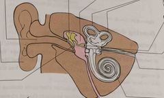
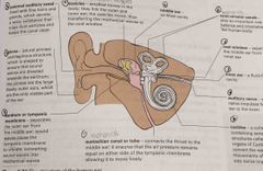
Pinna |
Shaped to ensure that sound waves are directed toward eardrum |
|
|
|
Eustachian canal |
Connects throat to middle ear and ensure that air pressure remains equal |
|
|
|
Tympanic membrane (eardrum) |
Sound waves causes it to vibrate converting it into mechanical waves |
|
|
|
Hearing |
Pinnae - external auditory canal - eardrum - ossicles - oval window - inner ear - organs of corti - auditory nerve |
|
|
|
External auditory canal |
Lined with fine hairs and glands which secrete waxy substances |
|
|
|
Tympanic membrane (eardrum) |
Sound waves causes it to vibrate concerting it into mechanical waves |
|
|
|
Ossicles |
Transfers mechanical waves into oval window |
|
|
|
Middle ear |
Air filled cavity |
|
|
|
Oval window |
Separates the middle ear from inner ear |
|
|
|
Inner ear |
Fluid filled cavity |
|
|
|
Semi- circular canals |
Play a role in balance |
|
|
|
Auditory nerve |
Carry nerve impulses to brain |
|
|
|
Cochlea |
Sensory structure called organs of corti which converge wave movements into nerve impulses |
|