Reading...
![]()
Play button
![]()
Play button
![]()
Use LEFT and RIGHT arrow keys to navigate between flashcards;
Use UP and DOWN arrow keys to flip the card;
H to show hint;
A reads text to speech;
50 Cards in this Set
- Front
- Back
|
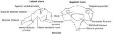
Typical Vertebrae
Body |
Cylindrical bone mass
Main weight-bearing part |
|
|
Typical Vertebrae
Intervertebral Disc |
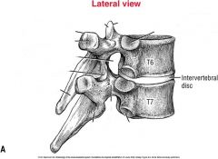
Thick ring of fibrocartilage
Vertebral column shock absorber |
|
|
Typical Vertebrae
Interbody Joint |
Formed by two vertebral bodies and intervertebral disc
|
|
|
Typical Vertebrae
Spinous Process |
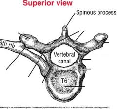
Most easily palpated through skin and muscle
Typically points posterior but may be inferior as well (especially thoracic) |
|
|
Typical Vertebrae
Transverse Process |
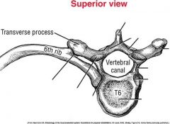
Lateral projections
Muscles and ligaments attachments |
|
|
Typical Vertebrae
Vertebral Canal |
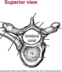
Houses and protects spinal cord
|
|
|
Typical Vertebrae
Pedicles |
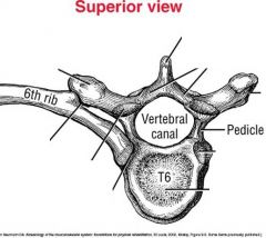
Short thick projections of bone connecting body of vertebrae to each transverse process
|
|
|
Typical Vertebrae
Laminae |
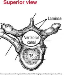
Thin plates of bone forming posterior wall of vertebral canal
Connect each transverse process to spinous process base |
|
|
Typical Vertebrae
Superior and Inferior Articular Facets |
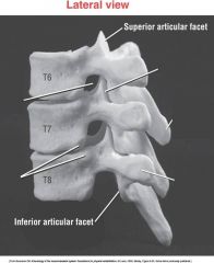
Inferior facets of one vertbera articulate with superior facets of vertebra below it
Together they make a facet (apophyseal) joint Help guide vertebral motion |
|
|
Typical Vertebrae
Intervertebral Foramina |
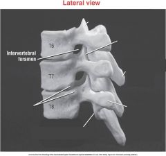
Between adjacent vertebrae
Passageways for nerve roots entering or exiting vertebral column |
|
|
Cervical Vertebrae
Typical (C3-C7) |
Transverse foramina: Holes for vertebral arteries heading to brain in the transverse processes
Bordered posterior-laterally by uncinate processes Most spinous processes are bifid |
|
|
Cervical Vertebrae
Atlas (C1) |
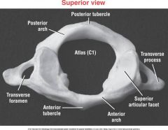
Two large concave superior facets sit on top of lateral masses to accept occipital condyles, forming atlanto-occipital j.
|
|
|
Cervical Vertebrae
Axis (C2) |
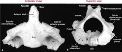
Functions as vertical axis of rotation for rotary movements between head and cervical region
|
|
|
Thoracic Vertebrae
|
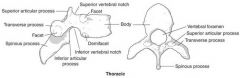
Inferiorly projected spinous processes
Large posterior-laterally projected transverse processes Facet (apophyseal) joints are aligned nearly in frontal plane |
|
|
Lumbar Vertebrae
|
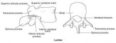
Facet joints in upper lumbar region are oriented close to sagittal plane, transition toward frontal plane in lower regions
|
|
|
Vertebral Segments and their planes
|
Cervical is triplanar
Thoracic is frontal Lumbar is sagittal |
|
|
Major ligaments
ligamentum flavum |
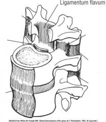
Limits flexion
|
|
|
Major ligaments
supraspinous and interspinous ligaments |
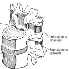
attaches between adjacent spinous processes from C7 to sacrum
Limits flexion |
|
|
major ligaments
Intertransverse ligaments |
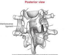
limits contralateral sidebending (lateral flexion)
|
|
|
major ligaments
anterior longitudinal ligament |
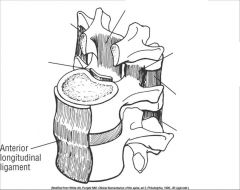
adds stability to vertebral column
Limits extension or excessive lordosis |
|
|
major ligaments
posterior longitudinal ligament |
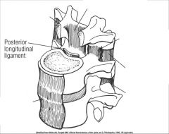
stabilizes vertebral column
limits flexion reinforces posterior annulus fibrosis |
|
|
craniocervical region
Set of three articulations |
atlanto-occipital joint, atlanto-axial joint, intracervical joint
most mobile area of entire vertebral column |
|
|
craniocervical region
flexion and extension |
85 degrees of cervical extension
45-50 degrees of cervical flexion |
|
|
craniocervical region
axial rotation |
allows visual field to approach 360 degrees
C1 and attached cranium rotate as fixed unit relative to axis |
|
|
craniocervical region
lateral flexion |
allows about 40 degrees of lateral flexion to each side
Motion guided by incline of facet joints |
|
|
thoracolumbar region
flexion and extension |
combined motion of thoracic and lumbar vertebrae allows about 85 degrees of forward flexion
Allows about 35-40 degrees of extension |
|
|
thoracolumbar region
axial rotation |
allows only about 35 degrees of horizontal plane rotation in either direction
|
|
|
thoracolumbar region
lateral flexion |
limited to about 45 degrees in either direction
|
|
|
lumbosacral junction
|
articulation between L5 and S1, weight transferred to pelvis
Facet joints of L5-S1 are oriented close to frontal plane to prevent lower spine from translating downhill |
|
|
sacroiliac joints
|
nutation is anterior rotation of sacrum relative to each ilium
counternutation is posterior rotation of sacrum relative to each ilium Primary function is to transfer forces of body weight to pelvis |
|
|
Innervation to craniocervical and trunk musculature
dorsal rami |
dorsal rami form short nerves that innervate most muscles of posterior neck and trunk
|
|
|
Innervation to craniocervical and trunk musculature
ventral rami |
form cervical, brachial, and lumbosacral plexus and innervate most muscles of anterior-lateral trunk and neck
|
|
|
anterior craniocervical (neck) muscles
superficial |
sternocleidomastoid
anterior, middle, and posterior scalenes |
|
|
anterior craniocervical (neck) muscles
deep |
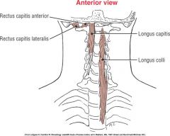
longus colli
longus capitis rectus capitis anterior rectus capitis lateralis |
|
|
anterior craniocervical muscles
sternocleidomastoid |
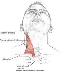
O: sternum and clavicle
I: mastoid process A: flexion of head and neck, contralateral rotation of head and neck, lateral flexion of head and neck N: spinal accessory nerve (cranial nerve XI) |
|
|
anterior craniocervical muscles
scalenes |
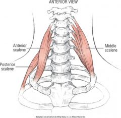
O: transverse processes of the cervical vertebrae
I: first and second ribs A: flexion of neck(anterior and middle scalenes, lateral flexion, assist with inspiration via elevation of first and second ribs |
|
|
posterior craniocervical muscles
suboccipital muscles |
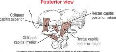
obliquus capitis superior
obliquus capitis inferior rectus capitis posterior minor rectus capitis posterior major |
|
|
posterior craniocervical extensors
superficial cervical extensors |
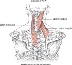
splenius capitis: extension, lateral flexion, and ipsilateral rotation of head and neck
splenius cervicis: extension, lateral flexion, and ipsilateral rotation of neck |
|
|
anterior muscles of trunk
matching pairs |
rectus abdominis
external oblique internal oblique transverse abdominis |
|
|
anteior muscles of trunk
rectus abdominis |
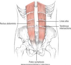
O: pubis
I: xiphoid process and costal cartilages of ribs 5-7 I: intercostal nerves T7-T12 A: flexion of trunk, posterior pelvic tilt, increase intra-abdominal and intrathoracic pressure |
|
|
anterior muscles of trunk
external oblique |
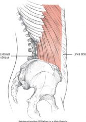
O: lower eight ribs laterally
I: iliac crest and linea alba A: flexion of trunk, posterior pelvic tilt, increase intra-abdominal and intrathoracic pressure, rotation of trunk to opposite side, lateral flexion of trunk N: Intercostal nerves T8-T12 |
|
|
anterior muscles of trunk
internal oblique |
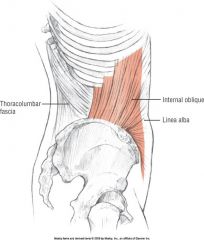
O: inguinal ligament, iliac crest, thoracolumbar fascia
I: ribs 9-12, linea alba, and rectus sheath A: flexion of trunk posterior pelvic tilt, increase intra-abdominal and intrathoracic pressure, lateral flexion of trunk, rotation of trunk to same side N: intercostal nerves T8-T12 |
|
|
anterior muscles of trunk
Transverse abdominis |
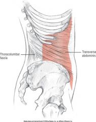
O: inguinal ligament, thoracolumbar fascia, cartilages of ribs 6-12
I: linea alba, contralateral rectus sheaths A: increase intra-abdominal pressure, increase tension in thoracolumbar fascia N: intercostal nerves T7-T12 |
|
|
Other functionally associated muscles of anterior trunk
iliopsoas |
combo of iliacus and psoas major, primary hip flexor and also plays role in other motions of trunk and pelvis
|
|
|
other functionally associated muscles of anterior trunk
quadratus lumborum |
attaches inferiorly to iliac crest, and superiorly to the 12th rib and transverse processes of L1-L4
bilateral activation of this muscle results in extension of lumbar spine |
|
|
posterior muscle groups of trunk
|
erector spinae
transversospinal muscles short segmental group |
|
|
posterior muscles of trunk
erector spinae |
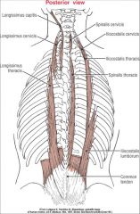
run up and down spine vertically to control extension and some fine tuning
consist of three thin columns of muscles: spinalis, longissimus, iliocostalis |
|
|
posterior muscles of trunk
transversospinal muscles |
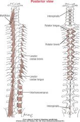
semispinalis, multifidus, rotators
lie deep to erector spinae, course in oblique direction from one vertebra's transverse processes to spinous process of another |
|
|
posterior muscles of trunk
actions of transversospinal muscles |
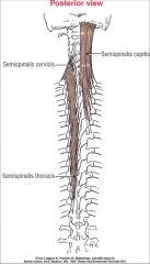
All are extensors of the vertebral column
Most can produce contralateral rotation the more horizontal and shorter the muscle the more potential to produce horizontal plane rotation |
|
|
posterior muscles of trunk
short segmental group |
intertransversarus, interspinales muscles
Assists with lateral flexion Effective at giving fine control over vertebral column, vertical stability in sagittal and frontal planes Essential for postural alignment with sensory feedback |