![]()
![]()
![]()
Use LEFT and RIGHT arrow keys to navigate between flashcards;
Use UP and DOWN arrow keys to flip the card;
H to show hint;
A reads text to speech;
144 Cards in this Set
- Front
- Back

What organelles are indicated by letters a-d |
A) nuclear envelope B) neucleolus C) nuclear pore D) intermediate filament |
|

E-H |
E-actin filament F- micro tubual G- intermediate filament H-centriole |
|

I-L |
I- cytoplasm J- lysosome K- plasma membrane L-perioxisome |
|
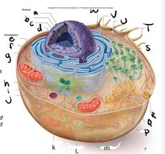
M-R |
M-mitochodria P-Golgi apparatus Q- vesicle R- Exocytosis |
|

S-W |
S-ribosomes T-microvilli U- smooth ER V- rough ER W- ribosomes |
|
|
Two parts of a cell |
1. organelles (tiny organs) 2. Cytosol ( complex gel-like Mass in which cell Structures can be found) |
|
|
Membrane Structures |
Endoplasmic reticulum Golgi complex Lysosomes, peroxisomes mitochondrial Chloroplast vesicles |
|
|
Non membrane Structures |
Ribosomes centrioles cytoskeleton |
|
|
most activities occur in _________. Including |
Cytoplasm. Including metabolic processes (glycolysis) and various processes (cell division. |
|
|
Molecules in cytosol |
Cytoskeletal filaments, dissolved molecules, salts, and water Fill the cell and areas bw organelles |
|
|
Macromollecular crowding |
Occurs due to cytosol’s network of fibres and high concentrations of dissolved macromolecules (i.e., proteins) |
|
|
What is the largest organelle |
Nucleus |
|
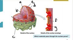
A-e |
A- chromatin B- neucleolus C- rough ER D- nuclear pore E- nuclear envelope |
|

F-G |
F- nuclear envelope G- nuclear pore |
|
|
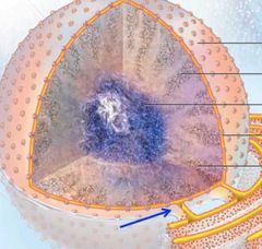
Nucleus |
protects DNA and keeps it from becoming damaged S tangled Within cytoplasm |
|
|
Nuclear membrane |
Controls What gets in and out of nucleus selectively Permeable 2 phospholipid biolayers w proteins |
|
|
Outer portion of nuclear membrane merges with |
rough ER membrane |
|
|
Inner portion of ER |
ANCHORS DNA molecules (chromatin) |
|
The arrow is indicating |
area where Nuclear membrane merges with ER membrane |
|
|
Chromatin Vs. Chromasome |
CHROMATIN – when cell is NOT DIVIDING → The DNA appears long and thin With small associated proteins called histones CHROMOSOME – when cell IS DIVIDING. The thin fibrous DNA coils into a thick complex (condenses) → Histone proteins maintain the chromosome structure. Can be seen with a light microscope |
|
|
Nuclear pores |
Cross BOTH nuclear membranes Create CHANNELS for free movement of small molecules and ions |
|
|
NeucleoPorins |
The proteins that make up the nuclear Pore complex The proteins that make up the nuclear Pore complex |
|
|
Nucleoplasm |
→ interior of nucleus no membrane bound Subcompartments. → contains protein complexes ie) RNA molecules, DNA |
|
|
Neucleolus |
→ assemble ribosomes before exporting to cytoplasm through nuclear pores |
|
|
Ribosomes |
→ Assemble proteins in cytoplasm (protein Synthesis) |
|
|
Endo membrane System (parts+ function) |
system of membrane bound organelles that : → modify new protein → build lipids (fats) → Package the completed molecules in vesicles and then sorts & ships them to various destinations → includes: ER, Golgi apparatus, vesicles, Lysosomes.
Does not include Chloroplast or mitochondria |
|
|
Rough Vs. Smooth ER |
Rough ER- Contains ribosomes which give rough appearance. modifies proteins smooth ER- no mebrane bound ribosomes. Synthesize lipids. |
|
|
What types of Cells have larger rough ER |
cells Specialized for protein Synthesis ie) cells Secrete digestive enzymes or anti-body secreting Plasma cells |
|
|
Endoplasmic reticulum produces ________ filled with Products move to _______ |
→ vesicles → Golgi |
|
|
Vesicles |
→ Vesicles FORM by “budding” from ER – patches of ER membrane bulge and break off → Vesicles move to the Golgi complex → The Golgi Apparatus (Golgi Body) ALSO forms vesicles – these then move out of cell (usually) |
|
|
4 functions of Golgi Body |
1. Sorting/ dispatching Station for Products of the ER →Prepares proteins and lipids for cell export. For SECRETION OUT OF THE CELL by exocytosis (e.g. digestive enzymes, mucous, etc…) 2. Major site of carbohydrate synthesis →Pectin and hemicellulose (plant cell wall components) 3. The modification of some proteins and lipids from the ER → Various changes are made to “mature” some proteins and lipids 4. Making Lysosomes → Vesicles containing various enzymes used to “digest” various components inside of cells → Stay inside the cell |
|
|
Lysosomes |
→ Vesicles - BUD from the Golgi membrane – BECOME lysosomes and STAY INSIDE cell → Contain digestive enzymes – to destroy things inside of cells → Interior of lysosomes is ACIDIC – to activate their internal digestive enzymes |
|
|
Peroxisomes |
→Vesicles - BUD from ER membrane – STAY INSIDE cell → Contain enzymes that break down fatty acids and amino acids |
|
|
Cytoskeleton |
network of Protein fibres and tubes extend throughout Cytoplasm → maintain Cell shape and Organization → intra + extracellular Movement → not Permanently rigid. Assemble+ disassemble based on cell activities |
|
|
Cytoskeleton Consists of |
microtubules - Organize cell interior intermediate filaments - add Strengths anchor filaments of actin+ myosin micro filaments - reinforce Cell Parts |
|
|
microtubules |
Cytoskeletal filament. →2S nm diameter → hollow tubule often grows from Centrosome located near nucleus → Composed of the protein tubulin → functions: Cell Shape, organize organelles, Chromosome Sorting, intracellular movement of Cargo, Cell motility |
|
|
intermediate Fillament |
→ cytoskeletal filament → 10 nm → twisted filament → Composed of different proteins ie) desmin, Keratin, lamin → functions: cell shape, Strength, Anchorage of cell + nuclear memberanes |
|
|
Actin filaments |
→ 7 nm diameter → Spiral filament → 2 Strands of actin → function: cell Shape, muscle Contraction, Cell movement, animal cell division, intracellular movement of cargo |
|
|
Structural Support for Cilia and Flagella formed by _ |
→ microtubules Covered by plasma membrane |
|
|
Cilia and flagella |
move unicellular organisim + Sperm Cillia move fluid over a surface ie) mucas through respiratory |
|
|
Micro tubules provide tracks to move _______ |
→vesicles bw Cell interior t plasma memberan → move+ Seperate Chromosomes during Cell division →motor proteins move vesicles and Chromosomes along Microtube tracks |
|
|
Microfilaments |
→ solid rods composed of actin →Microfilaments, with other proteins, form a 3-dimensional network just inside the plasma membrane - MICROVILLI →arranged parallel to each other → in Muscle cells → together with myosin filaments enable MUSCLE CONTRACTION as actin filaments slide past myosin filaments → In other cells, wide bands of actin filament networks push against the cell membrane - Causing CELL MOTILITY – I.e., migrating white blood cells |
|
|
intermediate filaments |
→ Made from intermediate proteins → occur Singlely, in parallel bundles and interlocking networks → Either alone or in combination with microtubules, microfilaments, or both → Provide STRUCTURAL SUPPORT in many cells and tissues |
|
|
Metabolism |
→intracellular Chemical reactions ie) Degredation, Synthesis, transformation of small organic molecules →Linear or Cyclic reactions |
|
|
Enzymes |
→play a vital role in metabolism → Mostly proteins, Catalytic → Optimum pH and temperature → Interact with substrate at the active site → Speed up Chemical reactions by bring Substrate molecules togetherso they interact. |
|
|
Coenzymes |
enzyme "helpers" →molecules that move hydrogen atoms & electrons to the sites of chemical reactions in cells →e.g., the nucleotides NAD+, FAD |
|
|
2 types of metabolic Pathways |
Anabolic process →Favour the SYNTHESIS of molecules for building up organs and tissues, Often CONDENSATION/DEHYDRATION reactions ex) “anabolic steroids” – drugs that increase proteins in cells - esp. in skeletal muscle cells Catabolic process→ Favour the BREAKDOWN of complex molecules into smaller more simple ones, Often HYDROLYSIS reactions (covered in more detail later) |
|
|
Photosynthesis |
→ Process by which carbon dioxide and water are converted (with the aid of light energy) INTO CARBOHYDRATES (mostly sugars - GLUCOSE) → By green plants, algae, some protists (pond organisms), and some bacteria (these are the primary producers) |
|
|
Primary Producers Store ___________ surplus as _________. Withdraw when energy is needed for _________ |
Glucose Starch Cellular respiration |
|
|
Primary consumers |
Feed on primary producers. Break down starch into glucose to support own metabolism. Break down further during cellular respiration. To “release” the energy that’s “trapped” in the chemical bonds within glucose And to “store” this energy in ATP molecules (in high energy phosphate bonds) |
|
|
Glucose is stored as |
Starch + cellulose in plants Glycogen in animals |
|
|
Cellular respiration |
PROCESS by which glucose (and some other sugars) is (are) BROKEN DOWN To yield carbon dioxide, water, and energy - Energy is in the form of ATP Cellular respiration releases the energy STORED in the CHEMICAL BONDS of glucose ( C6H12O6 ) to produce ATP (Cells use ATP as Energy) |
|
|
3 chemical pathways (cellular respiration) |
1. Glycolysis 2. Krebs Cycle (Tricarboxylic Acid Cycle, Citric Acid Cycle) – oxygen requiring 3. Electron Transport Chain – oxygen requiring |
|
|
MITOCHONDRIA |
provide cells with ENERGY through the BREAKDOWN of GLUCOSE and the FORMATION of ATP molecules during cellular respiration OXYGEN REQUIRING series of metabolic reactions Mitochondria require oxygen – O2 By inhaling air, organisms provide mitochondria with O2 Mitochondria also produce carbon dioxide – CO2 By exhaling, organisms remove the CO2 formed by mitochondria |
|
|
Cellular respiration final products |
Carbon dioxide – CO2 Water - H2O Energy (in the form of ATP) |
|
|
The mitochondria contains enzymes for ______ |
Krebs cycle Electron transport chain BOTH are oxygen-requiring processes |
|
|
ATP forms in the ______________ of the mitochondria |
Inner compartment |
|
|
Inner membrane has folds called ______ contain enzyme for |
CRISTAE Krebs cycle Electron transport chain |
|
|
Inner compartment |
The INNER SPACE bounded by inner membrane ATP forms IN the matrix |
|
|
OUTER compartment |
The SPACE BETWEEN inner and outer membranes Is the “Intermembrane Space” |
|
|
How cells make ATP |
Through the process of CELLULAR RESPIRATION → A SERIES of reactions that break down macromolecules (larger molecules) → Anabolic or catabolic? → Resulting in the formation of ATP → In particular, GLUCOSE is broken down and converted into INTERMEDIATE molecules |
|
|
HYDROGEN IONS (H+) |
are H atoms that lost their electron |
|
|
Glycosis |
→ Breakdown of GLUCOSE molecules to PYRUVATE (aka pyruvic acid) → Occurs in cell cytoplasm, OUTSIDE of mitochondria → Oxygen NOT required |
|
|
Final product of glycosis |
→ TWO pyruvate (these move to Step 2) → TWO NADH (not shown in figure) → Net energy yield is TWO ATP |
|
|
Go through 29-34 |
L08 |
|
|
Blood Glucose regulation |
→ takes place in Pancreas →Glucagon (HORMONE) Increases blood glucose → Insulin (HORMONE) Lowers blood glucose |
|
|
Functions of plasma membrane |
→ maintain Cell structural integrity → regulate movement in tout of cell → rocognition bw cells → communication bw cells → Sticking cells together to form tissues organs |
|
|
Plasma membrane |
Plasma Membrane: Phospholipid bilayer with attached or embedded proteins and cholesterol → Phospholipids: Polar heads, Non-polar tails, Form spherical bilayer when placed in water →Different proteins have different functions: Adhesion, Transport, Reception, Enzymes →Selectively permeable (semipermeable) |
|
|
MEMBRANE PHOSPHOLIPIDS are AMPHIPATHIC molecules |
AMPHIPATHIC: Presence of BOTH hydrophobic and hydrophilic regions HYDROPHOBIC: WATER-HATING, non-polar fatty acid tails, HYDROPHILIC: WATER-LOVING,polar heads |
|
|
Lipid Bilayer formation |
Phospholipid molecules organize into MICELLES & BILAYERS → VIA AGGREGATION of hydrophobic tails Lipid Bilayer is “SELF-SEALING” → Constant spontaneous re-arrangement of lipid molecules To ‘HIDE’ hydrophobic tails that become exposed |
|
|
Membrane Permeability |
INNER LIPID BILAYER OF HYDROPHOBIC TAILS creates a BARRIER TO WATER → Most materials dissolved in water UNABLE to pass across this lipid bilayer → Important materials that need to pass across this lipid bilayer MOVE THROUGH special protein complexes embedded in the bilayer called Transporter molecules |
|
|
Diffusion Accross bilayer |
BARRIER FUNCTION - impermeable to “SELECTED” solutes and ions (BLOCKS most hydrophilic molecule) Diffusion rate ACROSS lipid bilayer DEPENDS on (1) molecule size (2) it’s lipid permeability Small non-polar molecules: Readily diffuse across Uncharged polar molecules: Diffuse across if small enough, Some need transporter molecules Ions and charged molecules: Cannot diffuse across, Need transporter molecules |
|
|
Simple diffusion |
The RANDOM movement of a substance from a region of higher concentration TO a region of lower concentration Water moves across the plasma membrane by the simple diffusion process called OSMOSIS |
|
|
FACILITATED DIFFUSION |
The movement of a substance from a region of higher concentration TO a region of lower concentration WITH THE AID OF A MEMBRANE PROTEIN (transporter molecule) that acts as a channel or a “carrier” protein |
|
|
ACTIVE TRANSPORT |
movement of molecules across the plasma membrane from an area of lower concentration TO one of higher concentration With help from a Carrier Protein AND ENERGY, usually in the form of ATP |
|
|
ENDOCYTOSIS & EXOCYTOSIS |
Transporting LARGER MOLECULES Across Cell Membranes Endo - in Ex- out 3 Types of Endocytosis 1. Phagocytosis 2. Pinocytosis 3. Receptor-Mediated Endocytosis ↳ Ex., Low-density Lipoprotein (LDL) particles (contain cholesterol) |
|
|
slide 27 |
Lo9 |
|
|
3 types of cell junctions |
Tight junctions: hold cells together, prevent leak bw Cells Adhesion Junctions: Cement cells together Gap junctions: hold cells together, help Cells Communicate, connect Cytoplasm of connected Cells |
|
|
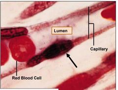
Tight Junctions |
Points of Contact bw plasma Membranes of adjacent cells ↳ tight Junction proteins ↳ Cells fused, form v tight Seal → multi-layer for protection → tight junction have sealing function (impermeable barrier) ex) intestinal cells → lumen fluids stay in lumen |
|
|
Lo9 |
33 |
|
|
Adhesion Junctions (Desmosomes, Anchoring Junctions) |
→Extremely TIGHT connections between adjacent cells → Plasma membranes do not touch n Material passes between cells →Held together by INTERCELLULAR protein filaments (cadherin family) ↳attached to cytoplasmic plaques, composed of protein material →Connect CYTOSKELETON FILAMENTS of adjacent cells (Intermediate Filaments) |
|
|
Adhesion junction provides __________ + examples |
Provide 1. Structural continuity 2. Tensile strength 3. Enable stretching of tissues and organs EX)skin, heart, gums, uterine cervix, etc |
|
|
Gap junction |
Adjacent cells connected by paired & joined “membrane channels” Form “CHANNELS” in between adjacent cell membranes ↳CYLINDRICAL ARRAYS (CONNEXON) of membrane-spanning proteins →Form OPEN CHANNELS between the cytoplasm of joined cells Allowing for material to PASS from one cell to its neighbour |
|
|
Gap junctions Function + example |
→Gap Junctions create TIGHT “cell-to-cell” CONNECTIONS →Gap Junctions also enable “cell-to-cell” COMMUNICATION ↳ Some ions, sugars, & small molecules ↳ Passing ‘signals’ between cells
e.g., cardiac cells in heart, between FOLLICLE CELLS and OOCYTE in ovary |
|
|
Intercalated disks |
TIGHTLY CONNECT adjacent cardiac cells. Contain numerous complexes of ‘adhesion proteins’ including gap junctions & desmosomes. |
|
|
Gap Junction |
(1) The follicle cells (granulosa cells) surrounding the oocyte are COUPLED to each other by gap junctions (2) In addition, the granulosa cells extend processes through the zonapellucida and make gap junctions with the oocyte (different type of Connexxons used) |
|
|
Cancer Cells |
→ lose Connection, ESCAPE normal growth mechanisms & over-proliferate →Cancer cells often LOSE their ability to normally connect to & communicate with other cells →spread throughout body to start new colonies - METASTASIS |
|
|
3 PRIMARY GERM LAYERS |
Ectoderm→ outer layer ↳ Nervous system, epidermis of skin, hair, nails, oil glands, sweat glands, mammary glands, and lining of mouth & rectum MESODERM – middle layer ↳Muscle, bone, connective tissue, dermis of skin, heart, kidneys, ovaries, and testes Endoderm ↳Lining of digestive & respiratory tracts & urinary bladder, pancreas, liver, thyroid gland, parathyroid glands |
|
|
4 mayor tissues |
Epithelial Connective Muscular Nervous |
|
|
Epithelial tissue |
↳Consists of TIGHTLY PACKED CELLS that form a CONTINUOUS layer that COVERS body surfaces →LINES internal & external surfaces features: UPPER surface EXPOSED to Body cavity Or external environment BOTTOM surface ATTACHED to the basement membrane Also known as basal lamina ↳CONNECTS the epithelium to the underlying connective tissue →In many epithelial cells, NUCLEI are somewhat basal (near basement) |
|
|
The bottom surface of Epithelial cells is attatched to |
basal lamina (basement membrane) |
|
|
Epithelia are SUPPORTED by |
connective tissue |
|
|
Connective tissue contains |
→variety of cells →extracellular matrix components |
|
|
fibroblast |
→predominant cell type in connective tissue →secretes abundant amounts of most of the extracellular matrix |
|
|
Structure of Epithelial |
→ONE SIDE of LAYER of Epithelial Cells (Epithelium) ↳ Exposed to space →OTHER SIDE of LAYER of Epithelial Cells (Epithelium) ↳ATTACHED to Basal Lamina (basement membrane) →SUPPORTED by Connective Tissue ↳attached to basement membrane |
|
|
Function of Epithelial tissue |
→PROTECTION: bw body + outside world. protects against moisture loss, mechanical trauma, toxic Substances + pathogen invasion →ABSORPTION: Digestive and respiratory linings →SECRETION:Hormones, mucus, enzymes, HCl, etc, Exocrine + Endocrine gland →EXCRETION & FILTRATION: Areas of kidney |
|
|
Exocrine glands |
secrete into DUCTS that empty to inside or outside of body (e.g., sweat glands, salivary glands, mammary glands...) |
|
|
Endocrine Glands |
lack ducts – secrete directly into the BLOODSTREAM (e.g., adrenal glands, thyroid, pancreas, testes, ovaries, etc...) |
|
|
Classification of Epitheliam Shape |
a. Squamous (flattened) b. Cuboidal (cubed) c. Columnar (column) d. Transitional (mixed) |
|
|
Classification of Epithelium Number of Layers |
a. Simple (one layer) b. Stratified (multilayered) c. Pseudostratified (one layer that looks like multilayers) |
|
|
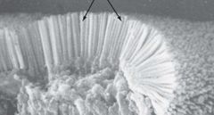
Surface Specializations |
a. Microvilli b. Cilia c. None |
|
|
Simple Squamous |
|
|
Simple squamous |
|
|
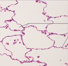
Simple Squamous epithelium, Description, function + location |
Description: Single layer of flattened cells with disc-shaped central nuclei and sparse cytoplasm; the simplest of the epithelia. Function: Allows passage of materials by diffusion and filtration in sites where protection is not important; secretes lubricating substances in serosae. Location: Kidney glomeruli; air sacs of lungs; lining of heart, blood vessels, and lymphatic vessels; lining of ventral body cavity (serosae) |
|
|
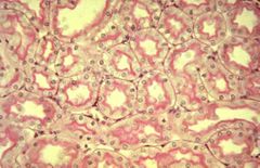
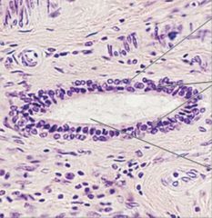
Simple Cobodial Epithelium, description, function + location |
Description: Single layer of cubelike cells with large, spherical centraI nuclei . Function : Secretion and absorption. Location: Kidney tubules; ducts and secretory portions of small glands; ovary surface. |
|
|
Simple Cubodial |
|
|
Simple Cubadial |
|
|
Simple Cubodial |
|
|
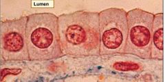
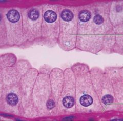
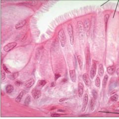
Simple Columnar Epitheliam |
Description: Single layer of tall cells with round to oval nuclei; some cells bear cilia; layer may contain mucus- secueting unicellular glands ( goblet cells). Function: Absorption; secntion of mucus, enzymes, and other substances; Ciliated type propels mucus (or reproductive cells) by ciliary action. Location: nonciliated type lines most of the digestive tract (stomach to anal canal), gallbladder, and excretory ducts of sale glands; ciliated variety lines small bronchi, uterine tubes, and some regions of the uterus. |
|
|
Simple columnar Epithelium |
|
|
Simple Columnar |
|
|
Simple Columnar unciliated with microvili |
|
|
simple columnar with Cilia |
|
|
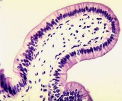
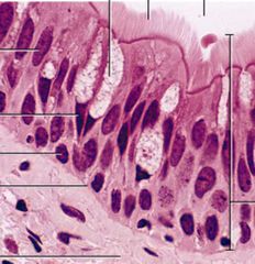
Pseudostratified Ciliated Columnar Epithelium |
Description: Single layer of cells of differing heights, some not reaching the free surface; nuclei seen at different levels; may contain mucus-secreting goblet cells and bear cilia. Function: Secretion, particularly of mucus; propulsion of mucus by Cilliary action Legation: nonciliated type in male's sperm-carrying ducts and ducts of large glands; ciIiated variety Iines the trachea most of the upper respiratory tract. |
|
|
Pseudostratified Ciliated Columnar Epithelium |
|
|
Pseudostratified ciliated Columnar Epithelium |
|
|
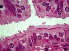
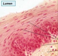
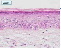
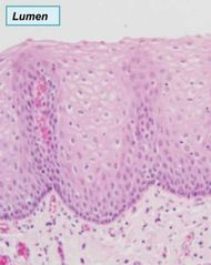
Stratified Squamous Epithelium |
Description: Thick membrane composed of several cell layers; basal cells are cuboidal or columnar and metabolically active : surface cells are flattened (squamous); in the keratinized type, the surface cells are full of keratin and dead; basal cells are active in mitosis and produce the cells of the more superficial layers. Function: Protects underlying tissues in areas subjected to abrasion. Location: Nonkeratinized type forms the moist linings of the esophagus, mouth, and vagina, urethra and anus; keratinized variety forms the epidedermis of skin a dry membrane. |
|
|
Stratified Squamous |
|
|
Stratified Squamous Epithelium Keratinized – Human Lip ↳ Outer Cells wo nucleus- dead Waterproof barrier |
|
|
Stratified Squamous Non-Keratinized → same as Keratinized wo Surface layer of dead cells |
|
|
Stratified Squamous Non-Keratinized Function |
Protection→ abrasion, pathogens, chemicals |
|
|
Stratified Cubodial Epithelium |
Description: Generally two layers of cubelike cells. Function: Protection Location: Largest ducts of sweat glands, mammary glands, and salivary glands. |
|
|
Stratified cubodial Epithelium |
|
|
Stratified Columnar Epithelium |
Description : Several ceII layers ; basal cells usually cuboidal; superficial ceIl is elongated and columnar. Function: Protection; secretion. Location : Rare in the body; small amounts in male urethra and in large ducts of some glands. |
|
|
Transitional Epithelium |
Description: Resembles both gratified squanous and stratified cuboidal; basal cells cuboidal or columnar; surface cells dome shaped or squamous-like, (spending on degree of organ sketch. Function: Stretches readily and permiits distension of urinary organ by contained urine. Location: Lines the ureters, bladder, and part of the urethra. |
|
|
Functions of Connective tissue |
→ Framework: can provide Support or bind organs together → Transport: Blood, oxygen, Carbon dioxide, nutrients, hormones → Storage: Triglycerides and Electrolytes → insulation: Heat loss → Protection: produce blood cells, White blood cell ingest bacteria. Antibodies in Plasma cells combat disease. → Repair: fractures, Sprains + wounds |
|
|
Characteristics of Connective tissue |
→ most abundant+ widely distributed type of tissue → vascular →connective tissue CELLS ARE WIDELY SPACED →Cells are WITHIN THE (MATRIX → non-cellular fluid Varying Consistancy from solid to fluid to semifluid) → may have fibres |
|
|
Fibroblasts, reticular cells |
→ migratory → Produce and secrete protein fibres → collagen fibres Strength→ resist stretching → Elastic fibre → flexible, maintain Shape after stretching →Reticular fibres → networks & support frameworks for soft organs Thin collagen fibres coated with glycoprotein |
|
|
Adipocytes |
→non-migratory → Energy/ insulation (protection) → Store tryglicerides (lipids, fats) |
|
|
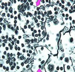
White blood cells |
→migratory →Neutrophils & lymphocytes, plasma cells – protection & defense |
|
|
Matrix |
→Determines the connective tissue properties → Composed of Protein fibres: Provide elasticity, strength, and structure i.e., collagen fibres, elastic fibres, reticular fibres → Composed of Ground Substance:Viscous & slippery material Cells are suspended in, ranges from solid, Semi fluid to fluid, Composed of an assortment of large molecules that bind cells together |
|
|
Soft Connective tissue (Structure, location t Function) |
Structure: Fibres - COLLAGEN, ELASTIC, Cells - FIBROBLASTS, adipocytes, few white blood cells Location:FILLS IN spaces, beneath skin dermis and around digestive, respiratory, & urinary tracts, between muscles & around blood vessels, nerves, joints Function: SUPPORTS EPITHELIUM & many internal organs, presence in lungs, arteries, & urinary bladder allows these organs to expand. forms part of protective covering for muscle, blood vessels, & nerves |
|
|
→ loose connective tissue areolar |
|
|
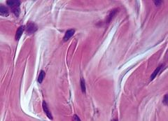
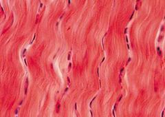
Dense fibrous tissue |
→ dense Packed collagen Fibres → Made up of fibrolast cells → located:Tendons (muscle-bone), Ligaments (bone-bone), Covering of skeletal muscle, Dense irregular forms parts of the Dermis of skin → Function→ depends on type + location ↳ Attachment, protection, Support, Strength, Elasticity |
|
|
Dense irregular |
|
|
dense regular |
|
|
Difference bw denseregular + dense irregular connective tissue |
→ regular dense connective tissue can withsand great tensile pressure When force is applied in one direction. → irregular able to withstand tension in multiple direction |
|
|
5 Specialized connective tissues |
Adipose Reticular Cartilage Bone Blood |
|
|
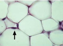
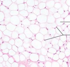
Adipose Tissue |
Fibres - few to none Cells – ADIPOCYTES - derived from fibroblasts that enlarge to store fat, occupy most of matrix Location- beneath the skin; some in loose connective tissue; around certain organs (heart, kidneys) Function: Store Energy protection + insulation |
|
|
adispose tissue |
|
|
addispose tissue |
|
|
reticular Connective tissue |
Fibres – RETICULAR FIBRES (wavy and curly) Cells – RETICULAR CELLS Location - Spleen, Kidney, Lymph Nodes, Bone Marrow |
|
|
Reticular connective tissue |
|
|
Reticular connective tissue |