![]()
![]()
![]()
Use LEFT and RIGHT arrow keys to navigate between flashcards;
Use UP and DOWN arrow keys to flip the card;
H to show hint;
A reads text to speech;
43 Cards in this Set
- Front
- Back
|
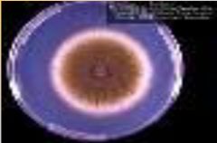
Aspergillus flavus - macroscopic view |
|

|
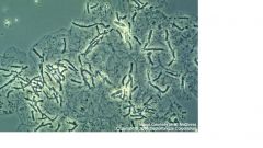
Malasezzia furfur |
|
|
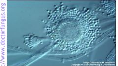
Aspergillus flavus - microscopically |
|

|
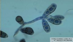
Curvularia lunata - macro |
|
|
Curvularia |
|
|
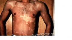
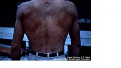
Tinea versicolor - the condition caused by Malassezia furfur - organism |
|
|
Causes tinea versicolor. Skin scrapings reveal spaghetti and meatballs appearance. Fluoresces under Woods light. Culture and ID not routinely done |
Malassezia furfur - organism |
|
|
Malassezia furfur |
|
|
Tinea versicolor - condition caused by Malassezia furfur - organism |
|
|
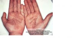
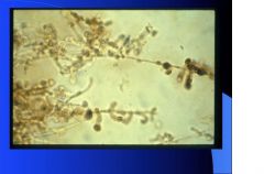
Causes tinea nigra - normally asymptomatic. It is a dimorphic fungus. Microscopic skin scrapings show darkly pigmented yeastlike cells and hyphal elements Culture and ID with Sabouraud's dextrose agar grows pigmented yeast and fungal hyphae seen microscopically |
Exophiala Werneckii - organism |
|

|
Exophiala Werneckii - organism |
|
|
Tinea nigra - condition caused by Exophiala Werneckii - organism |
|

|
Tinea nigra - condition caused by Exophiala Werneckii - organism |
|
|
Causes black piedra. Colonizes the hair shaft. Detected by micro exam of hair showing a hard black nodule surrounding the hair shaft.
|
Piedraia hortae - organism |
|
|
Piedraia hortae - organism |
|
|
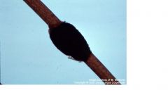
Black piedra - condition caused by Pedraia hortae - organism Masses of fruiting bodies of ascospores on human hair |
|
|
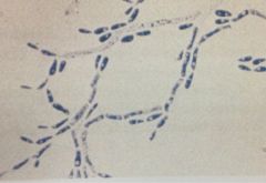
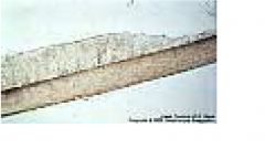
Causes white piedra, which colonizes around the hair shaft (white and creamy) Detected on micro exam of hairs showing a cream-colored soft, pasty growth surrounding the hair shaft Culture and ID by Sabouraud's dextrose agar presenting as a dimorphic fungus - septate hyphae, arthroconidia, and blastoconidia |
Trichosporon beigelii - organism |
|
|
Trichosporon beigelii - organism |
|
|
White piedra - condition caused by Trichosporon beigelii - organism |
|
|
what are fungal infections called? |
Mycoses |
|
|
fungi are eukaryotic, lack chlorophyll and absorb all nutrients from the environment, and cell walls are made of chitin |
general characteristics of fungi |
|
|
Fuzzy, fluffy, cottony, wooly, filamentous |
Hyphae (molds) |
|
|
Moist, creamy, wet, pasty cololnies |
Yeasts |
|
|
pathogenic fungi which can be both - "the bad boys" Grow as yeast at body temp and molds at room temp |
Dimorphic fungi |
|
|
Unicellular - 2-60 micrometers Reproduce by budding. If no septum formed, elongated bud called a pseudohypha (germ tube) is formed - this is the branching seen Resemble bacterial colonies on agar |
Yeasts |
|
|
grow by tubelike projections - true hyphae, which intertwine to form a cottony matt called mycelium. vegetative hyphae reaches into the agar for nutrients aerial hyphae reaches into the air for nutrients |
Molds |
|
|
aerial hyphae have specialized reproductive structures from which what occurs? |
sexual spores or asexual conidia occurs |
|
|
No cross walls Exampleis Phylum zygomycota |
Aseptate hyphae |
|
|
Most fungi have hyphae that occur this way |
Septate hyphae |
|
|
Individual oval to round cells which bud to form daughter cells |
Yeasts |
|
|
Fungi that exist as either yeast or mold. At room temp they grow as molds, at body temp they grow as yeast. Most are pathogenic |
Dimorphs |
|
|
Sabouraud's agar Dermatophyte agar Mycosel BHI |
Primarily fungal culture media |
|
|
Growth rate, colonial morphologic features, and microscopic morphological features are used to |
ID molds |
|
|
tease mount cellophane tape mount slide culture |
Methods of microscopic examination of molds |
|
|
fundamental microscopic units, tube-like projections |
hyphae |
|
|
asexual spores produced by specialized vegetative hyphae called conidiophores. May be macro or micro |
conidia |
|
|
flask or vase shaped extension from hyphae that may support conidia |
phialides |
|
|
fragmentation of the mycelium at the septum into cylinder or cask-shaped thick-walled spores |
arthroconidia |
|
|
thick-walled asexual spores formed by "rounding up" and enlargement of the terminal hyphal cells |
chlamydospores |
|
|
These colonize skin, hair, and nails. They produce keratinase, which hydrolyzes keratin |
dermatophytes |
|
|
these become filamentous when they invade tissues |
Candida |
|
|
Dimorphic, forming yeasts in the human body |
Systemic mycoses |
|
|
What organism produces a capsule which protects it from the immune system? |
Cryptococcus neoformans |