Reading...
![]()
Play button
![]()
Play button
![]()
Use LEFT and RIGHT arrow keys to navigate between flashcards;
Use UP and DOWN arrow keys to flip the card;
H to show hint;
A reads text to speech;
289 Cards in this Set
- Front
- Back
- 3rd side (hint)
|
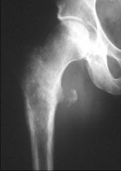
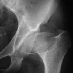
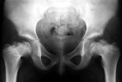
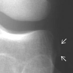
stage 1 avn radiographic findings
|
normal
|
|
|
|
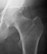
stage 2 avn radiographic findings
|
osteopenia/sclerosis, round head
|
|
|
|
stage 3 avn radiographic findings
|
flattening of head/crescent sign
|
|
|
|
stage 4 avn radiographic findings
|
secondary degeneration
|
|
|
|
DDX: hemosiderin in joint (3)
|
hemophilia; PVNS; old trauma
|
|
|
|
DDX: diffuse periostitis (4)
|
chronic venous stasis; HPOA; caffey disease; thyroid acropachy
|
|
|
|
caffey disease AKA
|
Infantile cortical hyperostosis
|
|
|
|
DDX: arthritis with subluxation (3)
|
RA; SLE; neuropathic
|
|
|
|
DDX: arthritis with osteoporosis (4)
|
RA; JRA; disuse/steroids; septic arthritis
|
|
|
|
DDX: arthritis without osteoporosis (4)
|
seronegative (psoriatic); gout; synovial osteochondromatosis; PVNS
|
|
|
|
DDX: arthritis with preservation of joint space (3)
|
PVNS; synovial osteochondromatosis; TB (Phemister's triad)
|
|
|
|
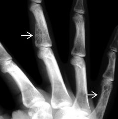
DDX: hook osteophytes (3)
|
Hemochromatosis; CPPD; acromegaly
|
|
|
|
target joints of CPPD (3)
|
MCPs, radiocarpal, patellofemoral
|
|
|
|
target joints of RA (3)
|
MCP, PIP, pan-knee
|
|
|
|
target joints of OA (3)
|
DIP, PIP, Medial > lateral in knee
|
|
|
|
ABCDE of arthritis
|
alignment; bone mineralization; cartilage; distribution; erosions
|
|
|
|
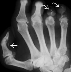
"juxtaarticular erosion with overhanging edge"
|
gout
|
|
|
|
DDX: soft tissue calcification (3)
|
HPT; connective tissue diseases: (scleroderma, SLE, dermatomyositis, polymyositis); trauma
|
|
|
|
DDX: IP-predominant arthitis (3)
|
OA; psoriatic; EOA;
|
|
|
|
sausage digit =
|
psoriatic arthritis
|
|
|
|
subchondral cysts, periarticular erosion and preserved joint space--affects one joint
|
PVNS
|
|
|
|
reducible subluxations, no erosions or joint space loss
|
SLE arthropathy
|
|
|
|
In osteomyelitis, a sequestrum is...
|
the dead bone
|
|
|
|
involucrum is...
|
the surrounding reactive bone in osteomyelitis
|
|
|
|
SAPHO is...
|
synovitis, acne, pustulosis, hyperostosis, osteitis
|
|
|
|
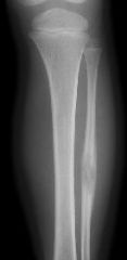
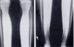
wimberger sign
|
syphlitic osteochondritis along the medial/proximal tibia in congenital syphilis
|
|
|
|
celery stalking...
|
congenital rubella (metaphyseal irregularity)
|
|
|
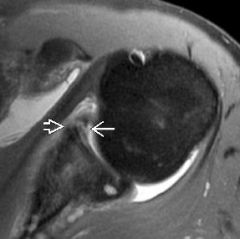
DX?
|
Bankart (Labrum and periosteum are torn)
|
|
|
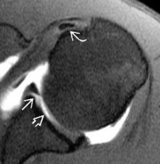
DX?
|
GLAD (Glenolabral Articular Disruption)
|
|
|
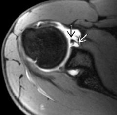
DX?
|
ALPSA (Anterior Labroligamentous Periosteal Sleeve Avulsion)
|
|
|
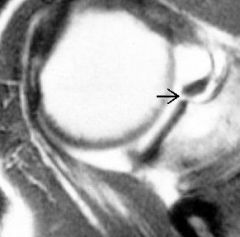
DX? (does not extend posterior to biceps anchor)
|
Sublabral foramen
|
|
|
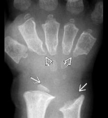
DX?
|
Mucopolysaccharidoses (fan-shaped metacarpals)
|
|
|
|
DDX: generalized osteopenia (4)
|
osteoporosis, osteomalacia, HPT, neoplasm
|
|
|
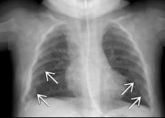
DX?
|
Rickets
|
|
|
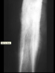
DX?
|
Melorheostosis
|
|
|
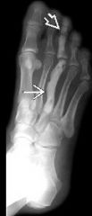
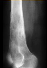
DX?
|
Ewing Sarcoma
|
|
|
|
DDX: metaphyseal lesion (3)
|
NOF, osteochondroma, sarcoma
|
|
|
|
DDX: epiphyseal lesion (4)
|
subchondral cyst, GCT, chondroblastoma [low T2], Brodie's Abscess
|
|
|
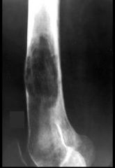
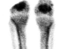
DDX? DX?
|
enostosis (bone island); blastic met
|
bone island cold on bone scan
|
|
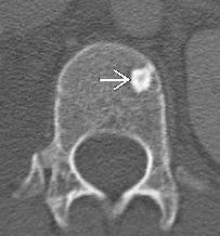
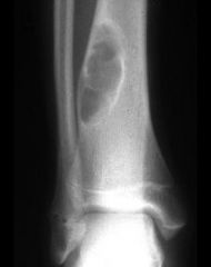
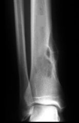
DDX? DX?
|
Osteoid Osteoma (oval lucency); Brodie Abscess
|
|
|
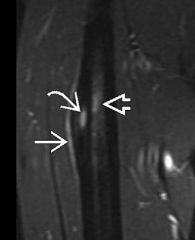
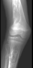
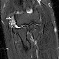
DX? Anatomy?
|
DeQuervain's Tenosynovitis; Abductor Pollicus Longus, Extensor Pollicus Brevis
|
|
|
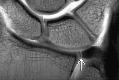
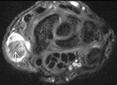
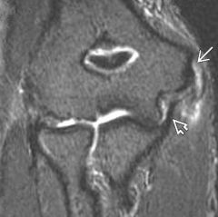
DX?
|
TFCC tear
|
|
|
|
Components of Essex-Lopresti Fracture (3)
|
Radial head fracture, interosseous membrane injury, DRUJ disruption
|
|
|
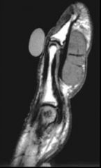
DX? explanation?
|
Gamekeeper's thumb with Stener lesion (balled up ulnar collateral ligament superficial to the adductor aponeurosis)
|
|
|
DX?
|
Extensor Carpi Ulnaris Tendonitis and subluxation
|
|
|
|
Extensor tendons of the wrist--I
|
APL and EPB
|
|
|
|
Extensor tendons of the wrist--II
|
Extensor Carpi Radialis Brevis/Longus
|
|
|
|
Extensor tendons of the wrist--III
|
Extensor Pollicus Longus
|
|
|
|
Extensor tendons of the wrist--IV
|
Extensor Digitorum/Indices
|
|
|
|
Extensor tendons of the wrist--V
|
Extensor Digiti Minimi
|
|
|
|
Extensor tendons of the wrist--VI
|
Extensor Carpi Ulnaris
|
|
|
|
Median nerve is closest to which extensor tendon group: I or VI?
|
I
|
|
|
|
Lister's Tubercle is between which Extensor Tendon Groups?
|
II and III
|
|
|
|
Expansile, circumscribed lytic lesion involving extremities and posterior elements of spine
|
osteoblastoma
|
|
|
DX? DDX?
|
Osteoblastoma (calcified matrix clinches);
ABC |
|
|
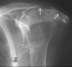
DX?
|
Osteosarcoma
|
|
|
|
Common location for osteosarcoma (part of body, part of bone, extension, location in >50 year old)
|
around knee, metaphysis, extension into epiphysis, axial skeleton/flat bones
|
|
|
DX?
|
Parosteal Osteosarcoma
|
|
|
|
Aggressive Osteosarcoma variant and main features?
|
Telangiectatic Osteosarcoma; minimal osteoid matrix formation, fluid-fluid levels/hemorrhage
|
|
|
|
Osteosarcoma with good prognosis?
|
Parosteal Osteosarcoma
|
|
|
|
risk factors for secondary osteosarcoma (4)
|
Paget’s, XRT, chronic bone infarct, chronic osteomyelitis
|
|
|
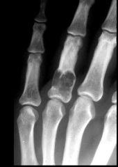
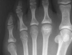
DX?
|
Enchondroma (no chondroid matrix in hand is okay)
|
|
|
DX?
|
Mafucci Syndrome (multiple enchondromas, hemangiomas, high risk of sarcoma)
|
|
|
|
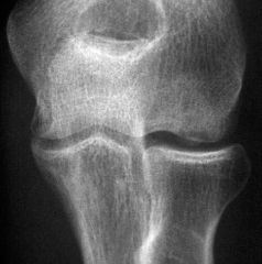
Well-defined lytic lesion centered in epiphyses of a skeletally immature patient
|
chondroblastoma
|
|
|
DX?
|
Chondroblastoma
|
|
|
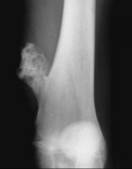
DX?
|
Osteochondroma
|
|
|
|
Complications of osteochondroma (4)
|
neurovascular impingement, fracture, bursitis, malignant degeneration
|
|
|
|
Signs of malignant degeneration of an osteochondroma (3)
|
pain, growth after skeletal maturity, cartilage cap > 1.5cm,
|
|
|
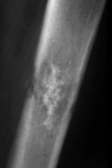
DX?
|
Malignant degeneration of an enchondroma (note cortical destruction)
|
|
|
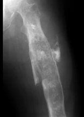
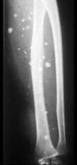
DX?
|
Path Fx through a chondrosarcoma
|
|
|
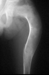
DX?
|
Fibrous Dysplasia (Shepherd's Crook Deformity)
|
|
|
DX?
|
Polyostotic fibrous dysplasia
|
|
|
DX?
|
Fibroxanthoma (NOF)
|
|
|
|
size difference between fibrous cortical defect and non-ossifying fibroma
|
>2 cm = NOF
|
|
|
DX?
|
Fibrosarcoma (note permeative cortical destruction and wide zone of transition)
|
|
|
DX?
|
Malignant Fibrous Histiocytoma (fibrosarcoma could look like this, too)
|
|
|
DX?
|
Soft tissue hemangioma
|
|
|
|
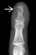
MR appearance of glomus tumor (nailbed)
|
T2/STIR bright, intense enhancement, distal erosion
|
|
|
|
DDX: fluid-fluid level in a bone lesion (2)
|
Telangiectatic osteosarcoma; ABC (secondary to GCT, CB, or OB)
|
|
|
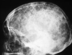
DX?
|
Multiple Myeloma
|
|
|
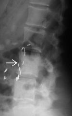
DX? DDX?
|
Plasmacytoma; Multiple myeloma, Metastasis, Benign (osteoporotic) compression fracture, Vertebral hemangioma (VH)
|
|
|
DX?
|
Aneurysmal Bone Cyst
|
|
|
|
Etiology of Primary ABC
|
Trauma
|
|
|
|
Etiology of secondary ABC
|
Tumor (GCT, OB, CB)
|
|
|
DX?
|
Solitary bone cyst with pathologic fracture and fallen fragment
|
|
|
|
Solitary bone cyst etiology and description
|
growth plate defect; central lucent lesion abutting growth plate while active
|
|
|
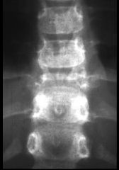
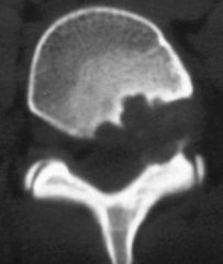
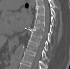
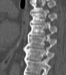
Finding? DDX (5)?
|
Vertebra Plana; (EG (#1 in kids), myeloma, mets, osteoporosis, Ewing sarcoma)
|
|
|
DX? DDX (3 yo F)?
|
EG vs Ewing Sarcoma
|
|
|
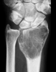
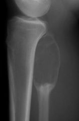
DX?
|
Giant Cell Tumor
|
|
|
|
Lytic epiphyseal lesion extending to subchondral bone without surrounding sclerosis--Occurs typically in skeletally mature patients
|
Giant Cell Tumor
|
|
|
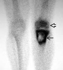
DX? (Name the sign)
|
Giant Cell Tumor (doughnut sign)
|
|
|
|
DDX for something that looks like a Giant Cell Tumor (5)
|
chondroblastoma, chondrosarcoma, mets, MM, post traumatic cyst, geode
|
|
|
|
Ewing Sarcoma: Peak age, most common location (2)
|
10-15yo; diaphysis and flat bones
|
|
|
DX? (low on T2)
|
Ewing Sarcoma (high nucleus/cytoplasm ratio)
|
|
|
|
Large, lobulated, expansile lytic lesion in distal tibial meta-diaphysis
|
adamantinoma
|
|
|
DX?
|
Adamantinoma
|
|
|
|
common lytic mets (3)
|
lung, renal, breast
|
|
|
|
common blastic mets (3)
|
prostate, breast, bladder
|
|
|
DX? Name the sign
|
Benign Peripheral Nerve Sheath Tumor (schwannoma); target sign is typically seen in neurofibroma
|
|
|
|
Findings favoring a malignant myxoma (3)
|
large >5cm, deep, increased heterogeneity
|
|
|
DX?
|
Primary bone lymphoma
|
|
|
DDX? (4)
|
Lymphoma, Paget, blastic metastases, low grade osteomyelitis
|
|
|
|
lesion at spinoglenoid notch results in:
|
pressure on the suprascaular nerve and denervation of the infraspinatus muscle
|
|
|
|
lesion at suprascapular notch results in:
|
pressure on the suprascaular nerve and denervation of the supra- and infraspinatus muscles
|
|
|
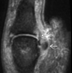
DX?
|
Capitellar Osteochondritis Dissecans
|
|
|
|
Osteochondrosis of the entire capitellum in a 5-11 yo child =
|
Panner's disease
|
|
|
|
mnemonic for elbow ossification
|
CITE (capitellum 1-2, internal/medial epicondyle 4, trochlea 8, external/lateral epicondyle 10)
Radial head 3-6 Olecranon 6-12 |
|
|
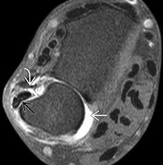
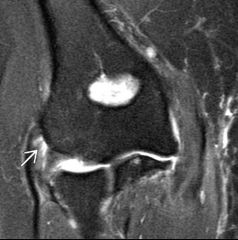
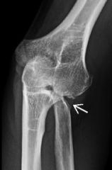
DX?
|
Medial epicondylitis
|
|
|
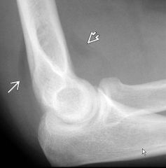
DX?
|
Lateral Epicondylitis
|
|
|
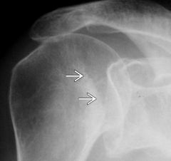
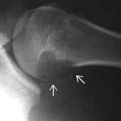
DX? (Name the sign)
|
Posterior glenohumeral dislocation (Trough sign)
|
|
|
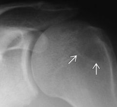
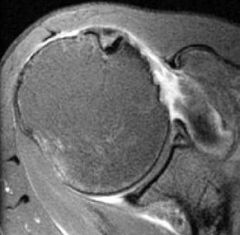
Finding? Significance?
|
Hill-Sachs Fracture; indicates prior anterior dislocation
|
|
|

Name the attachment site: A (Posterolateral intercondylar notch)
|
ACL origin
|
|
|

B: Medial tibial spine
|
ACL Insertion
|
|
|

C: Lateral proximal tibia @ joint line
|
Lateral capsular ligament
|
|
|

D: Intercondylar notch at anterior-mid aspect medial femoral condyle
|
PCL origin
|
|
|

E: Posterior mid tibial plateau, extraarticular
|
PCL insertion
|
|
|

F: Medial femoral condyle, immediately distal to adductor tubercle
|
MCL origin
|
|
|

G: Medial proximal tibia, adjacent to joint line
|
MCL insertion, deep meniscofemoral ligament (coronary ligament)
|
|
|

H: Anteromedial proximal tibia, approximately 5 cm distal to joint line
|
MCL insertion, superficial fibers
|
|
|

I: Lateral femoral condyle
|
LCL origin
|
|
|

J: Fibular styloid
|
Arcuate ligament (and popliteofibular, fabellofibular) insertion
|
|
|

K: Anterolateral tibial plateau, adjacent to joint line
|
ITB insertion (Gerdy tubercle)
|
|
|
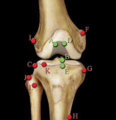
L: Fibular head & anterolateral tibia
|
Conjoint tendon (LCL & biceps femoris) insertion
|
|
|
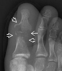
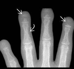
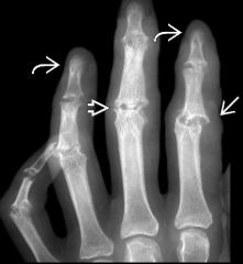
Findings? DX?
|
Sausage finger (Dactylitis) in Psoriatic arthritis
|
|
|
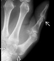
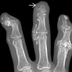
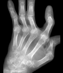
Findings? DDX (5)?
|
Arthritis Mutilans; RA, Psoriatic, Charcot, Juvenile Idiopathic Arthritis, Gout
|
|
|

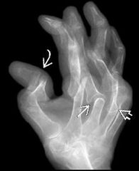
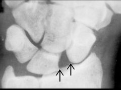
Finding? DDX (5)?
|
Arthritis Mutilans; RA, Psoriatic, JIA, Charcot, Gout
|
|
|
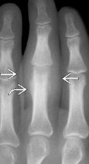
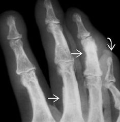
DX?
|
Erosive Osteoarthritis
|
|
|
DX?
|
Septic arthritis
|
|
|
DDX?
|
Epidermal Inclusion Cyst
|
|
|
DX?
|
Tuberculous Arthritis
|
|
|
|
DDX: Atrophic joint destruction (5)
|
RA, Charcot, Septic, TB, Fungal
|
|
|
|
Findings in MSK Sarcoid: (3)
|
STS, lace-like bone, punched-out erosions
|
|
|
DX?
|
Sarcoidosis
|
|
|
DX?
|
Leprosy (calcified digital nerve and acroosteolysis)
|
|
|
DDX (5), DX?
|
Acroosteolysis DDX: HPT, Thermal injury, Progressive systemic sclerosis, Traumatic amputation, Lesch-Nyhan
|
|
|
DX?
|
Progressive Systemic Sclerosis
|
|
|
DX?
|
enchondroma
|
|
|
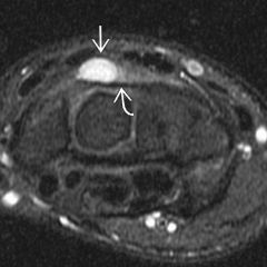
Dark on T1, no enhancement. DX? (Clue?)
|
Ganglion cyst. Look for tail pointing to origin)
|
|
|
DX? (Classic findings?)
|
Multicentric reticulohistiocytosis; red bumps in the skin (lipid-containing macrophages deposited in skin and around joints)
|
|
|
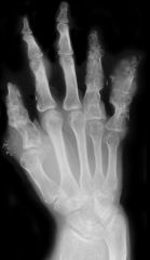
DX?
|
SLE arthiritis; Non-erosive severe deformity
|
|
|
|
Five forms of Juvenile Idiopathic Arthritis by prevalence:
|
Oligoarticular JRA (40%); Seronegative polyarticular JRA (25%); Still's disease (20%); juvenile-onset AS (10%); juvenile onset RA (5%)
|
|
|
|
Still's disease features: age, clinical (3), RF
|
<5yo, fever, anemia, hepatosplenomegaly; negative
|
|
|
|
oligoarticular JRA features: distribution, RF,
|
1-several large joints, RF negative
|
|
|
|
polyarticular JRA: distribution, RF
|
symmetric polyarticular, RF negative
|
|
|
|
coned epiphyses =
|
embolic disease (such as in meningococcemia)
|
|
|
DX? distance for abnormality? accentuated with ulnar or radial deviation?
|
Scapholunate ligament injury (4mm); radial deviation
|
|
|
DX?
|
Thyroid Acropachy
|
|
|
DX?
|
Scapholunate ligament tear
|
|
|
DX?
|
Scapholunate ligament tear
|
|
|
DX?
|
Scapholunate advanced collapse
|
|
|
DX?
|
Calcium Hydroxyapatite deposition disease at gluteus maximus insertion
|
|
|
DX?
|
Rotary subluxation of the scaphoid
|
|
|
DX?
|
Lunate dislocation
|
|
|
Stage, DX?
|
Perilunate dislocation (stage 2)
|
|
|
stage, DX?
|
Midcarpal dissociation (stage 3)
|
|
|
DX? Stage?
|
Lunate dislocation (stage 4)
|
|
|
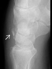
DX?
|
triquetral fracture
|
|
|
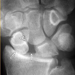
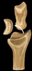
DX?
|
osteonecrosis of the proximal pole of a non-union scaphoid fracture
|
|
|
DDX?
|
heavy metal ingestion, physiologic lines
|
|
|
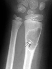
DX, classic features (4)
|
chondroblastoma; teenager, arises in epiphyses, +/- extends into metaphyses, sclerotic rim,
|
|
|
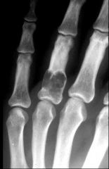
DX?
|
Sail sign
|
|
|
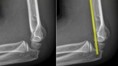
DX?
|
Anterior humeral line intersect anterior 1/3 of capitellum...supracondylar fracture
|
|
|
DX? Name the tendon group which is injured.
|
Lateral epicondylitis, common extensor tendons.
|
|
|
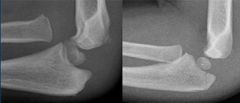
DX? (two separate patients)
|
Radial head subluxation (nursemaid's elbow)
|
|
|
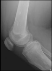
DX?
|
Nail Patella Disease
|
|
|
DX?
|
Hypertrophic Pulmonary Osteoarthropathy
|
|
|
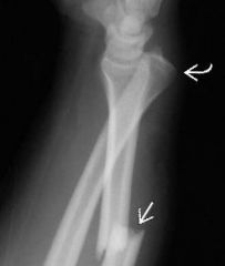
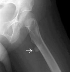
DX?
|
Galeazzi fracture-dislocation
|
|
|
|
Common site of tendon injury in general
|
musculotendinous junction
|
|
|
DX?
|
Triceps Tendon Tear
|
|
|
|
Parsonage-Turner Syndrome AKA...
|
Acute brachial neuritis
|
|
|
|
Increased signal in supraspinatus, infraspinatus, and deltoid muscles =
|
Parsonage-Turner Syndrome
|
|
|
DX?
|
Parsonage-Turner Syndrome
|
|
|
|
Focal painful denervation of the teres minor muscle
|
Quadrilateral space syndrome
|
|
|
|
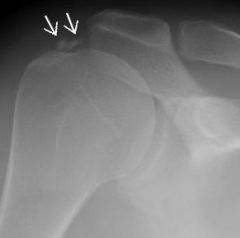
DDX for distal clavicular resorption (6)
|
chronic rotator cuff tear, post-operative, RA, HPT, AC separation (mimic), post-traumatic osteolysis
|
|
|
|
Benign DDX for parosteal osteosarcoma (1)
|
Juxtacortical Chondroma
|
|
|
DX?
|
Juxtacortical Chondroma
|
|
|
DX?
|
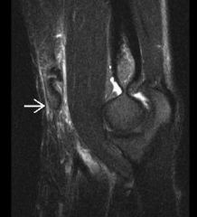
Distal biceps tendon rupture
|
|
|
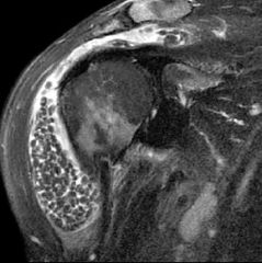
DX?
|
Subscapularis rupture
|
|
|
DX?
|
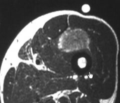
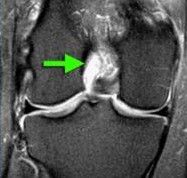
Synovial osteochondromatosis
|
|
|
|
elevated humeral head which articulates more with the acromion/clavicle than glenoid =
|
Rheumatoid Arthritis
|
|
|
DX?
|
RA with rotator cuff tear
|
|
|
|
Isolated glenohumeral degenerative changes DDX (3)
|
pyrophosphate arthropathy, neuropathic arthropathy, posttraumatic arthropathy
|
|
|
DX?
|
Calcific tendinitis
|
|
|
DX?
|
Tumoral Calcinosis
|
|
|
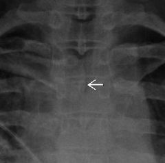
DX?
|
sternoclavicular dislocation
|
|
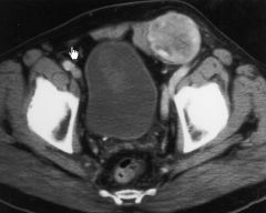
|
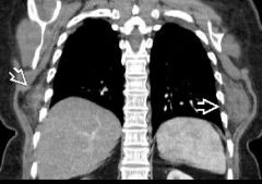
DX?
|
elastofibroma dorsi (benign fibroblastic proliferation)
|
|
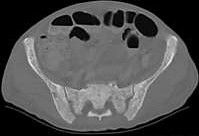
|
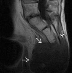
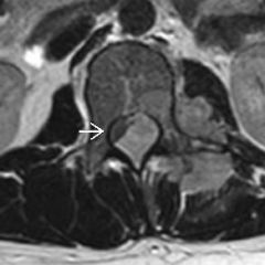
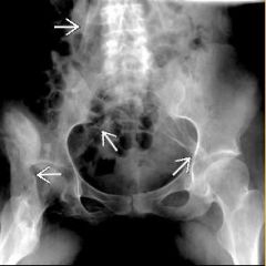
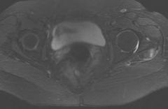
DX? DDX? (5)
|
sacral chordoma; chondrosarcoma, giant cell tumor, plasmacytoma, metastasis
|
|
|
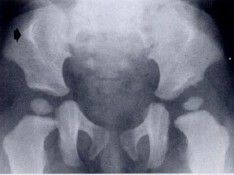
DX? (Finding)
|
Developmental dysplasia of the hip; (coxa magna)
|
|
|
|
DDX: Prostrusio acetabuli (5)
|
trauma, arthritis (RA, OA, AS, JIA), Paget's, renal osteodystrophy, fibrous dysplasia
|
|
|
DX?
|
Fibrodysplasia ossificans progressiva
|
|
|
|
hip dislocations: % anterior/posterior
|
85-90% posterior
|
|
|
|
Mazabraud's Syndrome =
|
fibrous dysplasia + intramuscular myxomas
|
|
|
|
ligamentous calcification in fluorosis (2)
|
sacrospinous and sacrotuberous
|
|
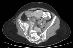
|
DX?
|
gluteus medius tendon tear
|
|
|
|
DDX unilateral sacroiliitis
|
reactive arthritis, psoriatic arthritis
|
|
|
|
bowel disease associated with AS (3)
|
Crohn, UC, whipple's
|
|
|
DX?
|
Extraskeletal osteosarcoma
|
|
|
DX?
|
Nail-Patella Syndrome
|
|
|
|
Nail-Patella Syndrome AKA (2)
|
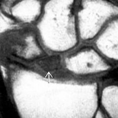
Fong's syndrome, osteoonychodysostosis
|
|
|
DDX?
|
Paget's, lymphoma, leukemia, metastases, MYELOFIBROSIS, mastocytosis
|
|
|
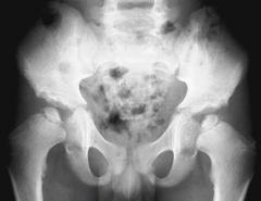
DX?
|
Hemangiomatosis
|
|
|
DX?
|
Multiple hereditary exostosis
|
|
|
|
Multiple Hereditary Exostosis: gender, genetics
|
males > females; autosomal dominant
|
|
|
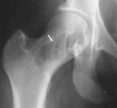
DX? Finding?
|
Sickle Cell Disease, bone within a bone
|
|
|
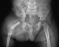
DX?
|
Proximal Focal Femoral Deficiency
|
|
|
|
Transient BM edema vs. occult fracture (2 features)
|
no inciting event, resolves within weeks
|
|
|
DX?
|
Multiple epiphyseal dysplasia
|
|
|
|
Spectrum of osteopetrosis (3 clinical groups), eponym for one; pathophysiology involves what cell type?
|
autosomal recessive/infantile fatal; intermediate (early detection, benign course); Autosomal dominant (adult detection) AKA Albers-Schoenberg; osteoclast dysfxn
|
|
|
|
Definition of Legg-Calve-Perthes disease
|
idiopathic osteonecrosis of the capital femoral epiphysis in a skeletally immature patient
|
|
|
DX?
|
Legg-Calve-Perthes Disease
|
|
|
DX?
|
osteopoikilosis
|
|
|

DX? Name the line.
|
slipped capital femoral epiphysis. Klein's Line
|
|
|
DX? (Classification scheme)
|
Intertrochanteric fracture (Evans-Jensen, based on number of fragments and displacement)
|
|
|
DX? (high T2 signal intensity within the lesion)
|
liposclerosing myxofibrous tumor of bone
|
|
|
Most likely DX?
|
Malignant fibrous histiocytoma
|
|
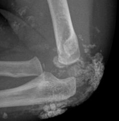
|
DX? DDX? (3)
|
myositis ossificans; sarcoma, surface osteosarcoma, osteochondroma
|
|
|
|
myoblastoma AKA; common locations; course
|
granular cell tumor; breast, chest wall, skin, subcutaneous tissues; benign
|
|
|
|
glomus tumor arises from
|
neuromyoarterial glomus
|
|
|
18 yo male with knee pain. DDX? Next step? DX?
|
Give contrast to prove cyst vs. mass lesion. Solid enhancement in this synovial sarcoma
|
|
|
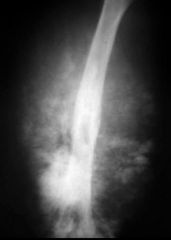
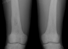
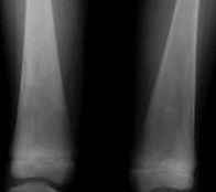
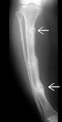
DX?
|
Engelmann's Disease (progressive diaphyseal dysplasia)
|
|
|
Description? DX?
|
Cortical and medullary sclerosis. Erdheim-Chester disease.
|
|
|
|
Erdheim-Chester disease AKA
|
lipid granulomatosis
|
|
|

DX? (AKA)?
|
Trevor disease AKA articular chondroma AKA dysplasia epiphysealis hemimelica
|
|
|
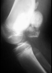
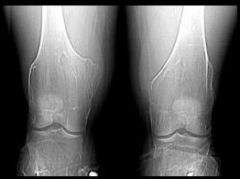
DX?
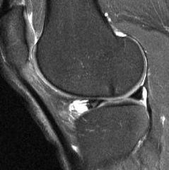
|
Hemophilia
|
|
|
DX?
|
Marrow infiltrative diseases (gaucher's)
|
|
|
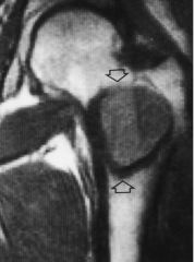
DX?
|
Discoid lateral meniscus
|
|
|
DX?
|
Chondromalacia patella
|
|
|
DX?
|
Pigmented villonodular synovitis
|
|
|
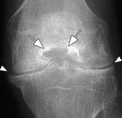
DX?
|
CPPD
|
|
|
DX? Name the sign...
|
Empty notch sign = ACL tear (complete)
|
|
|
DX? sign?
|
empty lateral notch sign, complete acl tear
|
|
|
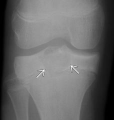
DX? Association?
|
Segond fracture (lateral capsular avulsion), associated with ACL tear.
|
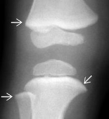
|
|

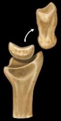
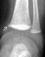
Salter Harris type?
|
I
|
|
|
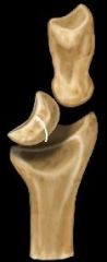
Salter-Harris type?
|
II
|
|
|

Salter-Harris type?
|
III
|
|
|

Salter-Harris type?
|
IV
|
|
|

Salter-Harris type?
|
V
|
|
|
|
Baker's cyst protrudes through...
|
semimembranosus and medial gastrocnemius tendons
|
|
|
Two signs? DX?
|
Double PCL, double delta; bucket-handle meniscal tear with flipped fragment
|
|
|
|
Components of O'Donaghue's terrible triad...mechanism...
|
ACL, MCL, Medial meniscus; valgus stress in flexion
|
|
|
|
repetitive traction injury at in the inferior pole of the patella in an adolescent
|
Sindig-Larsen-Johanssen syndrome
|
|
|
DX?
|
Transient lateral dislocation of the patella with medial patellar retinaculum tear
|
|
|
DX?
|
Lipoma Arborescens
|
|
|
DX?
|
Corner (metaphyseal) fracture, non-accidental trauma
|
|
|
|
SONK is now called...
|
Insufficiency fracture of the knee
|
|
|
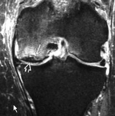
DX?
|
Insufficiency fracture of the knee (AKA SONK)
|
|
|
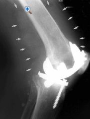
DX?
|
Metallosis
|
|
|
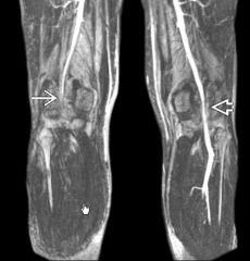
Normal at rest, this appearance during forced flexion. DX?
|
Popliteal entrapment
|
|
|
|
popliteal entrapment caused by?
|
medial head of gastrocnemius
|
|
|
DX? Most common locations (2)?
|
Meniscal cyst. AHLM, PHMM
|
|
|
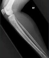
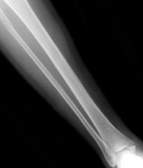
DX?
|
Pseudoarthrosis in NF1
|
|
|
|
Direction of tibial bowing in NF1?
|
anterolateral
|
|
|
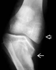
DX? Description?
|
Small, beaked medial tibial metaphysis in Blount's disease
|
|
|
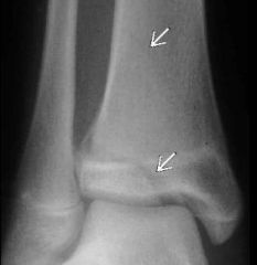
DX?
|
Maisonneuve fracture
|
|
|
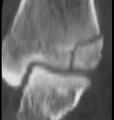
DX?
|
Juvenile Tillaux
|
|
|
DX?
|
Triplane fracture
|
|
|
DX?
|
Achilles tendinopathy
|
|
|
DX?
|
tibialis posterior tendon tear
|
|
|

DX?
|
Homolateral Lisfranc injury
|
|
|
Dx?
|
Divergent Lisfranc injury
|
|
|
Dx?
|
Medial swivel subtalar dislocation
|
|
|
DX?
|
Plantar Fasciitis
|
|
|
|
Normal thickness of plantar fascia?
|
3-4 mm upper limits
|
|
|
DX?
|
Plantar fibromatosis
|
|
|
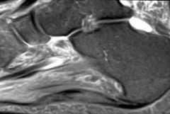
DX?
|
Sinus tarsi syndrome
|
|
|
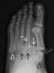
DX? sign?
|
Anteater sign in calcaneonavicular coalition
|
|
|
|
Three types of connection in coalition:
|
Synostosis (bone-bone); synchondrosis (cartilagenous); syndesmosis (fibrous)
|
|
|
|
Most common locations of tarsal coalition (2)
|
talocalcaneal, calcaneonavicular
|
|
|
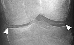
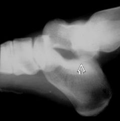
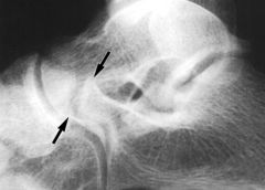
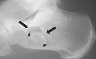
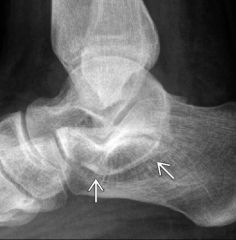
DX? Name the finding.
|
Talar beak, talocalcaneal coalition
|
|
|
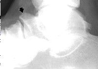
DX?
|
talipes equinovarus
|
|
|
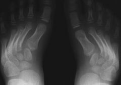
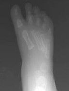
DX?
|
metatarsus adductus
|
|
|
DX?
|
intraosseous lipoma
|
|
|
DX?
|
intraosseous lipoma
|
|
|
DX?
|
JIA
|
|
|
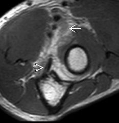
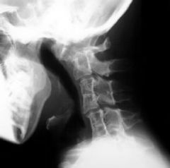
DX?
|
DISH
|
|
|
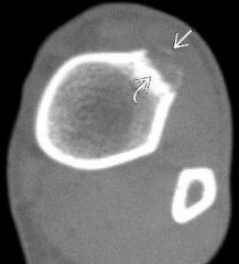
DX?
|
Giant cell tumor of tendon sheath
|
|
|
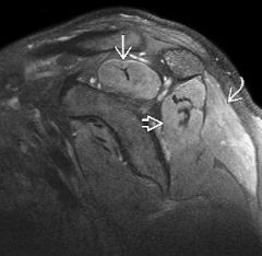
DX?
|
Freiberg Infraction
|
|
|

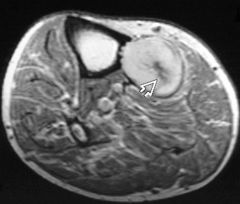
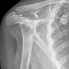
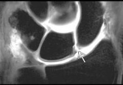
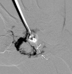
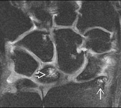
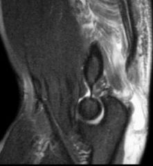
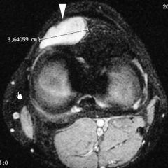
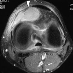
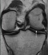
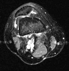
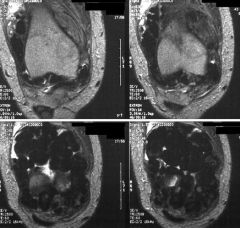
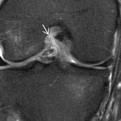
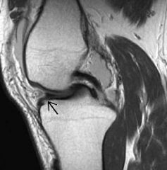
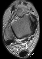
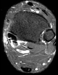
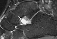
DX? Location of object pointed to by arrow?
|
Synovial osteochondromatosis, distended subcapsularis bursa
|
|
|
|
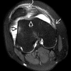
deep sulcus sign, which side, what cut-off for depth, associated meniscal tear?
|
lateral, 1.5mm, lateral
|
|
|
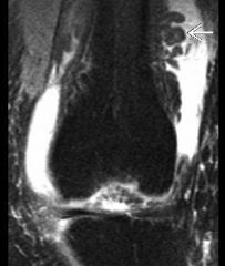
DX?
|
dermatomyositis
|
|
|
DX?
|
Rheumatoid arthritis with rice bodies
|
|
|
|
fracture associated with posterior glenohumeral dislocation
|
lesser tuberosity
|
|
|
|
dense joint effusion
|
hemophilia
|
|
|
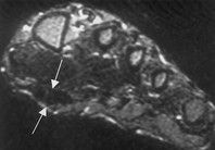
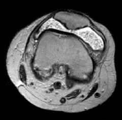
DX?
|
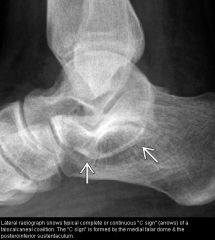
Talocalcaneal coalition
|
|
|
|
name of bone in sprengel deformity
|
omovertebral
|
|
|
|
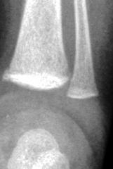
volar plate injury happens where
|
PIP joint in finger
|
|
|
|
dense end of a bone in a teenager
|
think osteosarcoma (osteoblastic)
|
|
|
|
thick ilioischial/iliopectineal lines
|
paget's
|
|
|
|
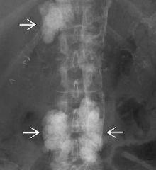
sternal lesion (3)
|
chondrosarcoma (100% calcify), mets, myeloma
|
|