![]()
![]()
![]()
Use LEFT and RIGHT arrow keys to navigate between flashcards;
Use UP and DOWN arrow keys to flip the card;
H to show hint;
A reads text to speech;
36 Cards in this Set
- Front
- Back
|
Describe rheumatoid arthritis in broad terms |
It is a symmetrical arthritis of the appendicular skeleton, sparing the axial skeleton, except for the cervical spine. |
|
|
Name 3 important features of rheumatoid arthritis
|
1. Periarticular joint swelling, Juxta-articular osteoporosis (progressing to generalized osteoporosis), Uniform loss of joint space.
|
|
|
Name 3 other features of rheumatoid arthritis
|
Any of Lack of bone formation, Marginal errosions (eg the ulnar styloid), Synovial cyst formation, Subluxations (volar subluxation of the MCPJ with ulnar deviation), Bilateral symmetrical distribution, Predelection for the hands, feet, knees, hips, cervical spine and elbows (in decreasing order of frequency).
|
|
|
What ossifies in a syndesmophyte?
|
Sharpey's fibers and perhaps the deep layers of the anterior longitudinal ligament.
|
|
|
What is the difference between a marginal osteophyte and syndesmophyte?
|
A syndesmophyte is vertical ossification of Sharpeys fibers connecting two adjacent vertebrae, whereas a marginal osteophyte is a horizontal extension of bone from the vertebral endplate.
|
|
|
Does a osteophyte have a medullary cavity?
|
yes
|
|
|
What are the two earliest signs of osteoarthritis?
|
Periarticular joint swelling and juxtarticular osteoporisis.
|
|
|
What are the specific locations of erosions in the wrist?
|
Waist of the scaphoid, capitate and articulation of the hamate with the 5th metacarpal and the radial and ulnar styloid.
|
|
|
What are the late changes of RA?
|
Generalized osteoporosis, uniform joint narrowing, large subchondral erosions, joint subluxation.
|
|
|
What is the cause of subluxation in RA?
|
inflammation leading to loss of integrity of the joint capsule and surrounding ligaments.
|
|
|
What is endstage RA called?
|
Arthritis mutilans.
|
|
|
Where should you look for early RA changes in the foot?
|
lateral head of the 5th metacarple
|
|
|
What tarsal bone changes occur in RA?
|
Uniform joint space loss and ankylosis. Ankylosis is not seen distal to the tarsals
|
|
|
What fraction of RA patients have hip involvement?
|
50%.
|
|
|
What are the signs of RA in the hip?
|
Uniform loss of cartilage --> resulting axial migration of the hip in the acetabulum, subchondral cyst formation, widespread osteoporosis with noteable absence of reparative bone and osteophyte formation.
|
|
|
Acetabulo protusio is seen in:
|
Rheumatoid arthritis,
|
|
|
How commonly are the knees involved with RA?
|
80% of the time
|
|
|
What is the appearance of RA in the knee?
|
Uniform loss of cartilage in all 3 compartments with noteably little reparative bone formation.
|
|
|
How is an intraosseous cyst formed?
|
Synovium breaks through the thinned cartilage and protrudes into the bone due to the ball-valve effect as the joint moves.
|
|
|
Bakers cyst is associated with which arthropathy?
|
Rheumatoid arthritis.
|
|
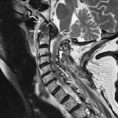
What is going on here |
RA. |
|
|
What is your differential for a lucent lesion in the calcaneum? |
simple bone cyst, interosseus lipoma or normal trabeculation (psudocyst) |
|
|
DDx of chondrocalcinosis |
1. Almost always CPPD, but somtimes 2. HPT / renal OD 3. hemochromatosis |
|
|
What is HADD? |
Hydroxyapatite deposition disease |
|
|
Define PVNS |
Pigmented villonodular synovitis is a proliferation of the synovium in a characteristic villonodular pattern with haemosiderin deposition. |
|
|
The synovial part of the SI joint is anterior/posterior ? |
anterior |
|
|
Isolated glenohumeral joint space loss DDx. |
diseases that result in isolated destruction of the cartilage - ie: 1. crystalline deposition disease (usually CPPD), 2. acromegaly 3. ochronosis |
|
|
What is ochronosis? |
Autosomal recessive metabolic disorder resulting in breakdown of collagen and cartilage. Also called alkaptonuria. |
|
|
Isolated loss of the subachromial space DDx. |
1. Chronic rotator cuff tear 2. Certain positions in subacromial impingement syndrome. |
|
|
How wide should the subacromial space be? |
7 mm (Brower) |
|
|
Resporption of the distal end of the clavicle DDx |
1. trauma 2. hyperparathyroidism 3. scleroderma |
|
|
Involvement of all 3 shoulder compartments DDx |
1. Rheumatoid arthritis 2. Psoriatic arthritis 3. Ankylosing spondylitis 4. Bleeding abnormality |
|
|
Shoulder pain without joint involvement DDx |
1. Hydroxyapatite deposition disease 2. Osteonecrosis |
|
|
What muscle is affected by ischio-femoral impingement? |
quadratus femoris |
|
Give 2 differentials |
Ilio-psoas bursitis or a paralabral cyst. Key is to demonstrate the tear communicating with the paralabral cyst. |
|

|
Radiolucent metaphyseal bands are found in childhood leukaemia. |