Reading...
![]()
Play button
![]()
Play button
![]()
Use LEFT and RIGHT arrow keys to navigate between flashcards;
Use UP and DOWN arrow keys to flip the card;
H to show hint;
A reads text to speech;
182 Cards in this Set
- Front
- Back
|
CHAPTER 4
|
ABDOMEN
|
|
|
THE MOST COMMON PROJECTION IN AN ANTEROPOSTERIOR (AP) SUPINE ABDOMEN IS CALLED
|
KUB
|
|
|
WHAT DOES KUB STAND FOR
|
KIDNEYS, URETERS AND BLADDER
|
|
|
WHAT PART OF THE BODY CAN HAVE A PROJECTION TAKEN WITHOUT THE USE OF CONTRAST MEDIA
|
KUB
|
|
|
PLAIN RADIOGRAPHS OF THE ABDOMEN (KUB) COMMONLY ARE TAKEN BEFORE ABDOMINAL EXAMINATIONS ARE PERFORMED WITH THE USE OF CONTRAST MEDIA TO RULE OUT _____________
|
CERTAIN PATHOLOGIES
|
|
|
In radiology, an examination that usually includes an erect kidney, ureter, and bladder (KUB) projection, a recumbent KUB projection, and a left lateral decubitus view of the chest to assess free air, infections, or obstruct
|
acute abdomen series:
|
|
|
CERTAIN ACUTE OR EMERGENCY CONDITIONS OF THE ABDOMEN MAY DEVELOP FROM THE CONDITIONS SUCH AS
|
BOWEL OBSTRUCTION, PERFORATIONS INVOVING FREE INTRAPERITONEAL AIR, EXCESSIVE FLUID IN THE ABDOMEN OR A POSSIBEL INTRABDOMINAL MASS
|
|
|
WHAT DOES INTRAPERITONEAL AIR MEAN
|
AIR OUTSIDE THE DIGESTIVE TRACT
|
|
|
ACUTE ABDOMINAL SERIES IS ALSO KNOWN AS ____________WHERE DIFFERENT POSITIONS ARE TAKEN TO DEMONSTRATE AIR-FLUID LEVELS AND AIR WITHIN THE ABDOMINAL CAVITY
|
A TWO WAY, THREE WAY ABDOMEN SERIES
|
|
|
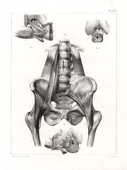
WHAT THE 3 MOST IMPORTANT MUSCLES IN THE ABDOMINOPELVIC CAVITY IS
|
DIAPHRAGM
RT/LT PSOAS MAJOR |
|
|
WHAT IS LOCATED IN THE ABDOMINAL MUSCLES AND IS AN UMBRELLA SHAPED MUSCLE THAT SEPARTES THE ABDOMINAL CAVITY FROM THE THORACIC CAVITY
|
DIAPHRAGM
|
|
|
WHAT MUST BE PERFECTLY MOTIONLESS DURING RADIOGPHY OF EITHER THE ABDOMEN OR THE CHEST. MOTION OF THE PATIENT OR THE BODY CAN BE STOPPED WHEN APPROPRIATE BREATHING INSTRUCTIONS ARE GIVEN
|
DIAPHRAGM
|
|
|
WHAT TWO MUSCLES ARE LOCATED ON EITHER SIDE OF THE LUMBAR VERTEBRAL COLUMN AND THE LATERAL BORDERS OF THESE TWO MUSCLES SHOULD BE FAINTLY VISBLE ON A DIAGNOSTIC ABDOMINAL RADIOGRAPH OF A SMALL TO AVERAGE SIZE PATIENT
|
TWO PSOAS MAJOR
|
|
|
HOW DOES A PSOAS MAJOR MUSCLE LOOK LIKE
|
http://www.bing.com/images/search?q=PSOAS+MAJOR+MUSCLE+RADIOLOGY++FILM&qs=n&form=QBIR&pq=psoas+major+muscle+radiology+film&sc=0-0&sp=-1&sk=#view=detail&id=1542C76C1BEB10BEFD44CDE81341C6924DEAD3F4&selectedIndex=2
|
|
|
WHAT ARE CONSIDERED ACCESSORY ORGANS LOCATED IN THE DIGESTIVE SYSTEM
|
LIVER
GALLBLADDER PANCREAS |
|
|
WHAT ORGAN IS LOCATED POSTERIOR TO THE STOMACH AND IS NOT WELL VISUALIZED
|
PANCREAS- A mixed exocrine and endocrine gland situated transversely across the posterior abdominal wall in the epigastric and hypochondriac regions
|
|
|
WHAT PART OF THE BODY IS ALSO PARTIALLY VISIBLE IN THE LEFT UPPER ABDOMEN POSTERIOR TO THE STOMACH
|
SPLEEN
|
|
|
THE SPLEEN IS PART OF WHAT BODY SYSTEM
|
LYMPHATIC SYSTEM
|
|
|
ORAL CAVITY
PHARYNX ESOPHAGUS STOMACH SMALL INTESTINE LARGE INTESTINE ARE SIX ORGANS OF WHAT SYSTEM |
DIGESTIVE SYSTEM
|
|
|
WHAT ORGANS ARE COMMON TO THE RESPIRATORY SYSTEM AND THE DIGESTIVE SYSTEM
|
ORAL CAVITY (MOUTH)
PHARYNX (OROPHARYNX AND LARYNGOPHARYNX) |
|
|
WHAT IS ANOTHER NAME FOR ORAL CAVITY
|
(MOUTH)
|
|
|
WHAT ARE THE 2 PARTS OF A PHARYNX
|
OROPHARYNX AND LARYNGOPHARYNX)
|
|
|
WHAT ORGAN IS LOCATED IN THE MEDIASTINUM OF THE THORACIC CAVITY
|
ESOPHAGUS
|
|
|
WHAT ARE THE THREE DIGESTIVE ORGANS WITHIN THE ABDOMINAL CAVITY
|
STOMACH
SMALL INTESTINE LARGE INTESTINE |
|
|
WHAT IS THE FIRST ORGAN OF THE DIGESTIVE SYSTEM THAT IS LOCATED WITHIN THE ABDOMINAL CAVITY
|
STOMACH
|
|
|
WHAT IS AN EXPANDABLE RESEVIOR FOR SWALLOWED FOOD AND FLUID AND ITS SIZE AND SHAPE ARE HIGHLY VARIABLE DEPENDING ON THE VOLUME OF ITS CONTENTS AND BODY HABITUS
|
STOMACH
|
|
|
WHAT HAS 3 PARTS AND CONTINUES FROM THE STOMACH AS A LONG TUBE LIKE CONVOLUTED STRUCTURE ABOUT 4.5 TO 5.5 METERS (15 TO 18 FEET) IN LENGTH
|
SMALL INTESTINE
|
|
|
DUODENUM
JEJUNUM ILEUM ARE APART OF WHAT ORGAN |
SMALL INTESTINE
|
|
|
WHAT IS THE FIRST PORTION OF THIS ORGAN THAT IS THE SHORTEST BUT WIDEST IN DIAMETER OF THE 2 SEGMENTS AND IT IS ABOUT 25 CM = 10 INCHES IN LENGTH. WHEN FILLED WITH CONTRAST MEDIUM THIS ORGAN LOOKS LOKE THE LETTER C
|
DUODENUM-letter C
|
|
|
WHAT IS THE PROXIMAL PORTION OF THE DUODENUM WHICH IS WELL SEEN ON THE BARIUM STUDIES OF THE UPPER GI TRACT AND IS CALLED THE
|
DUODENAL BULB
|
|
|
THE DUODENAL BULB IS ALSO KNOWN AS
|
CAP
|
|
|
________ FROM THE LIVER, GALLBLADDER AND PANCREAS DRAIN INTO THE DUODENUM
|
DUCTS
|
|
|
WHAT TWO PARTS OF THE SMALL INTESTINE IS CONSIDERED THE SMALL BOWEL AND LIES IN THE CENTRAL AND LOWER ABDOMEN
|
JEJUNUM
ILEUM |
|
|
THE FIRST 2/5 FOLLOWING THE DUODENUM IS CALLED THE ___________ AND THE DISTAL 3/5 IS CALLED THE ____________.
|
JEJUNUM
ILEUM |
|
|
THE ORIFICE (VALUE) BETWEEN THE DISTAL ILEUM AND THE CECUM PORTION OF THE LARGE INTESTINE IS CALLED
|
ILEOCECAL VALVE
|
|
|
WHAT IS SELDOMLY SEEN FILLING THE ENTIRE STOMACH OR SMALL INTESTINE ON THE PLAIN ABDOMINAL RADIOGRAPH OF A HEALTH, AMBULATORY ADULT
|
AIR
|
|
|
WHAT IS USED TO SHOW THE STOMACH, SMALL INTESTINE , AND proximal LARGE INTESTINE
|
RADIOPAQUE
|
|
|
RADIOPAQUE IS CALLED
|
BARIUM SULFATE
|
|
|
IN THE MIDABDOMEN AND LOWER ABDOMEN WHAT PART OF THE SMALL INTESTINE IS LOCATED
|
DUODENAL BULB
LONG CONVOLUTED LOOPS |
|
|
WHAT IS ATTACHED TO THE POSTEROMEDIAL ASPECT OF THE CECUM
|
APPENDIX (VERMIFORM APPENDIX)
|
|
|
WHAT IS THE VERTICAL PORTION OF THE LARGE BOWEL ABOVE THE CECUM
|
ASCENDING COLON
|
|
|
THE ASCENDING COLON JOINS THE TRANSVERSE COLON AT THE
|
RIGHT COLIC FLEXURE (HEPATIC)
|
|
|
THE TRANSVERSE COLON JOINS THE DESCENDING COLON AT THE
|
LEFT COLIC FLEXURE
|
|
|
WHAT ARE THE SECONARY NAMES FOR THE TWO COLIC FLEXURES
|
HEPATIC AND SPLENIC
|
|
|
RIGHT COLIC FLEXURE IS ALSO KNOWN AS
|
HEPATIC FLEXURE
|
|
|
LEFT COLIC FLEXURE IS KNOWN AS
|
SPLENIC FLEXURE
|
|
|
THE DESCENDING COLON CONTINUES TO THE S-SHAPED__________ COLON
|
SIGMOID COLON
|
|
|
WHERE IS THE SIGMOID COLON (also known as S shape colon) LOCATED IN THE
|
LOWER LEFT ABDOMEN - between the descending colon and the rectum
|
|
|
WHAT IS THE FINAL 15 CM (6INCH) OF THE LARGE INTESTINE
|
RECTUM
|
|
|
THE RECTUM ENDS AT THE
|
ANUS
|
|
|
WHAT MUSCLE AT THE END OF THE TERMINAL OPENS THE LARGE INTESTINE
|
SPHINCTER MUSCLE
|
|
|
THE SHAPE AND LOCATION OF THE LARGE INTESTINE VARY GREATLY WITH THE
|
TRANSVERSE COLON
|
|
|
THE TRANSVERSE COLON IS located high ON ___________AND LOW IN THE ABDOMEN ON SLENDER ____________AND ________TYPES
|
WIDE HYPERSTHEIC TYPES
SLENDER HYPOSTHENIC TYPES ASTHENIC TYPES |
|
|
WHAT ORGAN IS THAT PART OF THE LYMPHATIC SYSTEM along WITH THE HEART AND BLOOD VESSELS IS PART OF THE CIRCULATORY SYSTEM
|
SPLEEN---a part of two systems of the body
|
|
|
THE SPLEEN IS THAT PART OF WHAT SYSTEM
|
LYMPHATIC SYSTEM
|
|
|
WHAT ORGAN IS AN ABDOMINAL ORGAN THAT OCCUPIES A SPACE POSTERIOR AND TO THE LEFT OF THE STOMACH
|
SPLEEN
|
|
|
IN WHAT QUADRANT IS THE SPLEEN LOACTED
|
LEFT UPPER QUADRANT
|
|
|
WHAT ORGAN MAY BE VISUALIZED FAINTLY ON PLAIN ABDOMINAL RADIOGRAPHS, PARTICULARY IF THE ORGAN IS ENLARGED
|
SPLEEN
|
|
|
WHAT IS A FRAGILE ORGAN AND IS SOMETIMES LACERATED DURING TRAMA TO THE LOWER LEFT POSTERIOR RIB CAGE
|
SPLEEN
|
|
|
WHAT ARE THE ACCESSORY ORGANS OF DIGESTION OR THE DIGESTIVE SYSTEM LOCATED IN THE ABDOMINAL CAVITY
|
PANCREAS, LIVER, GALLBLADDER
|
|
|
WHAT ORGAN IS NOT SEEN ON A PLAIN RADIOGRAPH
|
PANCREAS
|
|
|
THE PANCREAS IS AN ORGAN AND AN ELONGATED GLAND THAT IS LOCATED POSTERIOR TO THE __________ AND NEAR THE POSTERIOR ABDOMINAL WALL BETWEEN THE DUODENUM AND THE SPLEEN
|
POSTERIOR TO THE STOMACH
|
|
|
THE _________ OF THE PANCREAS IS NESTLED IN THE C-LOOP OF THE DUODENUM AND THE BODY AND TAIL OF THE PANCREAS EXTEND TOWARD THE UPPER LEFT ABDOMEN
|
THE HEAD OF THE PANCREAS
|
|
|
THE REALATIONSHIP OF THE DUODENUM AND THE HEAD OF THE PANCREAS SOMETIMES IS REFERRED TO AS THE
|
THE ROMANCE OF THE ABDOMEN
|
|
|
THE PANCREAS IS PART OF THE ________ SECREATION SYSTEM AND IS ALSO PART OF THE __________ SECRETION SYSTEM.
|
ENDOCRINE (INTERNAL) AND EXOCRINE (EXTERNAL)
|
|
|
THE __________ PORTION OF THE PANCREAS PRODUCES CERTIAN ESSENTIAL HORMONES, SUCH AS INSULIN WHICH AIDS IN CONTROLLING THE BLOOD SUGAR LEVEL OF THE BODY
|
THE ENDOCRINE
|
|
|
AS PART OF ITS __________ FUCTIONS THE PANCCREAS PRODUCES LARGE AMOUNTS OF DIGESTIVE JUICES THAT MOVE TO THE DUODENUM THROUGH A MAIN PANCREATIC DUCT AS NEEDED FOR DIGESTION
|
EXOCRINE FUCTIONS
|
|
|
WHAT ORGAN IS THE LARGEST SOLID ORGAN IN THE BODY
|
THE LIVER ( ACCESSARY DIGESTIVE ORGAN)
|
|
|
THE LIVER ( ACCESSARY DIGESTIVE ORGAN) IS OCCUPYING MOST OF THE ____________QUADRANT
|
RIGHT UPPER QUADRANT
|
|
|
THE LIVER'S ( ACCESSARY DIGESTIVE ORGAN) FUCTION IS THE PRODUCTION OF _____________ WHICH ASSISTS IN THE DIGESTION OF FATS
|
BILE
|
|
|
IF BILE IS NOT NEEDED FOR DIGESTION IT IS STORED AND CONCENTRATED FOR FURTHER USE IN THE _______________________
|
GALLBLADDER--- ACCESSORY DIGESTIVE ORGAN
|
|
|
WHAT ACCESSORY DIGESTIVE ORGAN IS A PEAR SHAPED SAC LOCATED BELOW THE LIVER WHOS PRIMARY FUCTION IS TO STORE AND CONCENTRATE BILE AND TO CONTRACT THE RELEASE OF BILE WHEN STIMULATED BY THE APPRORIATE HORMONES
|
GALLBLADDER --- ACCESSORY DIGESTIVE ORGAN
|
|
|
WHAT NEEDS TO BE SEEN INSIDE AN ACCESSORY DIGESTIVE ORGAN IN MOST CASES BECAUSE IT CANNOT BE VISUALIZED WITH CONVENTIONAL RADIOGRAPHIC TECHNIQUES __________________
|
CONTRAST MEDIA
|
|
|
WHY IS IT THAT THIS ACCESSORY ORGAN-GALLBLADDER CANNOT BE VISUALIZED WITH NORMAL RADIOLOGICAL TECHNIQUES
|
BECAUSE THE GALLBLADDER AND BILARY DUCTS BLEND IN WITH THE OTHER ABDOMINAL SOFT TISSUES AND IN MOST CASES CANNOT BE VISUALIZED
|
|
|
ONLY ____________% OF ALL GALLSTONES CONTAIN ENOUGH CALCIUM TO ALLOW VISUALIZATION ON A PLAIN ABDOMINAL RADIOGRAPHIC IMAGE
|
10% TO 15%
|
|
|
WHAT TYPE OF IMAGE THROUGH VARIOUS LEVELS OF THE ABDOMEN DEMONSTRATE ANATOMIC RELATIONSHIPS OF THE DIGESTIVE ORGANS AND THEIR ACCESSORY ORGANS AS WELL AS THE SPLEEN
|
CROSS SECTIONAL (AXIAL) COMPUTED TOMOGRAPHY (CT)
|
|
|
USING THE SECTIONAL VIEW OF THE UPPER ABDOMEN AT THE LEVEL OF T/D 10 OR T/D 11 JUST BELOW THE DIAPHRAGM WHAT ACCESSORY ORGAN IS LOCATED AT THE LEVEL IN THE RIGHT UPPER ABDOMEN
|
THE LIVER
|
|
|
USING THE CROSS SECTIONAL VIEW WHAT ORGAN IS VISUALIZED POSTERIOR TO THE STOMACH IN THE LEFT UPPER ABDOMEN
|
SPLEEN ---IS NOT AN ACCESSORY ORGAN
|
|
|
AT THE CROSS SECTIONAL IMAGE OF T/D12 WHAT ACCESSORY ORGAN IS NOW SEEN ADJACENT TO A PORTION OF THE DUODENAL LOOP WHICH IS POSTERIOR TO THE DISTAL PART OF THE STOMACH
|
PANCREAS
|
|
|
AT A LOWER CROSS SECTION IMAGE T/D 12 THE ACCESSORY ORGAN PANCREAS IS NOW SEEN ADJACENT TO A PORTION OF THE DUODENAL LOOP POSTERIOR TO THE DISTAL PART OF WHAT ORGAN
|
STOMACH (IS NOT AN ACCESSORY ORGAN)
|
|
|
WHEN A BARIUM SWALLOW IS GIVEN THE DARK AIR FILLED PORTION OF THE STOMACH IS ON TOP (ANTERIORLY) INDICATING THAT THE PATIENT WAS LYING IN WHAT POSTION DURING THIS RADIOGRAPH
|
SUPINE
DARK=AIR WHITE =BARIUM |
|
|
THE SPLEEN (WHICH IS NOT AN ACCESSORY ORGAN) IS SEEN IN THE LEFT POSTERIOR ABDOMEN JUST LATERAL TO WHAT ORGAN ________________
|
LEFT KIDNEY
|
|
|
WHAT SYSTEM IS COMPOSED OF
2 KIDNEYS 2 URETERS 1 URNIARY BLADDER 1 URETHRA |
URNIARY SYSTEM
|
|
|
WHAT 2 ORGANS DRAINS BY WAY OF ITS OWN URETER TO THE SINGLE URINARY BLADDER
|
KIDNEY
|
|
|
WHAT ORGAN IS SITUATED ABOVE AND BEHIND THE SYMPHYSIS PUBIS AND SERVES TO STORE URINE
|
BLADDER
|
|
|
UNDER ________________ CONTROL IS THE STORED URINE PASSED TO THE EXTERIOR VIA THE URETHRA
|
VOLUNTARY CONTROL
|
|
|
WHAT 2 GLANDS OF THE ENDOCRINE SYSTEM ARE LOCATED AT THE SUPERAMEDIAL PORTION OF EACH KIDNEY
|
2 SUPRARENAL (ADRENAL) GLANDS
|
|
|
WHAT BEAN SHAPED ORGAN IS LOCATED AT THE SUPERAMEDIAL PORTION
|
THE KIDNEY
|
|
|
THE RIGHT KIDNEY IS USUALLY SITUATED A LITTLE LOWER THAN THE LEFT ONE BECAUSE OF THE PRESENSE OF WHAT
|
LARGE LIVER (ACCESSORY DIGESTIVE ORGAN) ON THE RIGHT
|
|
|
WASTE MATERIAL AND EXCESS WATER ARE ELIMINATED FROM THE BLOOD BY ________________ AND ARE TRANSPORTED THROUGH THE URETERS TO THE URNIARY BLADDER
|
KIDNEY
|
|
|
WHAT ORGANS ARE USUALLY SEEN FAIRLY ON A PLAIN ABDOMINAL RADIOGRAPH BECAUSE OF FATTY CAPSULES THAT SURROUNDS IT
|
KIDNEY
|
|
|
WHAT IS THE NAME OF THE CONTRAST MEDIUM EXAMINATION WHICH IS A RADIOGRAPHIC EXAMINATION OF THE URNARY SYSTEM, WHEREIN THE CONTRAST MEDIUM IS INJECTED INTRAVENOUSLY
|
IVU ALSO KNOWN AS EXCRETORY UROGRAM OR
INTRAVENOUS UROGRAM |
|
|
DURING WHAT EXAMINATION IS THE HOLLOW ORGANS OF THIS SYSTEM VISUALIZED WITH THE USE OF THE CONTRAST MEDIUM THAT HAS BEEN FILTERED FROM THE BLOOD FLOW BY THE KIDNEY
|
IVU INTRAVENOUS UROGRAM
OR EXCRETORY UROGRAM |
|
|
WHAT DOES PYELO REFERS TO
|
RENAL PELVIS OF THE KIDNEY
|
|
|
EXCRETORY OR INTRAVENOUS UROGRAM INCLUDES THE STUDY OF
|
ENTIRE URNIARY TRACT WHICH INCLUDES THE ENTIRE COLLECTING SYSTEM
|
|
|
ILLEC CREST IS AT WHAT LEVEL
|
L4 -L5
|
|
|
WHAT ARE THE TWO MAJOR BLOOD VESSELS OF THE ABDOMEN
|
LARGE ABDOMINAL AORTA
AND INFERIOR VENA CAVA |
|
|
WHAT ARE THE 4 IMPORTANT TERMS THAT DESCRIBE THE ANATOMY OF THE ABDOMINAL CAVITY
|
PERITONEUM
MESENTERY OMENTUM MESOCOLON |
|
|
MOST ABDOMINAL STRUCTURES AND ORGANS AND ABDOMINAL WALL OF THE ABDOMINAL CAVITY ARE COVERED BY A LARGE SEROUS DOUBLE WALLED SACLIKE MEMBRANE CALLED _______________________
|
PERITONEUM
|
|
|
THE TOTAL SURFACE AREA OF THE _________________ IS ABOUT EQUAL TO THE TOTAL SURFACE AREA OF THE SKIN THAT COVERS THE ENTIRE BODY
|
PERITONEUM
|
|
|
WHAT TWO TYPES OF PERISTONEUM EXIST
|
THE PARIENTAL AND VISCERAL
|
|
|
WHAT IS THE TWO LAYERED PERITONEUM THAT ADHERES TO THE CAVITY WALL
|
PARIETAL PERITONEUM
|
|
|
WHAT IS THE PORTION THAT COVERS THE AN ORGAN CALLED
|
VISCERAL PERITONEUM
|
|
|
THE SPACE OR CAVITY BETWEEN THE PARIETAL AND VISCERAL PORTIONS OF THE PERITONEUM IS CALLED
|
PERIOTONEAL CAVITY
|
|
|
THIS CAVITY IS NORMALLY FILLED WITH VARIOUS ORGANS. IF ALL THE LOOPS OF BOWEL AND THE OTHER ORGANS OF THE ABDOMINAL CAVITY WERE INCLUDED THE LITTLE ACTUAL SPACE WOULD BE LEFT IN WHAT CAVITY
|
PERITONEAL CAVITY
|
|
|
WHAT CAVITY CONTAINS SOME SEROUS LUBRICATING-TYPE FLUID WHICH ALLOWS ORGANS TO MOVE AGAINST EACH OTHER WITHOUT FRICTION
|
PERITONEAL CAVITY
|
|
|
WHAT IS AN ABNORMAL ACCUMULATION OF THIS SEROUS FLUID THAT CONSTITUES A CONDITION CALLED
|
ASCITES
|
|
|
THE LAYER OF _____________ PERITONEUM ONLY PARTIALLY COVERS CERTAIN ORGANS THAT ARE MORE CLOSELY ATTACHED TO THE POSTERIOR ABDOMINAL WALL
|
VISCERAL PERITONEUM
|
|
|
THE ASCENDING DESENDING COLON, THE AORTA, AND THE INFERIOR VENA CAVA ARE PARTIALLY COVERED THREFORE THIS LINING WOULD NOT BE CONSIDERED MESENTERY AND THESE STRUCTURES AND ORGANS ARE CALLED
|
RETROPERITONEAL
|
|
|
THE ____________ FORMS LARGE FOLDS THAT BIND THE ABDOMINAL ORGANS TO EACH OTHER AND TO THE WALLS OF THE ABDOMEN
|
THE PERITONEUM
|
|
|
what is the name of these double Folds that hold the small intestine in place
|
MESENTERY
|
|
|
MESENTERY is a double fold of _____________________ that extends interiorly from the posterior abdomen wall to completely enveloped a loop of small bowel
|
periosteum
|
|
|
what is the specific term for a double fold of peritoneum that loosely connects the small intestine to the posterior abdominal wall
|
mesentery
|
|
|
a specific type of double fold peritoneum that extends from the stomach to another organ is called
|
omentum
|
|
|
what part of the omentum extends from the lesser curvature of the stomach to portions of the liver
|
lesser omentum
|
|
|
what part of the omentum connects the transverse colon to the greatest curvature of the stomach inferiorly
|
greater omentum
|
|
|
what omentum drapes down over the small bowel then falls back on itself to form an apron along the anterior abdominal wall
|
greater omentum
|
|
|
if one were to surgically cut and enter the abdomen through the mid anterior wall the first structure encountered beneath the parietal peritoneal would be the
|
greater omentum
|
|
|
varying amounts of fat are deposited in the greater omentum which serves as a layer of __________________ between the abdominal cavity and the interior
|
insulation
|
|
|
the greater omentum is called _____________________ because of its location and the amount of fat contained therein
|
fatty apron
|
|
|
the peritoneum that attaches the colon to the posterior abdominal wall is called the
|
Mesocolon
|
|
|
The ascending, transverse, descending and sigmoid or pelvic are four terms of ________________ that exist each named according to that portion of the colon to which it is attached
|
MESOCOLON
|
|
|
what is a the visceral peritoneum that loosely connects the transverse colon to the posterior abdominal wall
|
transverse mesocolon
|
|
|
the major portion of the peritoneal cavity is known as the _____________ and is commonly referred as simply ______________ cavity
|
greater sac/peritoneal cavity
|
|
|
the smaller portion of the upper peritoneal cavity located posterior to the stomach is called the
|
Lesser sac
|
|
|
the lesser sac is known for the special name
|
omentum bursa
|
|
|
the mesentery connets one loop of small intestines to the posterior __________________ wall
|
abdominal wall
|
|
|
what type of colon is shown to be connecting the transverse colon to the posterior abdominal wall
|
transverse mesocolon
|
|
|
kidney, ureters, adrenal glands pancreas, duodenum, ascending and descending colon upper rectum, abdominal aorta and inferior vena cava are structures closely attached to the posterior abdominal wall that are known as
|
retroperitoneal organs
|
|
|
what structures are less mobile and move around less within the abdomen then do intraperitoneal organs
|
retroperitoneal organs
|
|
|
stomach small intestine transverse colon are known as what type of organs
|
retroperitoneal organs
|
|
|
what type of organs are the lower rectum urinary bladder and reproductive organs located under or beneath the periosteum in the true pelvis
|
infra peritoneal organs
|
|
|
what is the difference that exist between male and female peritoneal enclosure
|
the lower aspect of the peritoneal is closed sac in the mail and not in the female
|
|
|
the lower peritoneal sac lies above the urinary bladder totally separating the reproductive organs from those within the peritoneal cavity what gender is this
|
male
|
|
|
what gender has the uterus fallopian tubes (also known as uterine) and ovaries pass directly into the peritoneal cavity
|
female
|
|
|
within the abdominal cavity are partially or completely covered organs with peritoneum but are not retro peritoneal or infra peritoneal may be called
|
Intraperitoneal
|
|
|
Iiver gallbladder spleen stomach juejunum, ileum cecum and transverse and sigmoid colon are known as
|
Intraperitoneal organs
|
|
|
the body has how many quadrants and how many regions
|
four quadrants and nine regions
|
|
|
IF perpendicular planes at right angles were passed through the abdomen at ___________ it would divide the abdomen into four quadrants
|
umbilicus also known as navel
|
|
|
what crest is located at the level of intervertebral disc between l4 and l5
|
iliac crest
|
|
|
what cavity also can be divided into nine regions through the use of two horizontal or transverse planes into vertical planes
|
abdominopelvic cavity
|
|
|
what are the two transverse / horizontal planes that are used as the right and left lateral planes
|
Transpyloric plane/ transtubercular planeth
|
|
|
the two vertical planes are
|
right and left lateral
|
|
|
the ______________ plane is at the level of the lower border of L1 and the ______________ plane is at L5 is at the level of
|
Transpyloric plane/transtubercular
|
|
|
the ___________ and ____________ lateral planes are parallel to the midsagittal plane and are located midway between the anterior superior iliac spine (ASIS)
|
right / left
|
|
|
______________ borders and or organs within the cavity are not visible from the exterior because these soft tissues organs cannot be palpated directly
|
abdominal borders and organs
|
|
|
The xiphoid process is at the level of
|
T/D 9 AND T/D 10
|
|
|
The xiphoid process is the most distal or inferior process of the________________
|
sternum
|
|
|
The xiphoid process is a landmark that approximates the superior anterior portion of the diaphragm which is also known as the
|
Superior margin of the abdomen
|
|
|
_______________ is not a primary landmark for positioning the abdomen because of variation in body type
|
Superior margin of the abdomen
|
|
|
this landmark is used to locate upper abdominal organs such as gallbladder (accessory) organ and or stomach
|
Inferior costal (rib) margin (L2 -L3)
|
|
|
the _______________ is the uppermost portion of the curved border which can be palpated by pressing inward and downward along the mid lateral margin of the abdomen
|
Lliac crest ( L4 - L5)
|
|
|
what part of the abdomen is the uppermost or most superior portion of the crest and is the most commonly used abdominal landmark in corresponds to the level of which is just slightly below the level of the umbilicus on most people
|
Midabdomen (L4 - L5)
|
|
|
_______________ can be found by locating the iliac crest then palpating inferiorly until a prominent projection or bump is felt most prominent in females
|
ASIS (ANTERIOR SUPERIOR ILIAC SPINE)
|
|
|
this landmark is more easily Palpated on skiny patients but very firm, palpation generally is required to feel the movement of the trunk with one hand while rotating the leg internally and externally at the knee area with the other hand and it is about the same level as the upper border of the symphysis pubis
|
greater trochanter
|
|
|
What is the anterior junction joint of the two pelvic bones the most superior anterior portion can be palpated when the patient is in a supine position
|
Symphysis pubis
|
|
|
the symphysis pubis is also known as the
|
inferior margin of the abdomen
|
|
|
_____________ can be used to determine the lower margin on a PA abdomen with a patient in a prone position
|
Ischial tuberosity
|
|
|
abdominal radiographs are exposed on ________ with the diaphragm in a superior position for better visualization
|
expiration
|
|
|
chest radiograph are done with what breathing techniques
|
inspiration times 2
|
|
|
____________ is what pathological indication of abnormal accumulation of fluid in the peritoneal cavity of the abdomen cause by longstanding chronic conditions such as cirrhosis of the liver or metastatic disease to the peritoneal cavity
|
Ascites
|
|
|
Peristaltic action of the bowel is known as what type of movement
|
involuntary movement
|
|
|
CHAPTER 3
|
CHEST
|
|
|
WHAT IS LOCATED ON THE UPPER PORTION OF THE TRUNK BETWEEN THE NECK AND THE ABDOMEN
|
CHEST/THORAX
|
|
|
THE CHEST/THORAX IS DIVIDED INTO 3 SECTIONS
|
BONY THORAX, RESPIRATORY SYSTEM PROPER AND MEDIASTINUM
|
|
|
WHAT PART OF THE SKELETAL SYSTEM PROVIDES A PROTECTIVE FRAMEWORK FOR THE PARTS OF THE CHEST INVOVLED WITH THE BREATHING AND BLOOD CIRCULATION
|
BONY THORAX
|
|
|
what is the term used to describe the part of the chest consisting of the lungs and the remaining thoracic organs contained in the mediastinum
|
thoracic viscera
|
|
|
Anterior the Bony thorax consist of the ____________ which has three divisions
|
sternum
|
|
|
Manubrium, the body and the xiphoid process
|
Thoracic viscera
|
|
|
the bony thorax consist of 2 _______________ which connects to the sternum
|
2 clavicles
|
|
|
the sternum connects to the 2 _______________
|
2 scapulae
|
|
|
how many pair of rib circle the thorax
|
12 pair of ribs
|
|
|
the body has how many pair of thoracic vertebrae
|
12 thoracic vertebrae
|
|
|
for chest positioning the two landmarks are ____________ prominence and _____________ notch
|
Vertebra prominence / jugular notch
|
|
|
what is an important landmark for determining the position of a PA chest projection
|
vertebrae prominence cervical C 7
located at the base of the neck |
|
|
the _______________ is an important location for determining the AP chest projection
|
jugular notch also known as the manubrial notch or suprasternal notch
|
|
|
at what location is the jugular notch the Suprasternal notch or the manubrial notch
|
T/D 7
|
|
|
what is not a reliable landmark when doing chest positioning and is also located on T/D9 or T/10
|
Xiphoid process tip
certain body habitus will show the xiphoid process at T/D 11 or 12 |
|
|
what system is an exchange of gaseous substance is between the air we breathe and the Bloodstream
|
respiratory system
|
|
|
the pharaynx , trachea, Bronchi and lung are all a part ofwhat system
|
respiratory system
|
|
|
what is called the primary muscle of inspiration
|
diaphragm
|
|
|
half of a diaphragm is called
|
Hemi diaphragm
|