![]()
![]()
![]()
Use LEFT and RIGHT arrow keys to navigate between flashcards;
Use UP and DOWN arrow keys to flip the card;
H to show hint;
A reads text to speech;
25 Cards in this Set
- Front
- Back
|
Canal size |
At 1 mm from the apex, the range in small canals was ~ 200 to 400 microns. In large canals the range was ~300 to 500 microns. Green EN. JADA 1958. |
|
|
Accessory canals |
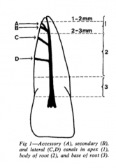
27.4% of the teeth studied (1140) had either lateral, secondary, or accessory canals(17% apical; 8.8% body, 1.6% base of the roots) DeDeus QD. J Endod 1975. |
|
|
Furcation canals |
Accessory canals were demonstrated in the furcation region 28.4% of the total sample; 29.4% in mandibular molars, and 27.4% in maxillary molars. Gutmann JL. J Periodontol 1978 |
|
|
Dens invaginatus |
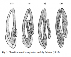
- 0.3 to 10% - Most common in maxillary lateral incisors - 79% Type I, 15% Type II and 5% Type III Hulsmann IEJ 1997, Alani and Bishop IEJ 2008, |
|
|
Isthmuses |
Weller et al JOE 1995: 4mm sections had isthmuses 100% of the time (MB root of max first molar). The concept of partial isthmus was introduced Teixeira et al IEJ 2004: Of the isthmuses present, 22% were complete and 37% partial in mandibular molars and 17.3% were complete and 11.7%partial in maxillary molars (Mand and max molars). |
|
|
Anatomy of the pulp chamber floor |
-Law of symmetry 1, 2, law of orifice location 1-3, law of color change. Krasner P, Rankow HJ. J Endod 2004 |
|
|
Deutsch and Musikant JOE 2004 (molars) |
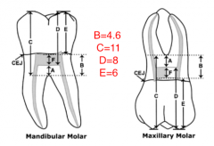
The pulp chamber ceiling was atthe level of the cementoenamel junction in maxillary, 98%, and mandibular, 97% of the specimens. |
|
|
Deutsch et al JOE 2005 (premolars) |
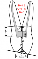
The only measurement that was statistically the same across maxillary molars, mandibular molars and bicuspids was measurement “B,” pulp chamber ceiling to furcation. |
|
|
Lee et al JOE 2007 (anterior teeth) |
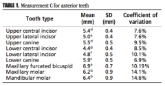
The distance from the lingual surface to the pulp chamber in anterior teeth varied from 4.4 to 5.9mm. |
|
|
C shaped root canals |
Fan et al 2008: The prevalence C-shaped roots in mandibular first premolar was 24%. Cooke and Cox 1979: case report. Melton et al 1991: original classification. Fan et al 2004: new classification. Zheng et al IEJ 2011: 39% prevalence in 2nd molars in Chinese population (Weine 1998: 7.6%) |
|
|
MMC |
Anthony and Vertucci 1974 Fabra-campos IEJ 1989: in vivo, 2.6% Nosrat et al JOE 2015: in vivo, 15% Azim et al JOE 2015: in vivo, 46% (troughing) |
|
|
Radix Entomolaris and paramolaris |
Carlsen and Alexandersen Scand J D R 1990 and 1991. Calberson et al IEJ 2004. |
|
|
Relationship of apex to foramen |
Kuttler 1955: : The average distancefrom the minor diameter to the apical foramen is 0.52 mm inyounger individuals and 0.66 mm in older individuals. The average deviation of apical foramen from apex was 68% in younger group and 80%in older group. Burch and Hulen 1972: 92.4% of the major foramina of all classes of teethdeviated from the anatomic apex. Theaverage distance between the foramen and the anatomic root apex was 0.59 mm |
|
|
Maxillary centrals, laterals and canines |
100% 1 canal Pineda and Kuttler 1972 Vertucci 1984 |
|
|
Maxillary first premolar |
9% 1 canal 85 % 2 canals (type II 13%, type III 72%) 6% 3 canals Carnes and Skidmore 1973 |
|
|
Maxillary second premolar |
40.3% 1 canal 58.6% 2 canals Bellizzi and Hartwell 1985 |
|
|
MB root of maxillary first molar |
96% 2 canals (type I 4.8%, type II 49.4%, type III 45.8%) Kulild and Peters 1990 56.8% 2 canals, 43.1% 1 canal Cleghorn et al 2006 |
|
|
MB root of maxillary second molar |
93.7% 2 canals (type I 4.8%, type II 49.4%, type III 45.8%) Kulild and Peters 1990 56.9% 3-3, 22.7% 3-4, 9% 3 roots 2 canals (MB and DB combine), 6.9% 2-2, 3.1% 1-1, 1.4% 4-4. Peikoff et al 1996 |
|
|
Mandibular central and lateral incisors |
60% 1 canal, 40% 2 canals, 98% 1 foramen Benjamin and Dawson 1974 |
|
|
Mandibular canines |
20% 2 canals Vertucci 1974 |
|
|
Mandibular first premolars |
Zillich and Dawson 1973: 22% 2 canals Cleghorn et al 2007: 2 canals 24% |
|
|
Mandibular second premolars |
Zillich and Dawson 1973: 12% 2 canals Cleghorn et al 2007: 9.1% 2 canals |
|
|
Mandibular first molar |
65% 3 canals 35% 4 canals Hartwell and Bellizzi 1982 M root: Type II -40%, Type III - 60% D root: Type II - 60%, Type III - 40% Skidmore and Bjorndal 1971 |
|
|
Mandibular second molar |
89% 3 canals 6% 4 canals Hartwell and Bellizzi 1982 |
|
|
Canal curvature |
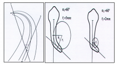
-Schneider 1971 -Pruett, Clement, Carnes 1997 |