![]()
![]()
![]()
Use LEFT and RIGHT arrow keys to navigate between flashcards;
Use UP and DOWN arrow keys to flip the card;
H to show hint;
A reads text to speech;
41 Cards in this Set
- Front
- Back
|
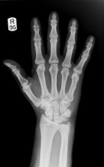
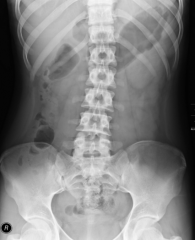
X-rays (Roentgen rays) |
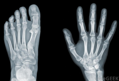
- Part of body positioned between x-ray and sensitized film-- creates a shadow of that body part. - The areas where x-rays strike the film directly appear black, whereas areas where the x-rays are blocked appear in shades of white or grey - More dense=more white |
|
|
Tissue density under X-ray |
- X-rays pass through body substances with varying ease: - Air is the least dense substance and exhibits the greatest transmission - Fat is denser than air - Water (soft tissue) is denser than fat - Metal (bone) is the most dense and transmits the least |
|
|
Radiolucent |
Radiolucent structures permit the passage of most x-rays (appear black on x-ray film) |
|
|
Radiopaque |
Radiopaque structures obstruct the passage of x-rays (appear white on x-ray film) |
|
|
Contrast Medium Technique |
- Radiopaque substances are used in diagnostic radiology to allow more accurate visualization of internal body parts and tissues in contrast to their adjacent structures. - Includes liquids, powders, gas, air, or pills that are administered orally, paternally, or via an enema. |
|
|
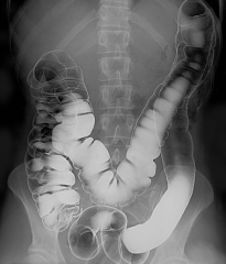
Barium Sulphate |
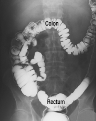
- Harmless, opaque, chalky compound available in a premixed, flavoured liquid or paste. - Upper GI series (barium meal/barium swallow)--oral ingestion of barium mixture to outline the esophagus, stomach, and small intestine - Lower GI series (barium enema)--outlines the colon after barium mixture is given through an enema |
|
|
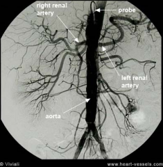
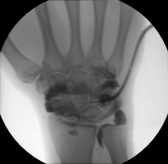
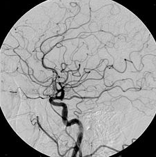
Angiography |
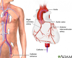
X-ray recording of blood vessels and the heart chambers following the injection of contrast medium through a catheter inserted into the appropriate vessel |
|
|
Arteriography |
X-rays are taken after dye has been injected into the aorta or into an artery in the groin |
|
|
Arthrography |
X-ray recording of a joint after injecting a contrast medium into the joint |
|
|
Bronchography |
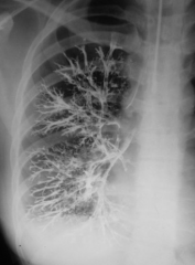
X-ray recording of the bronchial tree and lungs after instillation of a contrast medium into the bronchi via the trachea |
|
|
Cholangiography |
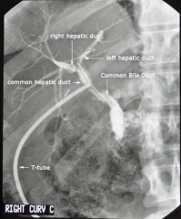
X-ray recording of the bile ducts after dye is injected intravenously or percutaneously, or is given orally and directed by the liver into the bile ducts |
|
|
Cholecystography |

X-ray recording of the gallbladder and bile ducts after oral ingestion of radiopaque granules or tablets, or IV injection of contrast |
|
|
Digital Subtraction Angiography (DSA) |
Imaging of blood vessels that have been injected with a contrast dye; two x-rays are taken (the first x-ray without contrast); a computer subtracts obscuring shadows from the image allowing only the vessels to be seen |
|
|
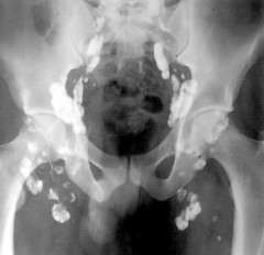
Hysterosalpingography |
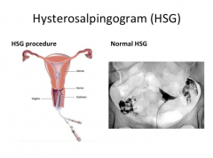
X-ray recording of the uterus and fallopian tube(s) after injecting a contrast medium through the vagina and into the uterus |
|
|
Lymphangiography (Lymphography) |
X-ray recording of the lymphatic vessels and lymph glands after the injection of a contrast medium into a vein |
|
|
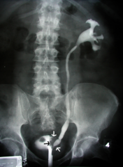
Intravenous Pyelography |
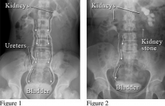
X-ray recording of the renal pelvis and urinary tract after contrast medium is injected into a vein |
|
|
Retrograde Pyelography |
X-ray recording of the renal pelvis and urinary tract after dye is injected through a catheter into the urethra, bladder, and ureters; warranted when the patient is allergic to the dye or if the patient has poor renal function making it impossible to use the IV dye |
|
|
Voiding Cystourethrogram (VCUG) |
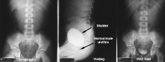
Same procedure used as for retrograde pyelography; bladder is filled with dye and x-rays are taken when the patient is voiding urine through the urethra |
|
|
Venography (Phlebography) |
X-ray recording of veins after contrast medium has been given intravenously |
|
|
Anteroposterior (AP) View |
X-ray beam is directed front to back; patient may be in a supine or standing position, having the back near the film and the front facing x-ray tube |
|
|
Posteroanterior (PA) View |
X-ray beam is directed from back to front; patient is usually in an upright position, having the back facing the x-ray tube and the front near the film |
|
|
Lateral View |
X-ray beam is directed from one side: - Right lateral (RL) view, the right side of the body is near the film and the x-ray tube is pointed toward the left side - Left lateral (LL) view, the left side of the body is nearest the film |
|
|
Oblique View |
X-ray tube is positioned at an angle that is not PA, AP, or lateral (an angle from the perpendicular plane); oblique views are used to show regions that would be hidden in routine views |
|
|
Axillary View |
Bean is directed toward the axilla(armpit) |
|
|
Mediolateral View |
Beam is directed from the midline toward the side of the part being filmed |
|
|
Supine Mediolateral View |
Beam is directed from the midline toward the side with the patient laying on their back |
|
|
Craniocaudal View |
Beam is directed from the superior to inferior levels ("head to toe") |
|
|
Nuclear Medicine Techniques |
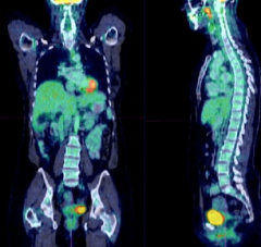
Nuclear medicine is the medical specialty that deals with the diagnosis and treatment of disease processes with the use of radioactive substances. Radionuclides (radioisotopes) are substances that give off high-energy particles or rays as they disintegrate |
|
|
Half-life |
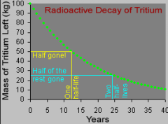
Information regarding the half-life of these particles is very important. Half-life is the time required for a radioactive substance to lose half of its radioactivity by disintegration. The half-life must be long enough to allow for diagnostic imaging, but as short as possible to minimize patient exposure to radiation |
|
|
Alpha particles |
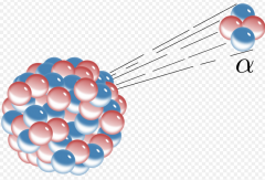
Low penetrating power |
|
|
Beta particles |
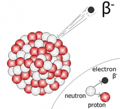
Penetrate a few millimetres of skin |
|
|
Gamma rays |
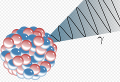
Have a greater penetrating ability than either alpha or beta particles, and thus are very useful in diagnosing and treating diseases |
|
|
In vitro (in the test tube) procedure |
- Involves analysis of blood and urine specimens using radioactive chemicals. - For example, a radioimmunoassay (RIA) is an in vitro procedure that combines the use of radioactive chemicals and antibodies to detect hormones and drugs in a patient's blood. This test can detect minute amounts of drugs in the urine and blood |
|
|
In vivo |
- Means "in the body". - A tracer (also referred to as a tag or label) is a radioactive isotope that is used in diagnostic x-ray techniques to allow a biological process to be seen. Types of tracers are radioactive iodine, and radioactive carbon |
|
|
Tracer Studies |
- The scanning of a tracer as it binds with specific substances and is followed with a scan or fluoroscope as it passes through various organs or systems |
|
|
Radiopharmaceutical (labeled compound) |
- A combination of a radionuclide and a drug or chemical. These can be used to diagnose a condition, and sometimes as a treatment |
|
|
Highly radiosensitive tumors include: |
- Ovarian tumors - Testicular tumors - Lymphomas - Wilm's tumor of the kidney - Retinoblastomas - Hodgkin disease |
|
|
Moderately radiosensitive tumors include: |
- Basal cell carcinoma of the skin - Squamous cell carcinoma of the skin - Adenocarcinoma of the prostate |
|
|
Highly radioresistant tumors include: |
- Sarcomas of the bone - Sarcomas of the connective tissue - Sarcomas of the muscle - Nerve tumors |
|
|
Moderately radioresistant tumors include: |
Moderately radioresistant tumors include: - Tumors of the pituitary gland - Tumors of the adrenal gland |
|
|
External Beam Radiation |
A machine directs a beam of photons from some distance (teletherapy) toward the tumor. The higher the energy of the photons, the greater the penetration of the beam |