![]()
![]()
![]()
Use LEFT and RIGHT arrow keys to navigate between flashcards;
Use UP and DOWN arrow keys to flip the card;
H to show hint;
A reads text to speech;
81 Cards in this Set
- Front
- Back
|
Selective Medium |
•culture medium that allows the growth of certain types of organisms,while inhibiting the growth of other organisms ***Sabouraud Dextrose Agar |
|
|
SDA-selects for fungus |
|
|
|
Differential Medium |
culture medium that allows one to distinguishbetween or among different microorganisms based on a difference in colonyappearance (color, shape, or growth pattern) on the medium. •Blood, Milk, Spirit Blue & Starch Agar •Gelatin, IMViC, Lysine Decarboxylase & SIM Tube •Nitrate, Thioglycollate & Urea Broth |
|
|
NeitherSelective/Differential |
•Mueller-Hinton, Nutrient, Tryptic SoyAgar• •Nutrient, Tryptone, Tryptic Soy Brothes |
|
|
SIM Tube |
differential mediaused to test for 1.Sulfur reduction: ifsulfur is reduced media turns black 2.Indole production: thesurface of the media will turn red in a positive reaction 3.Motility: if microbeis motile it will move out away from the stab line – if not it will stayconsistent with stab line |
|
|
DURHAM Tube (fermentation) |
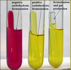
differential media used to detect carbohydrate fermentation and gas production acid production (as aresult of fermentation) is indicated bya yellow color the small invertedtube traps gas produced by the fermentationP “sugartubes” |
|
|
SPIRIT BLUE Agar |
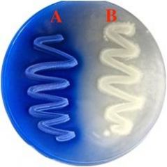
differentialmedia used to show the release of LIPASE
Clearing around thegrowth indicates a positive reaction A=negative reaction B=positive reaction |
|
|
Milk Agar |
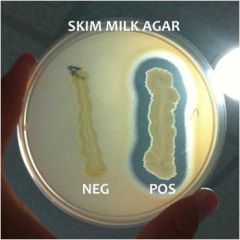
Differential mediathat shows the production of CASEINASE (enzyme the hydrolyzes casein, the proteinfound in milk) Clearing around thegrowth indicates a positive reaction |
|
|
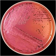
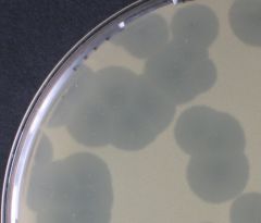
BLOOD Agar |
Gamma hemolysis Alpha hemolysis Beta hemolysis |
|
|
Gamma hemolysis |

No effect on the red cells within t/agar |
|
|
Alpha hemolysis |

Incomplete hemolysis-produces a greenish discoloration around the colony |
|
|
Beta hemolysis |
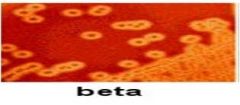
Enzymes lyse the blood cells completely, producing a clear area around the colony |
|
|
BLOOD Agar |
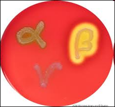
Alphahemolysis shows t/partialbreakdown of RBCs. Beta hemolysis shows t/completebreakdown of RBCs (indicative of a pathogen). Gamma hemolysis does not modify RBCsdue to t/absence of hemolysin. (Note to self- hemolysin is not an enzyme, more like a toxin w/c pops t/cellopen.) D |
|
|
SIMMONS CITRATE Agar |
Differential mediathat tests a microbes ability to use citrate as their sole source of carbon. CITRATE PERMEASE is the enzymeproduced in a positive reaction. A blue color producednear the surface indicates a positivereaction. |
|
|
LYSINE DECARBOXYLASE Tube |
|
|
|
LYSINE DECARBOXYLASE Tube |
•If an organism is able to ferment theglucose, acidic byproducts are formed, and the medium turns yellow. •Ifthe organism has decaboxylase, which removes the carboxyl group from an amino acid (LYSINE), thebyproducts are sufficient to raise the pH of the media so that the broth turnsback to purple. ••Onecan only be confident as to the presence or absence of decarboxylaseif on is certain the organism is able to ferment glucose. ••Anoverlay of mineral oil is added to the tube to create anaerobic conditions& promote fermentation. |
|
|
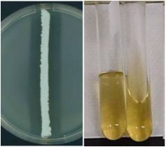
Thioglycollate |
Differential mediathat shows oxygen requirements based on growthpatterns. Obligate aerobes – grow only at thetop of the tube. Facultative or aerotolerant anaerobes – grow throughout the tube. Obligate anaerobes – grow only at thebottom of the tube Microaerophiles –grow in the middle of the tube. Resazurin – pink dye added sothat the O2 attach to the upper layer of the broth |
|
|
THIOGLYCOLLATE Tube |
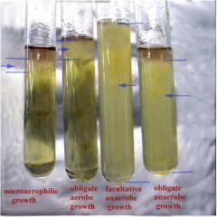
AddResazurin to bind O2 |
|
|
MSA |
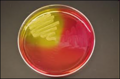
Mannitol Salt Agar Differential andselective media for gram + bacteria, high saltcontent inhibits other organisms Tests for mannitolfermenters Positive reaction (mannitol is beingfermented) – media turns yellow Negative reaction(mannitol not fermented) – no color change Selectsfor Staphylococcussp. & Bacillussp. |
|
|
Lactose Agars |
DESOXYCHOLATE EMB (eosin methyline blue) MacCONKEY |
|
|
DESOXYCHOLATE |
Deoxycholate agar: differential andselective media for entrics |
|
|
EMB |
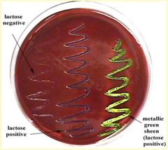
EosinMethylene Blue Differential and selective media for entrics (bacteria found in your gut) Inhibits growth of gram + bacteria Tests bacteria’sability to ferment lactose |
|
|
MacCONKEY |
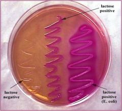
Differential and selective media for entrics Tests lactose fermenters = pink color |
|
|
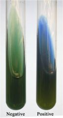
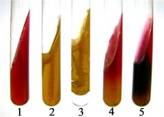
NITRATE Broth |
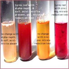
Media that tests forNITRATE REDUCTASES that anaerobically reduce nitrate Positivereaction –after adding reagents A and B a color change to red indicates reduction ofnitrates or nitrites *If there is no colorchange after reagents are added, zinc is added to the tube and will reduce anyremaining nitrate(color should change to red) Negative Reaction *If there is still nocolor change the nitrates were reduced beyond nitrites to ammonia or nitrogengas Positive Reaction *Ifcolorless after add zinc then complete positive |
|
|
UREA Broth |
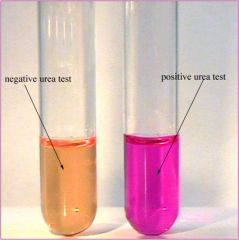
used to detect theenzyme UREASE that breaks down urea Positive reaction –broth turns pink/red (urea was broken down/urease is present) Negative reaction –no color change (urea not broken down/no urease present) |
|
|
STARCH Agar |
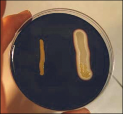
Differential mediawhich shows the production of AMYLASE, the enzyme that hydrolyzes starch *Iodine is poured on the plate after incubation and will bindto any intact starch producing a deep reddish brown color Positive reaction –indicated by a halo around the growth (amylase was produced – starch washydrolyzed) |
|
|
GELATIN Tube/GELATIN Agar |
Differentiates basedon an organisms ability to hydrolyze gelatin by enzyme GELATINASE Positive reaction –if the agar is in a tube the medium will turn to liquid if gelatin ishydrolyzed Negative reaction –if agar is in a plate, it is flooded with ammonium sulfate after incubation.The negative result is shows by a white precipitate around the streak indicating unhydrolyzed gelatin. |
|
|
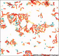
Gram Positive Cocci (can also be bacillus) |
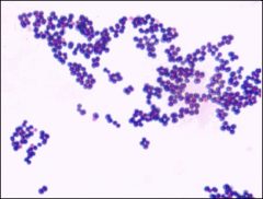
S. aureus |
|
|
Gram Negative Bacillus (can only be bacillus) |
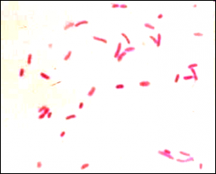
E. coli |
|
|
Vibrio |
cholera |
|
|
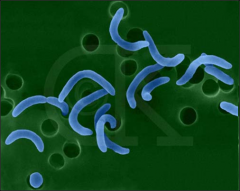
Spirochetes |
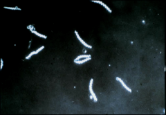
Syphilis |
|
|
Endospore (Stain) |
Greenendospores & red vegetativecells |
|
|
Capsule (Stain) |
Darkbackground, light capsule surrounding the cell |
|
|
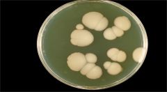
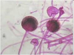
Aspergillus |

Dandelion |
|
|
Penicillium |
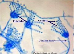
SkeletonHand645} |
|
|
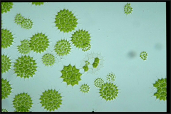
Rhizopus |
Tootsiepop |
|
|
Pediastrum |
Colonialgreen algae - spikey |
|
|
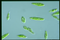
Spirogyra |
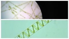
Filamentous/ spiral green algae |
|
|
Trypanosoma |
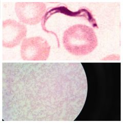
NOTE: Undulating membrane/blood-causes AfricanSleeping Sickness/Chagas Disease |
|
|
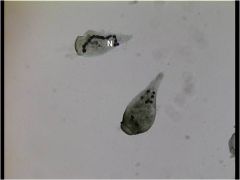
Balantidium |
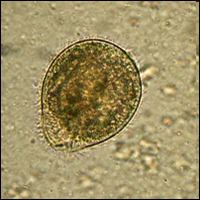
NOTE: Ciliated troph/contaminatedwater/stool sample |
|
|
MicrobialCulture |
apopulation of microbes growing in or on a medium |
|
|
Medium |
mediacontaining nutrients for the growth of microbes |
|
|
MicrobialColony |
agroup of microbes growing on a solid medium, all of the microbes having dividedfrom a single parent cell |
|
|
MixedCulture |
aculture containing two or more species of microbes |
|
|
PureCulture |
aculture containing only one species of microbes |
|
|
StockCulture |
aculture that you know the identity of the microbes growing in it |
|
|
DifferentialMedia |
allowsyou to differentiate microbes based on their metabolic activity |
|
|
SelectiveMedia |
allows the growth ofcertain microbes while inhibiting the growth of others |
|
|
KligerIron |
Two sugars – glucose & lactoseProteinSource of sulfur Yellow in the tube –indicates fermentation of glucose positive (butt) and lactose positive (slant)Cracks in agar –indicated production of “butt gas production” Slant is deep red –breakdown of (positive for) peptone (a protein) Black in the tube –sulfur reduction |
|
|
Ocularlens |
theeye piece, magnifies image 10x |
|
|
Objectivelens |
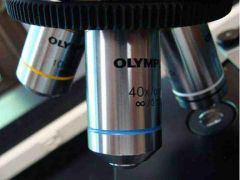
revolvinglenses that can magnify at differentlevels depending on which lens is in place |
|
|
Stage |

holdsthe specimen and moves up and down to bring it into focus |
|
|
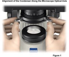
Condenser |
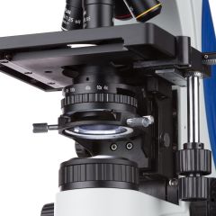
focusesthe light on the specimen yom |
|
|
IrisDiaphragm |
controlsthe intensity/brightness of the light |
|
|
Finefocus |

Finefocus |
|
|
TotalMagnification |
themagnification of the ocular lens multiplied by the objective lens |
|
|
Working distance |
theamount of space between the slide and the lens |
|
|
Par focal |
when aspecimen is in focus under one magnification, and the magnification is changed,the specimen will remain in focus |
|
|
Par center |
when aspecimen is in the center of the lens, and the magnification is changed it willstay centered |
|
|
Turbidity |
cloudy oropaque |
|
|
Flocculent |
having aloosely clumped texture |
|
|
Pellicle |
a thin membrane or film |
|
|
gram staining |
Use inoculating loop to make small pool of water on clean/dry slide. With loop mix culture into corresponding pool ofwater. Let the slide air dry, then heat fix the bacteria byquickly passing the slide through the flame three times Let the slide cool. Flood the slide with Gram Crystal Violet and let sitfor 30 sec before rinsing with distilled water Flood the slide with Gram Iodine for 1 min and rinse. Rinse slide with Gram Decolorizer (breaks down plasma membrane) for 10-20 sec and thenrinse with distilled water Flood the slide with Gram Safranin for 30 sec, rinsewith distilled water, and then allow to air dry |
|
|
Primary stain |
the stain that first stains the structure you want to see – Gram Crystal Violet |
|
|
Mordant |
makes a complex between the Crystal Violet dye and the iodine which is insoluble in water – Gram Iodine |
|
|
Secondary stain |
if the cell wall was degraded by the decolorizer (as it does in gram – bacteria), the cell will take up the color of this stain – Gram Safranin |
|
|
Endospore staining |
Differential staining technique. Red – safranin. Green – malachite green |
|
|
Dilution Series |
the purpose of a dilution series is to reduce the concentration of a culture in measured intervals, so that when a colony count is preformed the number of colonies can be accurately counted. **The number ofcolonies in the original solution is then estimated by multiplying the numberof colonies by the plating dilution factor and the tube dilution factor. #cells/ml=#colonies xplating DF x tube DF Tube dilution: 1mL =1/10thdilution Plate dilution: 0.1mL= 1/10thdilution |
|
|
Zone of inhibition |
displayed by areas of clearing of different sizes around a disc that is full of the chemical being tested. These discs are put on plates with bacteria on them. The amount of clearing around the disc indicates how well it prevented the bacteria from growing. (mueller hinton plates) The bigger the clearing, stronger the chemical/antibiotic. Resistant – bacteria grows back in the clearing A chemical usually thought to be a good antimicrobial agent may show no zone of inhibition because it may not be able to kill the specific bacteria it comes in contact with. |
|
|
Transformation Pglo |
Transformation is a process in which cells take up foreign DNA from their environment. Under properconditions, a cell that is incubated with plasmid DNA can absorb the plasmid into its cytoplasm |
|
|
Acrochaetium |
|
|
|
Euglena |
|
|
|
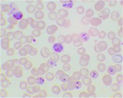
Plasmodium |
|
|
|
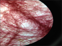
Blood Agar |
|
|
|
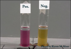
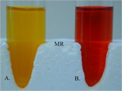
Methyl Red |
|
|
|
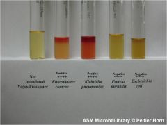
Voges Proskauer |
|
|
|
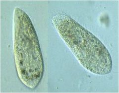
Paramecium |
|
|
|
Stentor |
|
|
|
Howto calculate total magnification |
calculated by multiplying the ocular lens magnification times the magnification of the objective lens being used (either: 4X, 10X, 20X, 40X, 100X) |
|
|
Plaque |
a clear area on an otherwise opaque field of bacteria that indicates the inhibition or dissolution of the bacterial cells by some agent, either a virus or an antibiotic |
|
|
Dilutionseries |
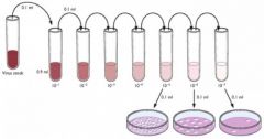
1ml transfered from 1st tube to 2nd tube 1ml taken from 2nd tube and added to 3rd tube, and so on..... |