![]()
![]()
![]()
Use LEFT and RIGHT arrow keys to navigate between flashcards;
Use UP and DOWN arrow keys to flip the card;
H to show hint;
A reads text to speech;
171 Cards in this Set
- Front
- Back
|
abduction |
movement away from the midsagittal (median) plane of the body or one of its parts
abduct = to take away |
|
|
adduction
|
movement toward the midsagittal (median) plane of the body
add = bring together |
|
|
medial |
pertaining to the midline of the body or structure
|
|
|
lateral
|
pertaining to a side |
|
|
superior (cephalad)
|
toward the head or upper portion of a structure
|
|
|
inferior (cuadal)
|
away from the head, or toward the tail or lower part of a structure
|
|
|
proximal
|
nearer to the center (trunk of the body) or to the point of attachment to the body
|
|
|
distal
|
further from the center (trunk of the body) or from the point of attachment to the body
|
|
|
anterior
|
front of the body
|
|
|
ventral
|
front of the body
|
|
|
dorsal
|
back of the body
|
|
|
posterior
|
back of the body
|
|
|
parietal
|
pertaining to the outer wall of the body cavity
|
|
|
visceral
|
pertaining to the viscera, or internal organs, especially the abdominal organs |
|
|
prone
|
lying on the abdomen face down |
|
|
supine
|
lying horizontally on the back, face up
|
|
|
inversion
|
turning inward or inside out |
|
|
eversion
|
turning outward
|
|
|
palmar
|
pertaining to the palm of the hand
|
|
|
plantar
|
pertaining to the sole of the foot
|
|
|
superficial
|
toward the surface of the body (external)
|
|
|
deep |
away from the surface of the body (internal)
|
|
|
symptom (Sx) |
a subjective indicator of disease
|
|
|
etiology
|
the study of the cause or origin of a disease or disorder
|
|
|
diagnosis (Dx)
|
the cause and nature of a disease
|
|
|
prognosis
|
the prediction of the course of a disease and its probable outcome
|
|
|
idiopathic
|
any disease whose cause is unkown |
|
|
sequelae
|
complications that arise directly from disease, injury, or tx (i.e. paralysis may be the sequela of a head injury) |
|
|
adhesion
|
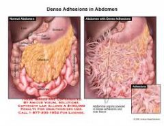
abnormally fibrous band that holds or binds together tissues that are normally separated
|
|
|
ascites
|
abnormal accumulation of fluid in the abdominal cavity |
|
|
edema
|
abnormal accumulation of fluid within tissue spaces (swelling)
|
|
|
febrile
|
pertaining to a fever
|
|
|
gangrene
|
death and decay of soft tissue, usually caused by circulatory obstruction, trauma, or infection
|
|
|
hernia
|
protrusion of any organ through the structure that normally contains it
|
|
|
inflammation
|
body defense against injury, infection, or allergy marked by redness, swelling, heat, pain, and sometimes, loss of function
|
|
|
mycosis
|
any fungal infection in or on the body
|
|
|
perforation
|
hole that completely penetrates a structure
|
|
|
peritonitis
|
inflammation of the peritoneum, the serous membrane that surrounds the abdominal cavity and covers its organs |
|
|
rupture |
sudden breaking or bursting of a structure or organ
|
|
|
sepsis
|
pathological state, usually febrile, resulting from the presence of microorganisms or their products in the blood stream |
|
|
suppuration
|
producing or associated with the generation of pus
|
|
|
infusion therapy
|
delivery of fluids directly into the blood stream via a vein for treating various disorders; also called IV therapy
|
|
|
ablation
|
removal of a part, pathway, or function by surgery, chemical destruction, electrocautery, freezing, or radio frequency (RF)
|
|
|
anastomosis
|
surgical joining of two ducts, vessels, or bowel segments to allow flow from one to another
|
|
|
cauterize
|
destruction of tissue by electricity, freezing, heat, or corrosive chemicals |
|
|
curettage |
scraping of a body cavity with a spoon–shaped instrument called a curette
|
|
|
incision and drainage (I&D)
|
incision made to allow the free flow or withdrawal of fluids from a wound or cavity |
|
|
laser surgery
|
use of a high intensity laser light beam to remove diseased tissues, stop bleeding blood vessels, or for cosmetic purposes
|
|
|
resection
|
removal of part or all of a structure, organ, or tissue
|
|
|
revision
|
surgical procedure used to replace or compensate for a previously implanted device or correct an undesirable result or effect of a previous surgery |
|
|
assessment techniques
|
sequence of procedures designed to evaluate the health status of a patient |
|
|
inspection
|
general observation of the patient as a whole, progressing to specific body areas
|
|
|
palpation
|
Gentle application of the hands to a specific structure or body area to determine size, consistency, texture, symmetry, and tenderness of underlying structures. |
|
|
percussion
|
tapping a structure with the hand or fingers to assess consistency and the presence or absence of fluids within the underlying structure |
|
|
auscultation
|
listening to the heart, bowel, and lungs with or without a stethoscope to assess the presence and quality of sound |
|
|
endoscopy
|
visual examination of a body cavity or canal using a specialized lighted instrument called and endoscope |
|
|
blood chemistry analysis
|
laboratory test, usually performed on serum, to evaluate various substances to determine whether they fall within a normal range
|
|
|
complete blood count (CBC)
|
panel of blood tests used as a broad screening for anemias, infections, and other diseases
|
|
|
organ–disease panels
|
series of blood tests used to evaluate a specific organ (liver panel) or disease (anemia panel)
|
|
|
computed tomography (CT)
|
imaging technique in which an x–ray emitter rotates around the area to be scanned and a computer measures the intensity of transmitted x–rays from different angles; formerly called computerized axial tomogrpahy |
|
|
fluroscopy |
technique in which x–rays are directed through the body to a fluorescent screen that displays internal structures on continuous motion
|
|
|
magnetic resonance imaging (MRI)
|
Technique that uses radio waves and a strong magnetic field rather than and x–ray beam, to produce highly detailed, multiplanar, cross–sectional views of soft tissues
|
|
|
nuclear scan
|
technique in which a radioactive material (radiopharmaceutical) called a tracer is introduced into the body (inhaled, ingested, or injected) and a specialized camera (gamma camera) is used to produce images of organs and structures |
|
|
position emission tomography (PET)
|
computed tomography records the positrons (positively charged particles) emitted from a radiopharmaceutical to produce a cross–sectional image of metabolic activity of body tissues to determine the presence of disease |
|
|
radiography
|
technique in which x–rays are passed through the body or area and captured on film to generate an image (also called x–ray)
|
|
|
single photon emission computed tomography (SPECT)
|
Radiological technique that integrates CT and a radioactive material (tracer) injected into the bloodstream to visualize the blood flow to tissues and organs |
|
|
ultrasonography (US)
|
high–frequency sound waves (ultrasound) are directed at soft tissue and reflected as "echoes" to produce an image on the monitor of an internal body structure; also called ultrasound, sonography, and echo
|
|
|
biopsy
|
removal of a representative tissue sample from a body site for microscopic examination, usually to establish a diagnosis
|
|
|
excisional biopsy
|
biopsy in which the entire lesion is removed
|
|
|
incisional biopsy
|
biopsy in which only a small sample of the lesion is removed |
|
|
integumentary system |
the skin makes up this system |
|
|
androgen |
generic term for an agent (usually a hormone, such as testosterone and androsterone) that stimulates development of male characteristics |
|
|
ductule |
very small duct |
|
|
homeostasis |
state in which the regulatory mechanisms of the body maintain an internal environment within tolerable levels, despite changes in the external environment |
|
|
synthesize |
forming a complex substance by the union of similar compounds or elements |
|
|
epidermis |
the relatively thin outer layer of skin which is thickest on the hands and soles of feet |
|
|
strata |
layers |
|
|
stratum corneum |
The outermost layer of the epidermis which is composed of dead, flat cells that lack a blood supply and sensory receptors. |
|
|
basal layer (of epidermis) |
Deepest layer of epidermis. It is the only layer of the epidermis which contains living cells where new cells are formed. These cells contain melanocytes which produce melanin. These cells die and become filled with keratin as they move outward to the stratum corneum. |
|
|
keratin |
a hard protein material which is relatively waterproof. It is in the epidermis where it prevents body fluids from evaporating and moisture from entering the body. |
|
|
melanocytes |
specialized cells which produce melanin in the basal layer of the epidermis. |
|
|
melanin |
a black pigment in epidermis which provides protection from UV radiation. Differences in skin color are attributed to the amount of this pigment in each cell. |
|
|
albino |
an individual who cannot produce melanin. Deficient of pigment in eyes, hair, and skin |
|
|
dermis |
the second layer of skin just below the epidermis. It is composed of living tissue and contains numerous capillaries, lymphatic vessels, and nerve endings. Also contains hair follicles, sebaceous (oil) glands, and sudoiferous (sweat) glands. |
|
|
cornium |
AKA dermis |
|
|
sebaceous gland |
oil gland |
|
|
sudoriferous gland |
sweat gland |
|
|
subcutaneous layer |
the most deep layer of the skin below the dermis. It is composed primarily of loose connective tissue and fat tissue interlaced with blood vessels. |
|
|
hypodermis |
AKA subcutaneous layer |
|
|
exocrine glands |
glands which secrete substances through ducts to an outer surface of the body rather than directly into blood stream |
|
|
sebum |
an oily secretion |
|
|
hair shaft |
the visible part of the hair |
|
|
hair root |
the part of the hair that is embedded in the dermis |
|
|
hair follicle |
the hair root, together with its coverings |
|
|
papilla |
the bottom of the hair follicle. It is a loop of capillaries enclosed in a covering. |
|
|
nail root |
where the nail is formed. It is composed of keratinized, stratified, squamous epithelial cells producing a very tough covering |
|
|
nail bed |
the layer of epithelium below the nail which the nail is attached to |
|
|
lunula |
the region where new growth of a nail occurs. it is the white half-moon-shaped portion of the nail. Its appearance is because the vascular tissue underneath does not show through. |
|
|
dermatology |
medical specialty concerned with diseases that directly affect the skin and systemic diseases that manifest their effects on the skin. |
|
|
dermatologist |
the physician who specializes in diagnosis and treatment of skin diseases |
|
|
lesions |
areas of tissue that have been pathologically altered by injury, wound or infection they may over an area of definite size of throughout the body |
|
|
neoplasms |
abnormal growths of new tissue that are classified as benign or malignant. |
|
|
Benign neoplasms |
noncancerous growths composed of the same type of cells as the tissue in which they are growing. They harm the individual only insofar as they place pressure on surrounding structures. |
|
|
malignant neoplasms |
AKA cancer. cells that tend to become invasive and spread to remote regions of the body. |
|
|
immunotherapy |
AKA biotherapy. a newer treatment for cancer that stimulates the body's own immune defenses to fight tumor cells. |
|
|
Pathologists |
physicians who grade and stage tumors to help in diagnosis and treatment planning, provide a possible prognosis, and aid comparison of treatment results when different treatment methods are used. |
|
|
Tumor grading |
cells from the tumor site are evaluated to determine the degree of loss of cellular differentiation and function. |
|
|
Tumor Node Metastasis (TNM) |
system used for staging tumors that allows comparison of statistics among cancer centers internationally. |
|
|
Basil cell carcinoma |
the most common type of skin cancer which is malignancy of the basal layer of the epidermis. It is commonly caused by overexposure to sunlight. |
|
|
papule |
solid, elevated lesion less than 1 cm in diameter that may be the same color as the skin or pigmented. examples: wart, pimple, ringworm, psoriasis, eczema |
|
|
nodule |
palpable, circumscribed lesion; larger and deeper than a papule. Extends into the dermal area. examples: intradermal nevus, tumor |
|
|
tumor |
solid, elevated lesion larger than 2 cm in diameter that extends into the dermal and subcutaneous layers examples: lipoma, steatoma, dermatofibroma, hemangioma. |
|
|
wheal |
elevated, firm rounded lesion with localized skin edema (swelling) that varies in size, shape, and color. paler in the center than its surrounding edges; accompanied by itching examples: hives, insect bites, urticaria |
|
|
macule |
flat, pigmented, circumscribed area less than 1 cm in diameter
examples: freckle, flat mole, or rash. |
|
|
vesicle |
elevated, circumscribed, fluid-filled lesion less than 0.5cm in diameter.
Examples: chickenpox, poison ivy, shingles |
|
|
pustule |
small, raised, circumscribed lesion that contains pus; usually less than 1 cm in diameter. Examples: acne, furuncle, pustular psoriasis, scabies |
|
|
bulla |
a vesicle or blister larger than 1cm in diameter
examples: second-degree burns, poison ivy, severe poison oak |
|
|
excoriation |
linear scratch marks or traumatized abrasions of the epidermis.
Examples: scratches, abrasions, chemical or thermal burns |
|
|
fissure |
small slit or crack-like sore that extends into the dermal layer. could be caused by continuous inflammation and drying |
|
|
ulcer |
an open sore or lesion that extends to the dermis and usually heals with scarring
Examples: pressure sore, basal cell carcinoma |
|
|
squamous cell carcinoma |
an invasive tumor with potential for metastasis and occurs most commonly in fair-skinned white men over age 60. It arises from skin that undergoes pathological hardening of epidermal cells. |
|
|
malignant melanoma |
malignant growth of melanocytes. The most lethal of the skin cancers. |
|
|
abscess |
localized collection of pus at the site of an infection (characteristically a staphylococcal infection) |
|
|
acne |
inflammatory disease of the sebaceous glands and hair follicles of the skin with characteristic lesions that include blackheads (comedos), inflammatory papules, pustules, nodules, and cysts. |
|
|
alopecia |
partial or complete loss of hair resulting from normal aging, an endocrine disorder, a drug reaction, anticancer medication, or a skin disease.
(baldness) |
|
|
Bowen disease |
form of intraepidermal carcinoma (squamous cell) characterized by red-brown scaly or crusted lesions that resemble a parch of psoriasis or dermatitis |
|
|
cellulitis |
diffuse (widespread), acute infection of the skin and subcutaneous tissue. |
|
|
chloasma |
pigmentary skin discoloration usually occurring in yellowish brown patches or spots |
|
|
comedo |
typical small skin lesion of acne vulgaris caused by accumulation of keratin, bacteria, and dried sebum plugging an excretory duct of the skin
(black head or whitehead) |
|
|
dermatomycosis |
infection of the skin caused by fungi |
|
|
ecchymosis |
skin discoloration consisting of a large, irregularly formed hemorrhagic area with colors changing from blue-black to greenish brown or yellow; commonly called a bruise. |
|
|
eczema |
chronic inflammatory skin condition that is characterized by erythema, papules, vesicles, pustules, scales, crusts, and scabs and accompanied by intense itching |
|
|
erythema |
redness of the skin caused by swelling of the capillaries |
|
|
eschar |
dead matter that is sloughed off from the surface of the skin, especially after a burn |
|
|
impetigo |
bacterial skin infection characterized by isolated pustules that become crusted and rupture |
|
|
keratosis |
thickened area of the epidermis or any horny growth on the skin (such as a callus or wart) |
|
|
lentigo |
small brown macules, especially on the face and arms, brought on by sun exposure, usually in middle aged or older person |
|
|
pallor |
unnatural paleness or absence of color in the skin |
|
|
pediculosis |
infestation with lice, transmitted by personal contact or common use of brushes, combs, or headgear |
|
|
petechia |
minute, pinpoint hemorrhage under the skin |
|
|
pressure ulcer |
inflammation, sore or skin deterioration caused by prolonged pressure from lying in one position that prevents blood flow to the tissues, usually in elderly and bedridden persons |
|
|
pruritus |
intense itching |
|
|
psoriasis |
chronic skin disease characterized by circumscribed red patches covered by thick, dry silvery, adherent scales and caused by excessive development of the basal layer of the epidermis |
|
|
purpura |
any of several bleeding disorders characterized by hemorrhage into the tissues, particularly beneath the skin or mucous membranes, producing ecchymoses or petechiae |
|
|
scabies |
contagious skin disease transmitted by the itch mite, commonly through sexual contact |
|
|
tinea |
fungal skin infection whose name commonly indicates the body part affected
(AKA ringworm) |
|
|
urticaria |
allergic reaction of the skin characterized by the eruption of pale red elevated patches called wheals or hives. |
|
|
verruca |
epidermal growth caused by a virus; AKA warts |
|
|
vitiligo |
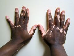
localized loss of skin pigmentation characterized by milk-white patches
|
|
|
chemical peel |
chemical removal of the outer layer of skin to treat acne scarring and general keratoses |
|
|
cryosurgery |
use of subfreezing temperature (commonly liquid nitrogen) to destroy or eliminate abnormal tissue, such as tumors, warts and unwanted, cancerous, or infected tissue |
|
|
debridement |
removal of necrotized tissue from a wound by surgical excision, enzymes, or chemical agents |
|
|
dermabrasion |
rubbing (abrasion) using wire brushes or sandpaper to mechanically scrape away (abrade) the epidermis |
|
|
fulguration |
tissue destruction by means of high-frequency electric current; also called electrodesiccation |
|
|
photodynamic therapy (PDT) |
procedure in which cells selectively treated with an agent called photo-sensitizer are exposed to light to produce a reaction that destroys the cells |
|
|
biopsy |
Representative tissue sample removed from a body site for microscopic examination |
|
|
frozen section (FS) biopsy |
Ultrathin slice of tissue from a frozen specimen for immediate pathological examination |
|
|
needle biopsy |
removal of a small tissue sample for examination using a hollow needle, usually attached to a syringe |
|
|
punch biopsy |
removal of a small core of tissue using a hollow punch |
|
|
shave biopsy |
removal of an elevated lesion using a surgical blade. |
|
|
Mohs |
Layers of cancer-containing skin are progressively removed and examined until only cancer-free tissue remains |
|
|
skin graft |
transplantation of healthy tissue to an injured site |
|
|
allograft |
transplantation of healthy tissue from one person to another person; also called homograft |
|
|
autograft |
transplantation of healthy tissue from one sire to another site in the same individual. |
|
|
synthetic skin graft |
transplantation of artificial skin produced from collagen fibers arranged in a lattice pattern |
|
|
xenograft |
transplantation (dermis only) from a foreign donor (usually a pig) and transferred to a human; also called heterograft. |
|
|
allergy skin test |
any test in which a suspected allergen or sensitizer is applied to or injected into the skin to determine a patient's sensitivity to it |
|
|
intradermal allergy skin test |
skin test that identifies suspected allergens by subcutaneously injecting small amounts of extracts of the suspected reaction |
|
|
patch allergy skin test |
skin test that identifies allergic contact dermatitis by applying a suspected allergen to a patch which is then taped on the skin, usually the forearm, and observing the area 24 hours later for an allergic response. |
|
|
scratch allergy skin test |
puncture or prick test |
|
|
culture and sensitivity |
laboratory test that grows a colony of bacteria removed from an infected area in order to identify the specific infecting bacterium and then determine its sensitivity to antibiotic drugs |