![]()
![]()
![]()
Use LEFT and RIGHT arrow keys to navigate between flashcards;
Use UP and DOWN arrow keys to flip the card;
H to show hint;
A reads text to speech;
31 Cards in this Set
- Front
- Back
|
What do the sensory receptor cells in the ear act as? |
Transducers which convert external sound signals into electrical impulses. |
|

|
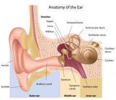
1. Semicircular ducts 2. Vestibular nerve 3. Cochlear nerve 4. Cochlea 5. Auditory tube 6. Inner Ear 7. Middle ear 8. Tympanic cavity 9. Eardrum 10. Auditory canal 11. Outer ear 12. Earlobe 13. Auricle 14. Stapes 15. Incus 16. Malleus 17. Temporal bone. |
|
|
What are the 3 main areas of the ear? |
1. Outer ear 2. Middle ear 3. Inner ear |
|
|
Describe the process of mechanotransduction. |
1. Sound waves indue vibrations in the tympanic membrane 2. Mechanical vibration in middle ear transmitted to cochlear in inner ear 3. Vibrations in cochlear fluids transmitted to sensory hair cells 4. Sterocilia deflect, transduction channels open, leading to K+ influx 5. Receptor potential triggers glutamate release, leading to action potential in cochlea nerve |
|
|
What is the consistency of the air in a sound wave? |
Zones of molecular compression - zones of compression and zones of low compression - zones of rarefraction |
|
|
What range of frequencies can the human ear hear? |
20 - 20,000 Hz |
|
|
What is amplitude in terms of compression and rarefraction zones? |
Difference between area of compression and area of rarefraction |
|
|
What are the main functions of the middle ear? |
1. Transmits sound vibrations from the tympanic membrane to the oval window and the middle ear. 2. Amplifies sound waves by 15-20x 3. Converts sound waves into mechanical vibrations |
|
|
What are the two main components of the inner ear? What is each filled with? |
Two fluid filled labyrinths 1. The Osseus labyrinth - contains perilymph 2. Membranous labyrinth - contains endolymph |
|

|
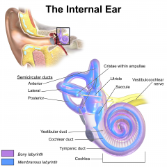
1. Cristae within ampullae 2. Utricle 3. Saccule 4. Vestibulocochlear nerve 5. Cochlea 6. Tympanic duct 7. Cochlear duct 8. Vestibular duct 9. Posterior semicircular duct 10. Lateral semicircular duct 11. Anterior semicircular duct 12. Semicircular ducts |
|
|
What component(s) of the inner ear make up the vestibular system? |
1. Semicircular ducts 2. Utricle 3. Saccule |
|
|
What component(s) of the inner ear make up the auditory system? |
The cochlear duct |
|
|
What is the cochlea? |
The cochlea is the organ of hearing. It is anatomically a tube within a tube. |
|
|
How are sound signals amplified from the middle to inner ear? |
The oval window is far smaller than the tympanic membrane. This amplifies the sound waves. |
|
|
Describe the sound transmission to the cochlea. |
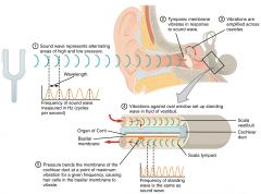
1. Sound waves coming through the external auditory canal move the tympanic membrane. 2. Middle ear bones move 3. Vibrates the membrane in the oval window 4. Causes oscillation of specific regions of the basilar membrane 5. Pressure bends the membrane of the cochlear duct at a point of maximum vibration for a given frequency, causing hair cells in the basilar membrane to vibrate. |
|

|
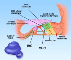
1. Outer Hair Cell stereocilia 2. Outer Hair Cell lateral membrane 3. Tectorial membrane 4. Auditory nerve 5. Inner hair cell 6. Outer hair cells 7. Deiter's cell / supporting cells 8. Basilar membrane |
|
|
What is the fluid which surrounds the organ of corti called? What is its ionic makeup? |
1. Endolymph (within the cochlear duct) 2. Low Na+ and high K+ |
|
|
How many rows of outer hair cells are there? Inner hair cells? |
3 rows of outer, 1 row of inner |
|
|
What is the function of supporting / deiters' cells? |
Support hair cells and connect them to the basilar membrane. This ensures efficient transmission of mechanical vibrations to the hair cells. |
|
|
How does the cellular composition of supporting cells support its function? |
Contain large transcellular cytoskeletal arrays. Made up of: i) microtubules ii) actin filaments iii) intermediate filaments |
|
|
What is the area which hair cells sit on called? How does its structure support its function? |
The cuticular plate. Strong actin network which serves as an anchor for stereocilia. |
|
|
Describe the basic morphological hallmarks of a stereocilia. |
1. Modified giant microvilli 2. Bundle of actin filaments which are interconnected by tip links. |
|
|
Describe briefly, a tip link. |
Two helically intertwined protofilaments which link the top of a stereocilium to an adjacent stereocilium. Tip links are involved in opening and closing of ion channels. |
|
|
Describe the process of stereocilia deflection. |
1. Mechanical vibrations in the basilar membrane. 2. Transmitted to the hair cells via the supporting cells 3. Shearing forces between stereocilia 4. Stereocilia deflection |
|
|
How do stereocilia become excited? |
1. Stereocilia deflect 2. Tip links become stretched - opening K+ ion channels 3. Depolarisation of stereocilia 4. Generation of receptor potential. 5. Release of neurotransmitter |
|
|
How do stereocilia become inhibited? |
1. When tip links become compressed K+ and Ca++ channels close. 2. Hyperpolarisation - K+ exit via basolateral channels 3. Inhibition of neurotransmitter release |
|
|
In which direction does stereocilia deflection occur towards? |
Always occurs towards the tallest stereocilia. |
|
|
Why does K+ enter the stereocilia when the channels open? |
K+ influx is driven by electrical potential differences. The endolymph has a potential of +85 mV and the hair cells -40mV |
|
|
What channel in hair cells pumps K+ out of the cell? When is it activated? |
The KCNQ4 channel which is activated on hair cell depolarisation. |
|
|
What nerve carried nerve impulses for hearing and balance to the brain? |
Vestibulocochlear cranial nerve |
|
|
What neurotransmitter is associated with hair cell depolarisation? |
Glutamate |